46
서 론경제적으로가장많은피해를끼치고있는어류및갑각류바 이러스성질병에대한치료대책은현재까지없으며
,
질병확산 에가장큰영향을미치는환경에대한정확한이해와분석을 통하여,
그위험성을최소화하는방법만이최선의예방법으로 생각되고있다.
국내의경우매년고수온기에
megalocyvirus
에 속하는돌 돔이리도바이러스(RBIV)
로인한 돌돔의대량폐사로 경제 적손실이매우크며(Jung and Oh, 2000),
저수온기에는viral haemorrhagic septicemia virus (VHSV)
로인한넙치의감염 및대량폐사가발생하고있는실정이다(Kim et al., 2009).
매 년주기적으로발생하고있는megalocytivirus, VHSV
와같은 바이러스성질병의경우발병이되기시작하면주변양식장으Article history;
Received 1 November 2012; Revised 28 December 2012; Accepted 4 February 2013
*Corresponding author: Tel: +82. 51. 629. 5941. Fax: +82. 51. 629. 5938 E-mail address: [email protected]
Kor J Fish Aquat Sci 46(1) 046-052, February 2013 http://dx.doi.org/10.5657/KFAS.2013.0046 pISSN:0374-8111, eISSN:2287-8815
ⓒ The Korean Society of Fishereis and Aquatic Science. All rights reserved
After an outbreak of viral disease in an aquafarm, release of virus (es) from infected fish into environmental seawa- ter has been suspected. In the present study, we utilized a negatively charged membrane (HA type) as an efficient method for concentration and detection of fish pathogenic viruses, specifically, megalocytivirus and viral hemor- rhagic septicemia virus (VHSV) present in field-collected seawater samples or inoculated into seawater artificially.
Positively charged viruses adsorbed onto the negatively charged membrane and were eluted with 1 mM NaOH (pH 10.5) following rinsing with 0.5 mM H
2SO
4(pH 3.0). Megalocytivirus and VHSV particles isolated using an- egatively charged HA membrane from seawater inoculated with each virus at a concentration of 10 viral particles/
mL were of sufficient quantity to show positive results in atwo-step PCR (or RT two-step PCR); however, despite it being negatively charged, a cellulose acetate (CA) membraneshowed negative results.In quantitative PCR, the detection limits of the HA membrane for megalocytivirus and VHSV in seawater were 1.20E+00 viral particles/
mL and 1.22E+01 viralparticles/mL, respectively. The calculated mean recovery yields from 1 L seawater spiked with known concentrations of megalocytivirus and VHSV particles were 28.11% and 23.00%, respectively. The concentrate of a 1-L sample of culturing seawater from the aquatank of flounder suffering from VHSV showed clear positive results in PCR when isolated with an HA, but not a CA, membrane. Thus, viral isolation using an HA membrane is a practical and reliable method for detection of fish pathogenic viruses in seawater.
Key words : Megalocytivirus, VHSV, Seawater, HA type membrane
Negatively Charged Membrane을 이용한 해수 중 어류질병바이러스의 검출
국립수산과학원 수산생물방역과, 1농림수산식품부 농림수산검역검사본부, 2부경대학교 수산생명의학과
Bo Young Jee, Kwang Il Kim1, Soon Jeong Lee, Ki Hong Kim2, Ji Woong Jin2 and Hyun Do Jeong2
*
지보영ㆍ김광일
1ㆍ이순정ㆍ김기홍
2ㆍ진지웅
2ㆍ정현도
2*Detection of Fish Pathogenic Viruses in Seawater Using Negatively Charged Membranes
Aquatic life disease control division, National Fisheries Research & Development Institute, Busan 619-902, Korea
1
Animal, Plant and Fisheries Quarantine and Inspection Agency, Ministry for Food, Agriculture, Forestry and Fisherises, Anyang 430-757, Korea
2
Department of Aquatic Life Medicine, Pukyong National University, Busan 608-737, Korea
로의전파가매우빠르며이에대한적절한관리방안이필요하 다
.
바이러스성질병에감염된어류나갑각류의대량폐사시환 경수중으로많은양의바이러스를배출하게되며배출된바이 러스는환경수내에서확산되어다른감수성종에대한재감 염가능성이높다
.
즉바이러스성어류질병은사육환경에바 이러스가존재하다가감수성이 있는어종에노출시발병되 는경우가일반적이다(Jones and Groman, 2001; EI-Matbouli and Soliman, 2011).
따라서질병발생지역의오염된환경사 육수그리고감염어류및갑각류로부터외부로유출된바이러 스의존재여부를확인하는 것은어류및갑각류의바이러스 성질병예방을위해매우중요하다고볼수있다.
그러나환경 수중소량으로존재하는바이러스의검출을위해서는최적화 된바이러스의효율적농축방법이필요하며, ethanol
침전법, PEG
침전법, ultracentrifugation
법,
한외여과막법및charged membrane
을사용한filtration
법이일반적으로알려져있다(Kitamura and Suzuki, 2000; Lewis and Meticalf, 1988; Oh et al., 2000; Katayama et al., 2002).
이중charged membrane
을사용하는흡착방법은Poliovirus, Norovirus, Hepatitis A virus (HAV)
등의enteric virus (Katayama et al., 2002; Liu et al., 2007)
및Adenovirus (Haramoto et al., 2007)
등의사람 에게감염되는바이러스에대하여보편적으로많이적용되어 왔으며,
수산생물질병제어를목적으로환경수중의바이러스 의농축과검출에대한연구로는White spot syndrome virus (WSSV), Cyprinid herpesvirus 3 (CyHV-3)
을대상으로한연 구가있다(Song et al., 2003, Minamoto et al. 2009).
하지만 국내에서만연하고있는megalovytivirus
와VHSV
등에대한 어류질병바이러스의환경수중에대한농축및검출에대한본 방법의적용가능성여부및응용에관한연구는아직이루어 지지않고있는실정이다.
따라서본연구에서는우리나라에서가장경제적으로큰손 실을입히고있는
DNA
바이러스인megalocytivirus,
그리고RNA
바이러스인VHSV
에대해negatively charged mem-
brane
을적용하여환경수중에미량으로존재하는바이러스검출을위한보다편리한바이러스농축기법을개발하고자하 였다
.
그리고농축법의검출한계분석및VHSV
발생양식장 의유입수와사육수에대한분석을실시하여현장적용에도접 근하였다.
재료 및 방법
바이러스의 배양
DNA virus
인megalocytivirus
와RNA virus
인VHSV
를각 각grunt fin (GF)
와chinook salmon embryo-214 (CHSE- 214) cell line
에서배양하였다. DNA virus
인megalocytivirus
의경우,
본go
연구실에서2000
년9
월에남해안에서샘플링한양식돌돔비장에서분리한
IVS-1 strain (Jeong et al., 2003;
GenBank accession numbers for ATPase gene : AF487899)
을GF cell
에접종하여배양하였다. VHSV
의경우2008
년2
월에 감포에서샘플링한양식넙치의신장조직을CHSE-214 cell
에접종하여20℃
에서배양하였다.
사용된VHSV
는G protein gene
분석으로type IVa
로확인하였다(data not shown).
접종한각
cell line
에서3-5
일배양한후cytopathic effect
(CPE)
가일어나부착된세포가떨어지면회수하여냉동과해동의과정을
3
번씩수행한후1,500 g
에서10
분간원심분리하 여상등액을분주하여-70℃
에보관하여사용하였다.
해수 중 어류질병 바이러스 검출 방법의 최적화
해수농축방법으로는Katayama
등(2002)
이제시한nega- tively charged membrane (0.45 μm pore)
을사용한방법을변 형하여해수내에존재하는바이러스를농축하고자하였다.
GF
와CHSE-214 cell
에서배양된megalocytivirus IVS-1 strain
과VHSV
의바이러스상등액을바이러스가존재하지않 는해수1 L
에1.00E+01 viral particles/mL
이되도록접종하 였다.
바이러스를접종한해수
1 L
를glass microfiber filters (GF/
C, 1.2 μm pore)
와Negatively charged membrane
으로여과 하여 바이러스가여과막에흡착되도록하였다. GF/Cmem- brane
은전여과지로사용되었으며, negatively charged mem- brane
은CA membrane (Cellulose acetate membrane, 0.45 μ m pore)
또는HA membrane (Nitrocellulose membrane, 0.45 μm pore)
을단일또는이중으로여과하여사용하였다.
이후
0.5 mM H
2SO
4를흘려주어양이온을씻어낸뒤여과막 에1 mM NaOH 10 mL
을첨가하여흡착된바이러스를회수 하였으며,
회수된시료를50 mM H
2SO
4로중화한뒤100×
TE buffer
로안정화시켰다. 10 mL
로농축시킨시료를Amicon Ultra-15 Centrifugal Filter Unit with Ultracel-30 membrane (Millipore)
을사용하여3,000 g
에서10
분이상원심분리하여200 μL
로농축하였다.
농축된200 μL
를nuclease-free
증류수 에1 mL
로현탁하여,
이중200 μL
로부터DNA
와RNA
를분 리하였다.
최적화된방법의모색을위해
GF
와CHSE-214 cell
에서배 양된megalocytivirus IVS-1 strain
과VHSV
의바이러스상 등액을바이러스가존재하지않는해수1 L
에1.00E+01 viral particles/mL
이되도록접종하였다.
그리고바이러스농축을 수행하였으며,
농축된시료로부터2-step PCR
을사용하여바 이러스검출을확인함으로써negatively charged membrane
에 따른효율을비교하였다.
DNA 및 RNA의 추출
농축한해수시료
1 mL
중200 μL
을사용하여DNA
와RNA
를각각분리하였다. Viral DNA
를분리하기위해AccuPrep
ⓇGenomic DNA Extraction Kit (Bioneer, Lorea)
을사용하였으 며, Viral RNA
를분리하기위해서는RNeasy
ⓇPlus Mini Kit (QiagenHiden, Germany)
를사용하였다.
각각제조사의pro- tocol
에따라서DNA
와RNA
를분리하여50 μL
의TE buffer
에현탁하였다.
분리된nucleic acids
는분광광도계(BioPho- tometer, Eppendorf)
를사용하여흡광도측정으로A260/A280 nm
값을구하여DNA
와RNA
의양을측정하였으며,
실험전 까지-20℃
에서보관하였다.
RT-PCR 및 2-step PCR
cDNA
합성을위하여M-MLV reverse transcriptase 1 μL (Promega), 5×buffer 2 μL, dNTP 2 μL, Random hexamer primers 1 μL (Promega), RNasin
ⓇRibonuclease inhibitor (Promega) 1 μL, extracted total RNA 1 μL
을넣고total vol- ume
이10 μL
이되게Nuclease-free water
을첨가한후, 42
℃
에서60
분, 99℃
에서5
분간반응시켰다.
여기서만들어진cDNA
는PCR
반응의template
로사용하였다.
1-step PCR (Applied Biosystems 2720 Thermal Cycler)
은아래와같은방법으로실시하였다. 10×PCR buffer 2 μ L, 200 μM
의각각의dNTP, 1 μM
의sense primer
와1 μM
의antisense primer (Table 1), Taq DNA polymerase (Taq DNA polymerase, Cosmo, Korea)
및template 1 μL (DNA
및cDNA)
를첨가한후distilled water
로최종액의volume
이20 μL
가되도록했다. PCR
혼합물은94℃
에서3
분간pre- denaturation
시킨후, 94℃
에서30
초denaturation, 55℃
에서30
초annealing, 72℃
에서30
초extension
의반응을30 cycle
또는35 cycle
수행한후72℃
에서7
분간post-extension
시켰 다. 2-step PCR amplification
은1-step PCR product 1μL
로사 용하여위와동일한방법으로실시하였다.
PCR
후증폭산물은0.5 μg/μL EtBr (Ethidium Bromide)
이첨가된
2% agarose gel
을이용하고, 1×TAE buffer (40 mM Tris-acetate, 1 mM EDTA)
를전기영동을위한완충액으로하 여전기영동을실시하였다. UV
검출기에서나타나는band
를 관찰하여그증폭여부를확인하였다.
qPCR에 사용될 표준검량곡선의 제조
Megalocytivirus
에감염된돌돔으로부터분리된IVS-1 iso- late
의genomic DNA
를,
그리고VHSV
에감염된 넙치로부 터분리된VHSV IVa subtype
의cDNA
를각각template
로하 여1-step PCR (30 cycle)
을각각의바이러스에대한특이적인primer set
와함께실시하였다(Table 1).
생성된PCR product (megalocytivirus
의경우163 bp, VHSV
의경우157 bp)
를GeneAll
ⓇExpin Gel SV kit (GeneAll Biotechnology, Korea)
를사용하여, agarose gel
로부터각각분리및정제하였다.
정 제된DNA
를pGEM-T Easyvecotor (Promega, USA)
에liga- tion
후E. coli DH5α
균주에transformation
시켰다. Plasmid DNA
는GeneAll
ⓇPlasmid SV Mini kit (GeneAll Biotechnol- ogy, Korea)
를 이용하여plasmid
를분리하여qPCR
을위한standard
로서사용하였다.
준비된
standard DNA
를Quant-iT
TMPicoGreen
ⓇdsDNA Reagent and Kits (Invitrogen Co., Carlsbad, CA, USA)
를사 용하여무게를측정하고,
이로부터plasmid copy
값을결정한 후DNA virus
인megalocytivirus
의경우2.00E+05-2.00E+01 copy,
그리고RNA virus
인VHSV
의 경우5.00E+04- 5.00E+00copy
로각각10-fold
씩단계희석하여표준검량곡 선을작성하는데사용하였다.
해수 내 오염된 어류질병바이러스의 정량적 분석
농축한해수시료에오염된megalocytivirus
와VHSV
의정 량적분석을위해qPCR (Rotor-Gene
TM6000, Corbett Re-
Table 1. Primers used in this study
Virus Primers Sequence (5' to 3') Product size (bp) Object Reference
Megalocytivirus
M1F GCTGCGCATGCCAATCATCT
401 1-step PCR
Kim et al., 2011
M1R ATGCGATGGAGACCCACTTG
M2F AATGACACCGACACCTCCTC
288 2-step PCR
M2R TGCGATGGAGACCCACTTGT
MC1F GAGGTGCGCATCCACTTC
163 qPCR Jun et al., 2009
MC1R CAAGATGATTGGCATGCG
VHSV
SF1 CACAGATCACTCAACGACC
559 1-step PCR
This study
SR1 GTGATCATGTGTCCTGGTG
SF2 GACTGGGACACTCCACTGTA
467 2-step PCR
SR2 CAAACCCCCTCTATGAAGTC
VqF TTTCTTGGTGATTCTGATCATCA
157 qPCR This study
VqR CCGAATCGGAACAAAGGAG
search, AUS)
을실시하였다. qPCR
을 실시하기위한 조건 은10×buffer 2 μL, 200 μM
의각각의dNTP, 1 μM
의각각 의primer, Hot start Taq (HS prime Taq DNA polymerase, Genet Bio, Korea)
및template (
시료로부터추출한DNA,
준 비한cDNA, 10-fold
씩희석된plasmid standard)
를0.2 mL
의tube
에넣은후최종적으로EvaGreen (Biotium, Korea) 1 μL
를첨가하였다. qPCR
은95℃
에서10
분간pre-denaturation
시 킨후, 95℃
에서10
초denaturation, 55℃
에서15
초annealing, 72℃
에서20
초extension
의반응을40 cycle
수행하였다. 40 cycle
이끝난후72-95℃
에서1℃/sec
의속도로T m (melting point)
값을측정하였다.
바이러스 검출의 검출 한계 분석 및 회수율 비교
바이러스가존재하지않는것으로확인된해수1 L
에배양된megalocytivirus IVS-1 strain
과VHSV IVa
를각각1.20E-01 viral particles/mL seawater
부터1.20E+02 viral particles/mL seawater
의농도로인위오염시켰다.
농도별로바이러스가오 염된해수시료를가장검출률이좋았던GF/C+HA membrane
을사용하여이중여과및농축하였으며,
농축시료1 mL
에존 재하는viral particles
을qPCR
로정량하여바이러스회수율을 산출하였다.
VHSV 감염 현장 시료에 대한 적용
2011
년2
월에VHSV
에감염된제주도,
감포의넙치양식장 에서사육수및유입수를채수하여GF/C+HA membrane
을사 용하여해수를농축하였으며분리된RNA
를대상으로2-step PCR
을실시하였다.
사육수를채취한양식장은VHSV
에감염 된넙치(10 cm)
가수온15±0.5℃,
사육수순환은16-18
회/ day
회전하여환수하고있는10 m×10 m×1 m
의수조에서양식되고 있었으며
, VHSV
에감염되어누적폐사율이50%
(
입식후15
일후)
정도일때채수를실시하였다.
결과 및 고찰
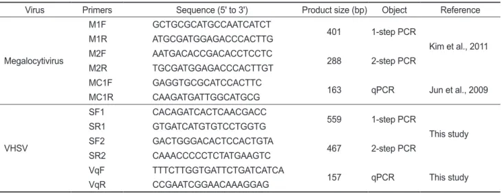
해수 중 어류질병 바이러스 검출 방법의 최적화 VHSV
배양상등액을1.00E+01 viral particles/mL seawater
농도로접종한1 L
의해수로부터VHSV
를검출시, GF/C
또 는CA
각하나의membrane
만을사용하였을경우2-step PCR
에서검출되지않았다.
그러나HA membrane
을함께사용한GF/C+CA+HA
또는GF/C+HA
여과를실시하여 바이러스 를농축후분리된total RNA
를대상으로실시된2-step PCR
에서VHSV
에특이적인band
를전기영동상에서확인할수있 었다(Fig. 1A).
또한, IVS-1
상등액을1.00E+01 viral particles/
mL seawater
농도로접종한1 L
의해수내의megalocytivirus
Fig. 1. Detection of viruses in 1 L seawater spiked with (A) VHSV and (B) IVS-1 (1.0E+01 viral particles/mL seawater, each) followed by concentration with different membranes. 1-step PCR and 2-step PCR was performed for left lanes 1-5 andright lanes 1-5, respectively. Lane 1, sterile seawater (GF/C+HA); lane 2, GF/C membrane; lane 3, CA membrane; lane 4, GF/C+CA+HA membrane; lane 5, GF/C+HA membrane.M, 100 bp DNA ladder.
N, No template control.
(A)
(B)
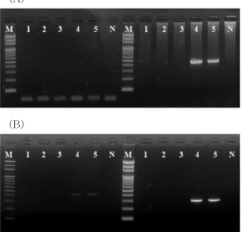
Fig. 2. Sensitivity test of HA method for detection of (A) VHSV and (B) IVS-1 in seawater. lane 1-5, concentrate of seawater spiked with VHSV or IVS-1 (0, 0.12, 1.2, 12, and 120 viral par- ticles/mL seawater respectively) ; M, 100 bp DNA ladder; N, no templatecontrol.
(A)
(B)
를
2-step PCR
을수행하여검출하고자하였을경우, VHSV
와마찬가지로GF/C
또는CA
각하나의membrane
만을사용 하였을때는검출이되지않았으나GF/C+CA+HA
또는GF/
C+HA membrane
을사용한바이러스농축을실시하였을때는 명확한megalocytivirus
의검출이가능하였다(Fig. 1B).
따라 서GF/C+CA+HA
또는GF/C+HA membrane
을사용하였을 때검출이가장잘되는것을확인할수있었으며,
이후의실험 에서는GF/C+HA membrane
을사용하여실험을실시하였다.
이러한결과는
1 L
의 해수에존재하는어류질병바이러스(1.00E+01 viral particles/mL)
의검출시2
가의음이온으로 구성된HA membrane
을사용하는것이1
가이온으로구성된CA membrane
을사용하는것에비해더적합함을의미한다.
또한, pore size
가1.2 μm
인GF/C
와HA membrane
을이중으 로사용함으로써해수중에존재하는부유물과유기물이HA
membrane
에흡착되는것을막을수있기때문에효율적으로해수여과및농축이가능하였다
.
바이러스 검출의 검출 한계 분석 및 회수율 비교
해수중의
VHSV
의검출한계를 분석하기위해VHSV
배양액을
1 L
의해수에1.22E+00 viral particles/mL seawater
이하 의농도로인위오염시켜1 mL
로농축한시료에서는2-step PCR
과qPCR
에서검출되지않았다(Fig. 2A, Table 2).
그리고1.22E+01 viral particles/mL
이상의농도로1 L
의해수에오염 되어있는시료를1 mL
로농축하였을때2-step PCR
에서검출 이가능하였으며,
이때농축시료1 mL
당3.28E+04, 2.38E+03 viral particles
로나타났다.
이를통하여1 L
의해수에농도별로 접종한VHSV
의회수율을산출하였을때26.89%, 19.10%
로 나타났다.
즉,
본방법에의한평균VHSV
회수율은23.00%
였으며
,
해수1 L
에존재하는VHSV
의검출한계는1.22E+01 viral particles/mL (1 mL
로농축시2.38E+03 viral particles)
이었다.
반면
megalocytivirus
의경우1 L
의해수중IVS-1
이1.20E- 01 viral particles/mL seawater
의농도로오염되어있을경우2-step PCR
과qPCR
에서검출되지않았으며, 1.20E+00 viral particles/mL seawater
의시료에서는2-step PCR
에서검출이 가능하였다.
또한1 L
의해수중에IVS-1
이1.20E+01 viral particles/mL seawater
이상으로오염되어있을경우1-step PCR
만으로도전기영동상에서IVS-1
에특이적인band
를확 인할수있었다.
그리고1.20E+00~1.20E+02 viral particles/
mL seawater
의농도로IVS-1
이오염된1 L
의해수를1 mL
로농축할경우,
농축시료mL
당각각3.07E+02, 3.29E+03, 3.76E+04 viral particles
의바이러스가회수되었으며,
이를통 하여회수율을산출하였을때25.58%, 27.42%, 31.33%
로나 타났다.
즉1 L
의해수를사용할경우IVS-1
에대한회수율 은평균적으로28.11%
이었으며,
검출한계는1.20E+00 viral particles/mL seawater
이었다(Fig. 2B, Table 3).
VHSV
의검출한계농도(1.22E+01 viral particles/mL sea- water)
와IVS-1
의검출한계농도(1.20E+00 viral particles/mL seawater)
를비교해볼때IVS-1
의 검출한계농도에 있어서IVS-1
이VHSV
에비해약10
배높은민감성을보였다.
본연구에있어
VHSV
를인위적으로해수에첨가한후GF/
C+HA membrane
으로농축하여결정한viral particle
값에있 어서, nucleic acid
분리과정중RNA
의안정성및cDNA
합성 수율문제에의하여소실량이DNA virus
에비하여높을수있으므로실질적인
VHSV
의양은본연구에서분석된양보다높을가능성이있다
.
또한,
실제로VHSV
에감염된넙치양식장 의유입수와사육수를대상으로GF/C+HA membrane
을사 용하여농축후바이러스의오염정도를분석한결과,
유입수 에서는바이러스를검출할수없었으나사육수에서는VHSV
의검출이가능하였다.
그러나넙치의VHSV
발병시감염방 법및어체의크기에따라누적폐사율이일정치않으며(Kim et al., 2009; Isshiki et al., 2001),
일반적으로바이러스감염시 질병의회복기에는바이러스의수중유출이매우감소하므로(Grant et al., 2011; Totland et al., 1996)
배출구위치에대비한유입구의위치
, VHSV
질병의진행정도등을고려한보다정밀한분석이필요할것이며
, megalocytivirus
감염양어장또한 비교분석할필요가있을것이다.
환경수중존재하는 바이러스의회수율을비교해볼때바 이러스별로회수율에대한차이가나타나며
, entericvirus
인poliovirus
의경우90%
이상의높은회수율을나타내는반면, HAV, Feline calicivirus (FCV)
의경우30-50%
의회수율을보 인다고보고하고있으며어류질병바이러스인Koi herpesvirus (KHV)
의경우3.9%
의낮은회수율을나타내고있다(Hara-
Fig. 3. Comparison of viral concentration method for seawater from two flounder farms suffering from VHSV. Influent seawaters from A farm (Jeju), B farm (Gampo) were concentrated by GF/C+CA (lane 1, 3) or GF/C+HA (lane 5, 7) membrane followed by 1-step PCR. Culturing seawater from A farm (Jeju), B farm (Gampo) were concentrated by GF/C+CA (lane 2, 4) or GF/
C+HA (lane 6, 8) membrane followed by 2-step PCR; M, 100 bp DNA ladder; N, no templatecontrol.
moto et al., 2009; Katayama et al., 2002).
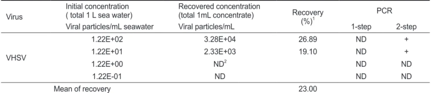
VHSV 감염 현장 시료에 대한 적용
2011
년2
월에VHSV
에감염된제주도,
감포의넙치양식장 에서사육수및유입수를채수하여GF/C+HA membrane
을사 용하여해수를농축하였으며,
분리된RNA
를대상으로2-step PCR
을실시하였다.
사육수를채취한양식장은VHSV
에감염 된넙치(10 cm)
가수온15±0.5℃,
사육수순환은16-18
회/ day
회전하여환수하고있는10 m×10 m×1 m
의수조에서 양식되고있었으며, VHSV
에감염되어누적폐사율이50%(
입 식후15
일후)
정도일때채수를실시하였다. VHSV
에감염된 넙치사육수1 L
시료를GF/C+HA membrane
을사용하여농 축할경우VHSV
를검출할수있었던반면, GF/C+CA mem- brane
을사용할경우검출할수없었다.
또한양식장의유입수 를대상으로GF/C+CA membrane
과GF/C+HA membrane
을 사용하여농축후바이러스의오염정도를분석한결과배출수 와는달리유입수에서는바이러스를검출할수없었다(Fig. 3).
본연구에서는어류질병바이러스의확산에가장큰영향을 미치는환경에대한정확한이해와분석을통하여그위험성 을최소화하기위해
,
국내에서만연하고있는megalocytivirus,
VHSV
을대상으로해수중바이러스농축방법의최적화작업및그검출한계점과바이러스회수율에대한분석을실시하 였다
. GF/C
와HA membrane
을사용한해수중바이러스농축법을어류질병바이러스인
megalocytivirus
와VHSV
를대상 으로적용하였을때회수율이28.11%, 23.00%
로각각나타나KHV
에비하여우수한결과를보여주어해수중의여러어류질병바이러스의검출에도본방법을적용할수있음을입증하 였다고할수있다
.
사 사
본연구는국립수산과학원
(
수산동물방역및검역체계구축, RP-2012-AQ-104)
의지원에의해운영되었습니다.
참고문헌
El-Matbouli M and H Soliman. 2011.Transmission of Cyprinid herpesvirus-3 (CyHV-3) from goldfish to naïve common carp by cohabitation. Research in Veterinary Science 90, 536-539. http://dx.doi.org/10.1016/j.rvsc.2010.07.008 Grant AAM, Jakob E, Richard J and Garver KA. 2011. Con-
centration of infectious aquatic rhabdoviruses from fresh- water and seawater using ultrafiltration. J Aquat Anim Health 23, 218-223. http://dx.doi.org/10.1080/08997659.20 11.644412
Haramoto E, Katayama H, Oguma K and Ohgaki S. 2007.
Quantitative analysis of human enteric adenoviruses in aquatic environments. J Appl Microbiol 103, 2153-2159.
Table 2. Recovery of virus from 1 L seawater spiked with VHSV Virus Initial concentration
( total 1 L sea water) Recovered concentration
(total 1mL concentrate) Recovery
(%)1 PCR
Viral particles/mL seawater Viral particles/mL 1-step 2-step
VHSV
1.22E+02 3.28E+04 26.89 ND +
1.22E+01 2.33E+03 19.10 ND +
1.22E+00 ND2 ND ND
1.22E-01 ND ND ND
Mean of recovery 23.00
1Recovery(%)=(Recovered viral concentration × vol/ Spiked viral concentration × vol)×100.
2ND, not detected.
Table 3. Recovery of virus from 1 L seawater spiked with IVS-1 Virus Inoculated concentration
(total 1 L sea water) Recovered concentration
(total 1mL concentrate) Recovery
(%)1 PCR
Viral particles/mL Viral particles/mL 1-step 2-step
IVS-1
1.20E+02 3.76E+04 31.33 + +
1.20E+01 3.29E+03 27.42 + +
1.20E+00 3.07E+02 25.58 ND +
1.20E-01 ND2 ND ND
Mean of recovery yield 28.11
1Recovery(%)=(Recovered viral concentration × vol/Spiked viral concentration × vol)×100.
2ND, not detected .
http://dx.doi.org/10.1111/j.1365-2672.2007.03453.x Haramoto E, Kitajima M, Katayama H, Ito H and Ohgaki S.
2009. Development of virus concentration methods for de- tection of koi herpesvirus in water. J Fish Dis 32, 297-300.
http://dx.doi.org/10.1111/j.1365-2761.2008.00977.x Isshiki T, Nishizawa T, Kobayashi T, Nagano T and Miyazaki
T. 2001. An outbreak of VHSV (viralhemorrhagicseptice- mia virus) infection in farmed Japaneseflounder
Paralich- thysolivaceus
in Japan. Dis Aquat Org 47, 87-99. http://dx.doi.org/10.3354/dao047087
Jeong JB, Jun LJ, Yoo MH, Kim MS, Komisar JL and Jeong HD. 2003. Characterization of the DNA nucleotide se- quences in the genome of red sea bream iridoviruses iso- lated in Korea. Aquaculture 220, 119-133. http://dx.doi.
org/10.1016/S0044-8486(02)00538-0
Jones SRM and Groman DB. 2001. Cohabitation Transmis- sion of Infectious Salmon Anemia Virus amongFreshwater- Reared Atlantic Salmon. J Aquat Anim Health 13, 340-346.
http://dx.doi.org/10.1577/1548-8667(2001)013<0340:CTO ISA>2.0.CO;2
Jun LJ, Jeong JB, Kim JH, Nam JH, Shin KW, Kim JK, Kang JH and Jeong HD. 2009. Influence of temperature shifts on the onset and development of red sea bream iridoviral disease in rock bream
Oplegnathusfasciatus
. Dis Aquat Org 84, 201-208. http://dx.doi.org/10.3354/dao02041Jung SJ and Oh MJ. 2000. Iridovirus-like infection associated with high mortalities of striped beakperch,
Oplegnathusfas- ciatus
(Temmincket Schlegel), in southern coastal areas of the Korean peninsula. J Fish Dis 23, 223-226. http://dx.doi.org/10.1046/j.1365-2761.2000.00212.x
Katayama H, Shimasaki A and Ohgaki S. 2002. Development of a virus concentration method and its application to detec- tion of enterovirus and norwalk virus from coastal seawa- ter. Appl Environ Microbiol 68, 1033-1039. http://dx.doi.
org/10.1128/ AEM.68.3.1033-1039.2002
Kim JW, Cho MY, Jin JW, Kim KH, Jeong HD and Kim KI.
2011. Detection of Megalocytivirus in shellfish using PCR with various DNA extraction methods. J Fish Pathol 24, 65-
Kim WS, Kim SR, Kim D, Kim JO, Park MA, Kitamura SI, 73.
Kim HY, Kim DH, Han HJ, Jung SJ and Oh MJ. 2009.
An outbreak of VHSV (viral hemorrhagic septicemia virus) infection in farmed olive flounder
Paralichthysoli- vaceus
in Korea. Aquaculture 296, 165-168. http://dx.doi.org/10.1016/j.aquaculture.2009.07.019
Kitamura S and Suzuki S. 2000. Occurrence of marine birna- virus through the year in coastal seawater in the Uwasea.
Mar Biotechnol 2, 188-194. http://dx.doi.org/10.1007/
s101269900025
Lewis GD and Metcalf TG. 1988. Polyethylene Glycol Pre- cipitation for Recovery of Pathogenic Viruses, Including Hepatitis A Virus and Human Rotavirus, from Oyster, Water, and Sediment Samples. Appl Environ Microbiol 54, 1983–1988.
Liu J, Wu Q and Kou X. 2007. Development of a Virus Con- centration Method and its Application for the Detection of Noroviruses in Drinking Water in China. J Microbiol 1, 48- Minamoto TM, Honjo N, Uchii K, Yamanaka H, Suzuki AA, 52.
Kohmatsu Y, Iida T and Kawabata Z. 2009. Detection of cyprinid herpesvirus 3 DNA in river water during and after an outbreak. Vet Microbiol 135, 261-266. http://dx.doi.
org/10.1016/j.vetmic.2008.09.081
Oh MJ, Kim SR, Jeong SJ, Kim HR, Kim HY and Yeo IK.
2000. A simple method for the concentration of fish patho- genic virus in seawater. J Fish Pathol 13, 61-66.
Song J, Choo YJ and Cho JC. 2008. Quantification of White Spot Syndrome Virus (WSSV) in seawater using real-time PCR and correlation analysis between WSSV and environ- mental parameters. Kor J Microbiol 44, 49-55.
Totland GK, Hjeltnes BK and Flood PR. 1996.Transmission of infectious salmon anaemia (ISA)through natural secretions and excretions frominfected smolts of Atlantic salmon