[논 문] 한국재료학회지 http://dx.doi.org/10.3740/MRSK.2011.21.8.415 Kor. J. Mater. Res.
Vol. 21, No. 8 (2011)
415
†Corresponding author
E-Mail : [email protected] (D. -S. Bae)
Synthesis and Characterization of SnO
2Nanoparticles by Hydrothermal Processing
Ho-Jung Kim, Jeong Hun Son and Dong-Sik Bae
†School of Nano & Advanced Material Engineering, College of Engineering, Changwon National University, Gyeongnam 641-773, Korea
(Received June 3, 2011 : Received in revised form July 11, 2011 : Accepted July 18, 2011)
Abstract
Tin (IV) dioxide (SnO2) has attracted much attention due to its potential scientific significance and technological applications. SnO2 nanoparticles were prepared under low temperature and pressure conditions via precipitation from a 0.1 M SnCl4·5H2O solution by slowly adding NH4OH while rapidly stirring the solution. SnO2 nanoparticles were obtained from the reaction in the temperature range from 130 to 250oC during 6 h. The microstructure and phase of the synthesized tin oxide particles were studied using XRD and TEM analyses. The average crystalline sizes of the synthesized SnO2 particles were from 5 to 20 nm and they had a narrow distribution. The average crystalline size of the synthesized particles increased as the reaction temperature increased. The crystalline size of the synthesized tin oxide particles decreased with increases in the pH value. The X-ray analysis showed that the synthesized particles were crystalline, and the SAED patterns also indicate that the synthesized SnO2 nanoparticles were crystalline. Furthermore, the morphology of the synthesized SnO2 nanoparticles was as a function of the reaction temperature. The effects of the synthesis parameters, such as the pH condition and reaction temperature, are also discussed.Key words
Hydrothermal, Nanoparticles, SnO2.1. Introduction
Recently, the synthesis of nanometer-sized particles has been investigated extensively because of their novel elec- trical, optical, magnetic, and chemical properties.
1)The effect of particle size on the electronic and optical properties of these nanosized particles during the growth of the crystallite from the molecular level to the bulk material is an area of fundamental interest.
2)Semiconductor nanomaterials have attracted much atten- tion because of their potential scientific significance and technological applications. Tin (IV) dioxide (SnO
2) is a semiconducting material with a band gap of 3.6eV at 300 K.
3)It is of considerable technological importance with a number of applications, especially for combustible and toxic gas detection, thin film coatings, and sensor devices.
4-9)The material shows high quasimetallic electrical conductivity but retains good optical transparency in the visible region.
3,10-11)These properties can be used for transistors
12)or photovol- taic devices.
13-15)Hydrothermal processes have a potential for the direct preparation of crystalline ceramic powders and offer a low- temperature alternative to conventional powder synthesis techniques in the production of oxide powders.
16)This pro-
cess can produce fine, high-purity, stoichiometric particles of single and multi-component metal oxides. Furthermore, if process conditions such as solute concentration, reaction temperature, and reaction time are carefully controlled, a desire shape and size of the particles can be produced.
17-18)These powders could be sintered at low temperature without calcination and milling steps.
19)The object of this study was to prepare SnO
2nanoparticles by hydrothermal method.
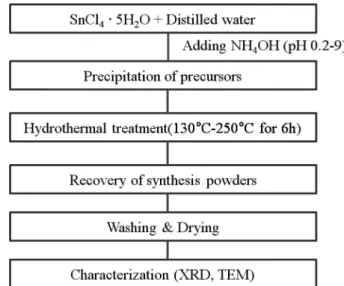
Fig. 1. Experimental flow chart of synthesized Tin oxide powders by hydrothermal processing.
416 Ho-Jung Kim, Jeong Hun Son and Dong-Sik Bae
2. Experimental Procedure
The preparation SnO
2nanoparticles were schematically illustrated in Fig. 1. Tin (IV) dioxide precursors were precipitated from 0.1 M SnCl
4·5H
2O solution by slowly adding NH
4OH with rapid stirring. pH value of starting solutions varied between 0.2 and 9. After 30 min of stirring the solution was placed in a 1000 ml stainless steel pressure vessel. Hydrothermal treatment was carried out at 130- 250
oC for 6h and cooled naturally to room temperature. The reaction products were washed five times by repeated centrifugation, and then dried at 80
oC for 12h in air.
The recovered powders were analyzed for phase compos- ition using X-ray diffraction (XRD, Philips X’pert MPD PW3040, Holland) over the 2 θ range from 20
oto 80
oat the scan speed of 2
omin
−1. The morphology of the synthesized particles was observed using transmission electron micro- scope (TEM, Jeol 2000FXII, Japan). For TEM studies, samples were prepared by adding drops of freshly prepared cluster solution on a carbon film supported on a Cu grid.
3. Results and Discussion
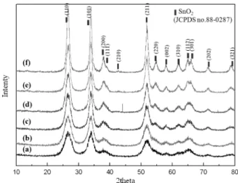
Fig. 2 shows X-ray diffraction patterns of the synthesized SnO
2nanoparticles as a function of reaction temperature.
From the X-ray analysis, all the diffraction peaks can be readily indexed to tetragonal SnO
2with cassiterite structure (JCPDS no. 88-0287). At lower temperature, very small nanosized SnO
2powders are synthesized. As the reaction temperature is increased, the crystallinity and the size of
SnO
2are increased.
The morphology of the obtained SnO
2nanopaticles was characterized by transmission electron microscope (TEM) observations. Fig. 3 shows TEM images and SAED patterns of the SnO
2nanopaticles. As shown in Fig. 3, the average crystalline sizes of the synthesized SnO
2nanoparticles increased with reaction temperature increased from 130°C to 250°C. With increasing hydrothermal temperature, the crystallinity of the synthesized nanoparticles is increased.
At lower temperature of 130°C, the synthesized SnO
2Fig. 3. TEM images (inset SAED patterns) of the synthesized SnO2 nanoparticles by hydrothermal reaction at pH 0.2 for 6h as a function of reaction temperature: (a) 130°C, (b) 170°C, (c) 210°C and (d) 250°C.
Fig. 2. X-ray diffraction patterns of the synthesized SnO2 nanoparticles by hydrothermal reaction at pH 0.2 for 6h as a function of reaction temperature: (a) 130°C, (b) 150°C, (c) 170°C, (d) 190°C, (e) 210°C and (f) 250°C.
Synthesis and Characterization of SnO2 Nanoparticles by Hydrothermal Processing 417
nanoparticles size is about 5 nm. At higher temperature of 250°C, the nanoparticles size is about 20 nm. The SAED patterns indicate the prepared SnO
2nanopaticles were well crystalline.
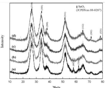
Fig. 4 shows the X-ray diffraction pattern of the synthe- sized SnO
2nanoparticles as a function of pH value. All the diffraction peaks can be readily indexed to tetragonal SnO
2with cassiterite structure (JCPDS no. 88-0287). With in- creasing pH value, the crystallinity of the synthesized nano- particles is decreased. The diffraction peaks are markedly
broadened, which indicates that the crystalline sizes of sam- ples are very small.
Fig. 5 shows TEM images and SAED patterns of the SnO
2nanopaticles. As shown in Fig. 5, the SnO
2nanopar- ticles are spherical, uniform, and there are almost no aggregates in large visual fields. The synthesized SnO
2particles size is about 5~10 nm. The SAED patterns indicate the prepared SnO
2nanopaticles were well crystalline.
4. Conclusion
Spherical SnO
2nanoparticles have been successfully syn- thesized by hydrothermal process using tin (IV) chloride pentahydrate (SnCl
4·5H
2O) as tin dioxide precursor. Nano- sized SnO
2particles were obtained in the temperature range of 130°C - 250°C and pH value of 0.2-9 for 6h. The average crystalline sizes and distribution of the synthesized SnO
2particles was about 5-20 nm and narrow, respectively.
The average crystalline size of the synthesized particles increased with reaction temperature increased. From X-ray analysis and TEM, the synthesized particles were crystal- line. The size and morphology of the synthesized SnO
2nanoparticles can be controlled as a function of starting solution pH and reaction temperature.
Acknowledgment
This research was financially supported by Changwon National University (2010).
Fig. 4. X-ray diffraction patterns of the synthesized SnO2 nanoparticles by hydrothermal reaction at 130°C for 6h as a function of pH value:
(a) pH 0.2, (b) pH 2, (c) pH 5 and (d) pH 9.
Fig. 5. TEM images (inset SAED patterns) of the synthesized SnO2 nanoparticles by hydrothermal reaction at 130°C for 6h as a function of pH value: (a) pH 0.2, (b) pH 2, (c) pH 5 and (d) pH 9.
418 Ho-Jung Kim, Jeong Hun Son and Dong-Sik Bae
References
1. R. Pool, Science, 248, 1186 (1990).
2. Y. Wang and N. Herron, J. Phys. Chem., 95, 525 (1991).
3. B. Falabrettia and J. Robertson, J. Appl. Phys. 102, 123703/
1 (2007).
4. P. G. Harrison and M. J. Willett, Nature, 332, 337 (1988).
5. W. Göpel, Sensors: A Comprehensive Survey - Vol. 9, p.
8, ed. W. Göpel, J. Hesse and J. N. Zemel, VCH:
Weinheim, Germany (1995).
6. M. Law, H. Kind, B. Messer, F. Kim and P. Yang, Angew.
Chem. Int. Ed., 41, 2405 (2002).
7. B. Wang, L. F. Zhu, Y. H. Yang, N. S. Xu and G. W.
Yang, J. Phys. Chem. C, 112, 6643 (2008).
8. G. X. Wang, J. S. Park, M. S. Park and X. L. Gou, Sensor.
Actuator. B Chem., 131, 313 (2008).
9. H. Wang, J. Liang, H. Fan, B. Xi, M. Zhang, S. Xiong, Y.
Zhu and Y. Qian, J. Solid State Chem., 181, 122 (2008).
10. R. E. Aitchison, Aust. J. Appl. Sci., 5, 10 (1954).
11. Y. S. He, J. C. Campbell, R. C. Murphy, M. F. Arendt and J. S. Swinnea, J. Mater. Res., 8, 3131 (1993).
12. X. Duan, Y. Huang, R. Agarwal and C. M. Lieber, Nature, 421, 241 (2003).
13. L. Vayssieres and M. Graetzel, Angew. Chem. Int. Ed., 43, 3666 (2004).
14. Y. Fukai, Y. Kondo, S. Mori and E. Suzuki, Electrochem.
Comm., 9, 1439 (2007).
15. G. R. R. A. Kumara, K. Tennakone, I. R. M. Kottegoda, P. K. M. Bandaranayake, A. Konno, M. Okuya, S. Kaneko and K. Murakami, Semicond. Sci. Tech., 18, 312 (2003).
16. S. Hirano, Am. Ceram. Soc. Bull., 66, 1342 (1987).
17. W. J. Dawson, Am. Ceram. Soc. Bull., 67, 1673 (1989).
18. S. B. Cho, S. Venigalla and J. H. Adair, Science, Technology, and Applications of Colloidal Suspensions Ceramic Transactions, p.139, ed. J. H. Adair, J. A. Casey, C. A. Randall and S. Venigalla, American Ceramic Society, Indiana, USA (1997).
19. K. Haberko and W. Pyda, Advances in Ceramics, Science and Technology of Zirconia II, p.774, ed. M. Ruhle, N.
Claussen and A. H. Heuer, Americal Ceramic Society, USA (1983).