Tuberc Respir Dis 2009;67:275-280
CopyrightⒸ2009. The Korean Academy of Tuberculosis and Respiratory Diseases. All rights reserved.
사이질 폐병의 최신지견: 특발사이질 폐렴을 중심으로
연세대학교 원주의과대학 내과학교실
리원연Clinical Year in Review of Interstitial Lung Diseases: Focused on Idiopathic Interstitial Pneumonia
Won-Yeon Lee, M.D., Ph.D.
Department of Internal Medicine, Yonsei University Wonju College of Medicine, Wonju, Korea
Interstitial lung disease (ILD) is a group of diseases characterized by pulmonary interstitial inflammation. Finally the inflammation results in pulmonary fibrosis and impairment of oxygen transportation. The causes of idiopathic interstitial pneumonia (IIP) are unknown. Diagnosis of IIP is not easy, especially distinguising between nonspecific interstitial pneumonia and usual interstitial pneumonia (UIP). First line treatments of IIP include corticosteroids and immune modulators, which have limited effect. Currently, several drugs are being researched to prevent and treat fibrosis. Newer drugs that may useful to treat pulmonary fibrosis include endothelin receptor antagonist, recombinant soluble TNF receptor antagonist, and cotrimoxazole. The causes of IIP are largely unknown, treatment is not specific, and prognosis is poor. Recent studies are underway to investigate the pathogenesis and treatment of IIP and pulmonary fibrosis. As the pathogenesis of IIP is elucidated, better treatments will emerge.
Key Words: Idiopathic interstitial pneumonia, Nonspecific interstitial pneumonia, Usual interstitial pneumonia, Pulmonary fibrosis
Address for correspondence: Won-Yeon Lee, M.D., Ph.D.
Department of Internal Medicine, Yonsei University Wonju College of Medicine, 162, Ilsan-dong, Wonju 220-050, Korea Phone: 82-33-741-1233, Fax: 82-33-741-0928
E-mail: [email protected] Received: Jun. 12, 2009
Accepted: Jun. 14, 2009
서 론
사이질 폐병(interstitial lung disease, ILD)은 폐 사이질 의 염증을 특징으로 하는 다양한 질환군으로 섬유화가 동 반이 될 수 있고 질환이 진행되면 산소운반에 장애가 생기 고 폐에 반흔을 남기게 된다. ILD는 원래 조직학적 형태로 분류되기 시작하여 다양한 임상양상을 고려하여 임상적 인 분류를 하게 되었고 2002년에 ATS/ERS에서 재분류를 하였다(Table 1, Figure 1). 이 분류에서는 ILD 대신에 미 만성 폐실질 질환(diffuse parenchymal lung disease, DPLD)이라는 용어를 제시하였다1. 이것은 주로 기도나
혈관을 침범하는 질환으로부터 DPLD를 구별하여 DPLD 는 폐실질이 주 손상 부위라는 점을 강조하기 위함이다2. 새로운 분류지침에 의하면 DPLD는 다음과 같이 네 가지 군으로 나뉜다. 1) 알려진 원인에 의한 DPLDs (e.g., 약 물, 결체조직질환, 환경/직업 노출), 2) 육아종성 DPLDs (e.g., 유육종증), 3) 기타 드문 DPLDs (e.g., lymphangio- leiomyomatosis, pulmonary Langerhans cell histiocy- tosis, 폐단백증, 호산구폐렴), 그리고 4) 특발사이질폐렴 (idiopathic interstitial pneumonia, IIPs).
앞에 기술하였던 것과 같이 ILD는 매우 다양한 질환들 을 포함한 광범위한 질환군으로 특발사이질폐렴(IIP)을 중심으로 최근 발표되고 있는 관심사에 대하여 요약해 보 고자 한다.
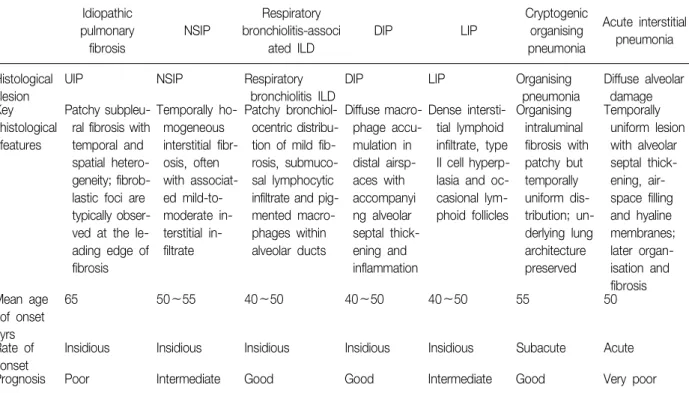
Table 1. Key features of the idiopathic interstitial pneumonias, as defined by the ATS/ERS consensus classification
Idiopathic
pulmonary fibrosis
NSIP
Respiratory bronchiolitis-associ
ated ILD
DIP LIP
Cryptogenic organising pneumonia
Acute interstitial pneumonia
Histological lesion
UIP NSIP Respiratory
bronchiolitis ILD
DIP LIP Organising
pneumonia
Diffuse alveolar damage Key
histological features
Patchy subpleu- ral fibrosis with temporal and spatial hetero- geneity; fibrob- lastic foci are typically obser- ved at the le- ading edge of fibrosis
Temporally ho- mogeneous interstitial fibr- osis, often with associat- ed mild-to- moderate in- terstitial in- filtrate
Patchy bronchiol- ocentric distribu- tion of mild fib- rosis, submuco- sal lymphocytic infiltrate and pig- mented macro- phages within alveolar ducts
Diffuse macro- phage accu- mulation in distal airsp- aces with accompanyi ng alveolar septal thick- ening and inflammation
Dense intersti- tial lymphoid infiltrate, type II cell hyperp- lasia and oc- casional lym- phoid follicles
Organising intraluminal fibrosis with patchy but temporally uniform dis- tribution; un- derlying lung architecture preserved
Temporally uniform lesion with alveolar septal thick- ening, air- space filling and hyaline membranes;
later organ- isation and fibrosis Mean age
of onset yrs
65 50∼55 40∼50 40∼50 40∼50 55 50
Rate of onset
Insidious Insidious Insidious Insidious Insidious Subacute Acute
Prognosis Poor Intermediate Good Good Intermediate Good Very poor
특발폐섬유화(IPF, UIP) vs.
비특이 사이질 폐렴(NSIP)
Idiopathic pulmonary fibrosis (IPF)는 원인이 불분명 한 만성 섬유화성 사이질 폐렴으로 조직학적으로 usual interstitial pneumonia (UIP)와 연관이 되어 있으며 병변 은 폐에만 국한되는 것을 특징으로 하는 질환이라고 정의 하였다(2002 ATS/ERS DPLD classification)1. 2002년의 분 류에서 nonspecific interstitial pneumonia (NSIP)를 조직 학적으로 다른 질환으로 분류하였으나 NSIP는 IPF와 구분 이 될 만한 특이 임상소견을 가지고 있지 않아 NSIP가 독 립적인 질환인지에 대한 의구심이 제기되고 있다. 그 이 유로 여러 가지 다른 원인 혹은 질환에 의하여 같은 조직 학적 변화를 가져올 수 있다. 그 예로 결체조직 질환이나 과민성 폐렴, 석면증, 유육종증이 진행되면 조직학적으로 UIP의 소견을 가진다. 또한 결체 조직질환, 과민성폐렴, 약물 유발폐병, 급성호흡곤란증후군의 회복과 crytogenic organizing pneumonia에서는 NSIP의 조직학적 소견을 보일 수 있다. UIP의 중요한 소견인 섬유모세포 병터(fib- roblastic foci)는 드물기는 하지만 NSIP에서 보일 수 있으 며 IPF를 가진 동일한 환자의 다른 부위의 폐조직에서 UIP와 NSIP가 각각 자주 발견이 된다.
고해상도 전산화 단층촬영(HRCT)과 함께 조직학적으 로 진단된 48명(NSIP 23명, IPF 25명)을 대상으로 후향적 으로 34개월에서 155개월 동안 추적 관찰한 연구에서도 처음에 HRCT에서 NSIP의 특징적인 소견을 보였던 환자 18명 중 5명이 특징적인 UIP의 소견으로 발전하는 것을 보고하였다. 이런 결과는 특발 NSIP가 임상적-방사선학적- 병리학적으로 UIP의 형태로 발전할 수 있고 현재의 진단 방법으로는 NSIP가 IPF로 진행할 가능성을 예측할 수는 없다는 것을 의미한다3. 그러나 최근 Travis 등4은 이전에 보고된 NSIP 305예를 재평가한 연구에서 67예를 명확한 (definite) 또는 가능한(probable) NSIP로 분류하였고 이 들의 특징을 기술하였다. 이들의 평균연령은 52세, 아시 아국가 46%, 여성 67%, 비흡연자 69%였다. 이 연구에서 저자들은 NSIP를 임상적, 방사선학적, 조직학적으로 독립 된 질환으로 결론을 내렸으며 예후는 5년 생존율 80%, 10 년 생존율 73%였다고 보고하였다. 또한 NSIP를 독립적인 질환으로 고려하는 것은 예후를 결정하거나 면역억제치 료에 대한 반응을 예측하는 데 임상적으로 도움이 될 수 있으나 NSIP가 UIP와 동반이 되어 있는 보고가 많아 이 둘 간의 상관관계에 대하여는 장기적인 관찰이 필요할 것 이다. 특발 NSIP에 대한 또 다른 연구로 국내에서 보고한 83명의 조직학적으로 진단된 환자의 임상경과, 폐기능의
Figure 1. DPLDs consist of disorders of known causes (e.g., collagen vascular disease or environmental or drug-related causes) and disorders of unknown causes. The latter include IIPs, granulomatous lung disorders, and other forms of interstitial lung disease.The most important distinction among the IIPs is that between IPF and NSIP. Other IIPs include DIP, RBILD, AIP, COP, and LIP. AIP: acute interstitial pneumonia; COP: formerly known as Bronchiolitis obliterans organ- izing pneumonia (BOOP); COP: cryptogenic organizing pneumonia; DIP: desquamative interstitial pneumonia; DLPD: dif- fuse parenchymal lung disease; IIP: idiopathic interstitial pneumonia; IPF: idiopathic pulmonary fibrosis; LIP: lymphocytic interstitial pneumonia; NSIP: nonspecific interstitial pneumonia; RBILD: respiratory bronchiolitis-associated interstitial lung disease; UIP: usual interstitial pneumonia. The ATS/ERS Classification of Diffuse Parenchymal Lung Disease 2002.
변화, 그리고 예후인자에 대한 후향적 연구가 있다5. 이 연구에서는 53개월의 추적기간 중 섬유화 NSIP 환자군의 22%가 NSIP와 연관되어 사망하였으며 생존율이 비교적 좋음에도(5년 74%) 불구하고 섬유화 NSIP는 재발률로 인 한 입원율이 36%였다. 추적관찰 기간 중 80% 이상의 환 자에서 폐기능이 호전되거나 안정적이었다. 초기 HRCT 소견에서 젖빛유리음영과 폐경화의 범위가 큰 경우와 벌 집음영소견을 보인 경우 예후가 불량하였고 또한 10%의 환자는 결체 조직 질환이 발생함을 보고하였다.
역 학
최근 들어 미국과 영국에서는 IPF의 발병이 이전에 비 하여 증가하고 있음을 시사하는 보고가 있으며 이것은 폐 섬유화로 인한 사망률의 증가로도 미루어 짐작할 수 있다.
1992년과 2003년을 비교하였을 때 남성은 30%, 여성은 40%가 증가하였으며 사망자 중 60%는 폐섬유화 자체가 사망원인이었다. 이것은 방광암, 급성골수성백혈병, 다발 성 골수종보다 높은 사망률이다.
미국의 경우 IPF로 인한 사망은 10만 명당 75.7명으로 남자, 백인, 85세 이상에서 가장 높았다. 또한 직업과 연관 하였을 때 목조 건물이나 이동식 주택(OR, 5.3), 금속광산
(OR, 2.2), 금속 철골구조제작(OR, 1.7)과 관련된 직업을 가진 사람에서 사망률이 높았다. 이것은 흡연 이외에 이 전에 제시되었던 목재나 금속 먼지가 폐섬유화와 연관이 된다는 보고를 뒷받침한다6.
IPF의 병인
1980년대 중반부터 IPF의 병인의 이해가 되기 시작했 으며 초기의 가설은 염증세포의 유입과 섬유화 유발물질 의 분비가 중요하며 이에 의해 시작되는 강화되고 조절되 지 않는 치유반응에 의하여 섬유화가 일어난다고 믿어져 왔다. 이런 염증과 관련된 병인의 가설은 ‘direct hypoth- esis’, ‘vascular hypothesis’, ‘plasticity hypothesis’, ‘mat- rix hypothesis’, ‘growth factor hypothesis’ 등이 있다. 그 러나 최근의 활발해진 연구에서 염증성 반응과 관계없이 섬유화는 진행된다는 증거들이 나타나며 다양한 병인을 제시하고 있다7.
IPF에서 섬유화의 발생을 일으키는 원인은 아직 알지 못한다. 그러나 IPF의 시작과 진행에 관여하는 다양한 가 능성 있는 손상인자들의 증거가 늘어나고 있다. 흡연, 산 화 스트레스, 환경오염 등이 IPF의 가능성 있는 원인으로 제시되고 있으며 바이러스의 호흡기 감염이 또 다른 원인
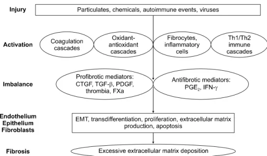
Figure 2. A new model for the pathogenesis of idiopathic pulmonary fibrosis: injury activates multiple inflammatory, cell signalling and repair pathways. Activation of these cascades causes an imbalance in pro- and antifibrotic mediators.
In turn, these mediators activate multiple cell types, causing changes in cellular functioning and cell-cell interactions that ultimately result in progressive fibrosis. Th: T-helper cell; CTGF: connective tissue growth factor; TGF-b: transforming growth factor-b; PDGF: platelet-derived growth factor; FXa: factor Xa; PG: prostaglandin; IFN-c: interferon-c; EMT: epi- thelial-mesenchymal transition7.
으로 제시되고 있다.
손상 후 IPF의 병인에 다양한 경로의 기전이 관여할 것 이라는 주장이 제기되고 있으며 동물모델의 섬유화에서 밝혀진 기전으로는 응고 연쇄반응, 산화-항산화 경로, 세 포자멸사, 성장인자, 폐표면 활성제, 기질조절인자 등이 관여한다고 알려져 있다. 이런 연쇄 반응은 다양한 세포 에 기능적, 형태적 변형을 시작하게 한다. 이런 세포로는 상피세포, 섬유모세포, 내피세포, 염증세포와 최근에 알려 진 circulating fibroblast progenitor cell 등이 있다. 앞에 기술한 중요한 경로의 상대적인 역할과 서로 다른 세포형 태 사이의 상호작용이 인간에서 섬유화의 병인에 관여한 다는 증거들이 점점 나오고 있다(Figure 2). 이런 다양한 경로의 병인모델은 앞으로 연구방법이나 치료방법의 개 발에 반영이 되어야 할 것으로 생각된다8.
폐섬유화의 치료
전통적으로 폐섬유화의 치료에 가장 먼저 고려되는 약 물은 스테로이드와 면역억제제일 것이다. 그러나 IPF의 경우에는 치료의 효과에 대하여 아직도 논란이 있으며 항 산화제로서 N-acetyl cystein이 치료제로서의 가능성을 보
여주고 있으나 보조적인 측면이 많으며 생존율에 대한 효 과는 아직 모르는 상태이다9,10. 최근 폐섬유화의 병인이 연구되며 새로운 약제들이 치료에 시도되고 있으며 En- dothelin receptor antagonist, Recombinant soluble TNF receptor antagonist, cotrimoxazole 등이 최근에 치료효 과가 있을 것으로 연구되고 있는 약제들이다.
1. Endothelin receptor antagonist
Endothelin-1 (ET-1)은 강력한 평활근세포의 증식과 섬 유모세포로부터 콜라젠의 합성을 증가시키고 사이질의 콜라젠 분해효소를 감소시키는 강력한 성장인자로 ET-1 이 증가가 IPF의 발생에 역할을 할 것으로 생각해왔다.
Bosentan은 ET-A, B 수용체를 모두 길항하는 약제로 폐 동맥 고혈압의 치료제 쓰이기 시작했고 동물실험에서 폐 의 콜라젠 축적을 감소시키는 것을 보여주었다. Bosentan 은 안면 홍조, 간기능 이상, 하지부종, 두통, 빈혈 등의 부작용이 나타날 수 있으나 IPF 환자에서 이루어진 예비 연구에서는 심하지 않았다. 최근 폐동맥 고혈압이 없는 (RVSP <50 mmHg) 158명의 IPF 환자에서 bosentan의 효과와 안정성을 보기 위하여 1년 동안 투여하여 시행한 임상연구에서(BUILD-1 trial) 1차 평가 지표인 6분 보행검
사가 위약과 비교하여 더 나은 결과를 얻지 못했다. 그러 나 폐기능의 악화, 입원율, 사망률(relative RR, 38%; p=
0.078), 호흡곤란이나 삶의 질 지표에서 긍정적인 경향을 보였다11. 현재 BUILD-3가 진행 중이다.
2. Recombinant soluble TNF receptor antagonist
Etanercept는 가용성 TNF 수용체로 TNF-a의 길항제로 작용한다. TNF-a는 염증유발 작용을 가진 주 cytokyne으 로 섬유모세포의 분화를 자극하며 콜라젠의 합성을 증가 시킨다. 2상 임상연구로 88명의 IPF 환자에게 Etanercept 를 48주간 투여 후 측정한 FVC, DLCO, (A-a)DO2가 위약 에 비하여 변화는 없었으나, 다른 폐기능 검사 항목과 삶 의 질 지표 수치에서 긍정적인 경향을 보였다12.
3. 경구 cotrimoxazole
진행성 폐섬유화 환자에서 예비임상 시험으로 3개월 동 안 경구 cotrimoxazole을 투여하고 6주 동안 호흡재활을 시행한 연구에서 왕복운동검사와 폐기능 검사, 호흡곤란 지수가 긍정적인 결과를 보였다. 비록 예비 시험이지만 항생제 또는 항진균제가 보조적인 치료제로 쓰일 수 있는 가능성을 보여주었다13.
4. 호흡재활
다른 만성 호흡기 질환과 마찬가지로 DPLD 환자에서 도 호흡재활이 운동능력, 건강상태, 삶의 질을 증가시킨다 는 새로운 증거들이 있다. 그러나 이런 연구들은 대상환 자 수가 적으며 6개월이 후에는 장점이 없다는 점에서 아 직 제한적이다14,15.
5. 급성악화
IPF의 급성악화는 또 다른 치료관점에서 해결해야 할 문제로 원인을 모르는 것으로 되어 있으며 2년 동안 10%
이상에서 발생하며 3∼6개월 동안 60∼70%가 사망을 한 다16. 이 경우 고해상도 단층촬영에서 새로운 간유리음영 이 증가하며 범위와 형태가 예후와 연관이 있다17. 그러나 불행하게도 아직 효과적인 치료가 없어 새로운 치료에 대 한 시급한 연구가 필요하다.
예후, 사망률의 예측 인자
예후 인자로 흡연이 생존율을 증가시키는 것같이 보고
되었으나 최근에 절대금연자, 과거흡연자, 현재흡연자를 비교한 연구에서 생존율을 증가시키지 않으며 초기의 IPF로 진단된 건강 흡연자에 의한 오류였던 것으로 밝혀 졌다18. 또 다른 예후를 결정하는 인자로 기관지폐포 세척 술의 역할이 관심이 되고 있었으며 이전의 연구에서는 기 관지폐포세척액의 림프구 분획이 20% 이상이면 항염증치 료에 반응이 있으며 예후가 좋다는 보고를 하였으며19 최 근 연구에서는 중성구 분획이 낮을수록(<3%) 예후가 좋 으며 중성구 분획이 두 배로 증가할수록 진단 후 1년 이내 사망률이 30%씩 증가한다고 보고하였다20.
결 론
특발 사이질폐렴은 아직 원인이나 병인이 제대로 밝혀 져 있지 않으며 예후가 악성종양에 비할 정도로 나쁘지만 특별한 치료 방법도 없는 경우가 대부분이다. 최근 들어 병인에 대한 연구가 많이 이루어지고 있으며 새로운 치료 약제에 대한 임상연구에서 조금은 희망적인 결과가 보고 되고 있으나 향후 더 많은 연구가 필요할 것이다.
참 고 문 헌
1. Demedts M, Costabel U. ATS/ERS international multi- disciplinary consensus classification of the idiopathic interstitial pneumonias. Eur Respir J 2002;19:794-96.
2. Morgenthau AS, Padilla ML. Spectrum of fibrosing dif- fuse parenchymal lung disease. Mt Sinai J Med 2009;
76:2-23.
3. Silva CI, Muller NL, Hansell DM, Lee KS, Nicholson AG, Wells AU. Nonspecific interstitial pneumonia and idio- pathic pulmonary fibrosis: changes in pattern and dis- tribution of disease over time. Radiology 2008;247:251- 59.
4. Travis WD, Hunninghake G, King TE Jr, Lynch DA, Colby TV, Galvin JR, et al. Idiopathic nonspecific inter- stitial pneumonia: report of an American Thoracic Society project. Am J Respir Crit Care Med 2008;177:
1338-47.
5. Park IN, Jegal Y, Kim DS, Do KH, Yoo B, Shim TS, et al. Clinical course and lung function change of idio- pathic nonspecific interstitial pneumonia. Eur Respir J 2009;33:68-76.
6. Pinheiro GA, Antao VC, Wood JM, Wassell JT. Occupa- tional risks for idiopathic pulmonary fibrosis mortality in the United States. Int J Occup Environ Health 2008;
14:117-23.
7. Maher TM, Wells AU, Laurent GJ. Idiopathic pulmonary fibrosis: multiple causes and multiple mechanisms? Eur Respir J 2007;30:835-39.
8. Bringardner BD, Baran CP, Eubank TD, Marsh CB. The role of inflammation in the pathogenesis of idiopathic pulmonary fibrosis. Antioxid Redox Signal 2008;10:287- 301.
9. Rogliani P, Mura M, Assunta Porretta M, Saltini C. New perspectives in the treatment of idiopathic pulmonary fibrosis. Ther Adv Respir Dis 2008;2:75-93.
10. Kim R, Meyer KC. Therapies for interstitial lung disease:
past, present and future. Ther Adv Respir Dis 2008;2:
319-38.
11. King TE Jr, Behr J, Brown KK, du Bois RM, Lancaster L, de Andrade JA, et al. BUILD-1: a randomized place- bo-controlled trial of bosentan in idiopathic pulmonary fibrosis. Am J Respir Crit Care Med 2008;177:75-81.
12. Raghu G, Brown KK, Costabel U, Cottin V, du Bois RM, Lasky JA, et al. Treatment of idiopathic pulmonary fib- rosis with etanercept: an exploratory, placebo-con- trolled trial. Am J Respir Crit Care Med 2008;178:948-55.
13. Varney VA, Parnell HM, Salisbury DT, Ratnatheepan S, Tayar RB. A double blind randomised placebo con- trolled pilot study of oral co-trimoxazole in advanced fibrotic lung disease. Pulm Pharmacol Ther 2008;21:
178-87.
14. Nishiyama O, Kondoh Y, Kimura T, Kato K, Kataoka K, Ogawa T, et al. Effects of pulmonary rehabilitation in patients with idiopathic pulmonary fibrosis. Respirol- ogy 2008;13:394-9.
15. Holland AE, Hill CJ, Conron M, Munro P, McDonald CF. Short term improvement in exercise capacity and symptoms following exercise training in interstitial lung disease. Thorax 2008;63:549-54.
16. Agarwal R, Jindal SK. Acute exacerbation of idiopathic pulmonary fibrosis: a systematic review. Eur J Intern Med 2008;19:227-35.
17. Akira M, Kozuka T, Yamamoto S, Sakatani M. Comput- ed tomography findings in acute exacerbation of idio- pathic pulmonary fibrosis. Am J Respir Crit Care Med 2008;178:372-8.
18. Antoniou KM, Hansell DM, Rubens MB, Marten K, Desai SR, Siafakas NM, et al. Idiopathic pulmonary fib- rosis: outcome in relation to smoking status. Am J Respir Crit Care Med 2008;177:190-4.
19. Ryu YJ, Chung MP, Han J, Kim TS, Lee KS, Chun EM, et al. Bronchoalveolar lavage in fibrotic idiopathic inter- stitial pneumonias. Respir Med 2007;101:655-60.
20. Kinder BW, Brown KK, Schwarz MI, Ix JH, Kervitsky A, King TE Jr. Baseline BAL neutrophilia predicts early mortality in idiopathic pulmonary fibrosis. Chest 2008;
133:226-32.