세포 및 동물모델에서의 알코올에 의해 유발된 간손상에 대한 지구자 추출물의 보호효과
유양희1*․정국영2*․이유현3․전우진1․이부용2†
1전남대학교 식품영양학과
2포천중문의과대학교 대체의학대학원
3수원대학교 식품영양학과
Hepatoprotective Effects of Hovenia dulcis Fruit on Ethanol-Induced Liver Damage in vitro and in vivo
Yanghee You1*, Kuk-Yung Jung2*, Yoo-Hyun Lee3, Woojin Jun1, and Boo-Yong Lee2†
1Dept. Food and Nutrition, Chonnam National University, Gwangju 550-757, Korea
2Graduate School of Complementary & Alternative Medicine, College of Medicine, Pochon CHA University, Gyeonggi 463-836, Korea
3Dept. Food Science and Nutrition, Suwon University, Gyeonggi 445-743, Korea
Abstract
The hepatoprotective effect of ethanol extract from Hovenia dulcis fruit (HD) against ethanol-induced oxidative damage was investigated. Ethanol-induced reactive oxygen species (ROS) generation and liver damage on HepG2/2E1 cells were protected by 100 μg/mL ethanolic extract from HD. Male C57BL/6 mice were divided into 3 groups; control (NC), ethanol (ET), ethanol plus 1 g/kg body weight ethanolic extract of HD (ET-HD).
The activities of serum alanine amintransferase (ALT), aspartate aminotransferase (AST), and alkaline phosphatase (ALP) were significantly increased in ethanol-treated group. However, ET-HD group showed protective effect by lowering serum activities. The ET group markedly decreased the activities of catalase (CAT), superoxide dismutase (SOD), and glutathione-s-transferase (GST) with the reduced level of glutathione (GSH) in liver. On the other hand, ET-HD group increased the activities of SOD and GST, and the level of GSH.
Lipid peroxidation level, which was increased after ethanol administration, was significantly reduced in ET-HD group. Based upon these results, it could be assumed that ethanolic extract of HD protected the liver against ethanol-induced oxidative damage by possibly inhibiting the suppression of antioxidant activity and reducing the rate of lipid peroxidation in vitro and in vivo. Therefore, extract of Hovenia dulcis fruit might be used as a protective agent for ethanol-induced hepatic damages.
Key words: alcohol, oxidative damage, hepatoprotective effect, HepG2/2E1, Hovenia dulcis
†Corresponding author. E-mail: [email protected]
†Phone: 82-31-725-8371, Fax: 82-31-725-8350
*These authors contributed equally to this work.
서 론
과량의 알코올 소비는 알코올성 지방간, 알코올성 간염 및 간경변과 같은 알코올-관련 질병의 간손상을 유발할 수 있다(1,2). 섭취된 알코올은 알코올탈수소효소, cytochrome P450 2E1(CYP 2E1) 및 catalase에 의해 분해되어 아세트알 데히드로 대사된다. 과량의 알코올 대사에서 과도하게 생성 된 아세트알데히드는 acetaldehyde-protein 부산물과 지질 과산화물의 생성을 유발시켜 간독성에 관여한다(3). 또한 알 코올 대사에 의해 유도되는 CYP 2E1, p450 reductase, NADPH oxidase, aldehyde oxidase, xanthine oxidase 등은 생체 내 반응산소종의 생성을 낳고 과량의 알코올 대사에서
이러한 효소들은 다량의 자유유리기를 생성함으로써 생체 내 산화스트레스를 유발하며 알코올성 간 손상의 주요 원인 이 된다(4). 만성적 알코올 섭취는 글루타치온 등과 같이 자 유유리기를 중화시키는 항산화제의 생성이나 작용을 저해함 으로써 산화스트레스를 촉진하여 간세포 손상을 일으킨다(5).
지구자는 갈매나무과에 속하는 헛개나무(Hovenia dulcis) 의 열매로서 동북아시아지역이 원산지이며, 한국의 중부이 남 지역을 비롯하여 일본, 중국 등지에서도 일부 자생하거나 재배되고 있다. 본초강목에 술을 썩히는 작용이 있다고 기록 되어 있으며, 한의학에서는 알코올성 간염, 간경화, 지방간, 황달 등의 주로 간장의 기능을 높여주고 간에 쌓인 독을 풀 어주는 효능이 있다고 알려져 있다(6,7).
지구자에 관한 연구로는 항알레르기 효과(8), 사염화탄소 에 의해 유도된 간독성 보호효과(9), 급성 알코올 섭취에 따 른 마우스 수면시간 및 혈중 알코올 농도의 저하에 관한 연 구가 있을 뿐(10), 알코올 유도에 의한 간 손상 보호에 관한 연구 자료는 매우 부족하다. 따라서 본 연구에서는 지구자 추출물을 대상으로 알코올로 유발된 간 손상에 대한 보호효 과를 세포 및 동물모델에서 검토하고자 하였다.
재료 및 방법
재료
본 실험에서는 2006년도에 수확하여 건조된 지구자를 대 효제약사(Suwon, Korea)로부터 공급받아 마쇄한 후, 수집 한 분말시료 50 g에 80% 알코올 1 L를 넣고 250oC에서 3시 간 동안 환류냉각하면서 추출하였다. 추출한 용액은 여과지 (Whatman No. 6)를 사용하여 여과하고, 여액을 40oC에서 vacuum rotary evaporator로 감압농축한 후 지구자 알코올 추출물(HDE)를 수득하고 이를 -70oC에 냉동 보관하면서 사 용하였다.
세포배양
본 실험에 이용한 HepG2/2E1 세포는 20% fetal bovine serum(FBS)과 0.5%(V/V)의 streptomycin(50 g/mL)과 penicillin(50 IU/mL)을 첨가한 IMDM 배지를 사용하였으 며, 배양은 37oC, 5% CO2, 95% humid air로 조절된 배양기를 사용하였다.
실험동물의 군 분류 및 사육
실험동물은 생후 8주령 C57BL/6 수컷 마우스 30마리를 (주)중앙실험동물로부터 구입하여 1주일 동안 사육환경에 적응시킨 후 무작위로 10마리씩 3군으로 나누었다. 각 군은 대조군(NC), 알코올군(ET), HDE 1 g/kg body weight 투여 군(ET-HD)으로 나누고, 알코올은 5 g/kg body weight로 ET군과 ET-HD군에 1주일 동안 매일 1회 경구투여 하였다.
사육실의 온도는 23±3°C, 상대습도 55±15%, 환기횟수 10
~20회/hr, 조명 cycle은 12시간 간격으로 하였으며 조도는 150~300 Lux로 조절하였다. 실험기간 동안 물과 사료는 제 한 없이 공급하였다.
세포생존율 측정
HepG2/2E1 세포를 5×10⁴ cell로 24 well plate에 배양하 고 20시간 후 2% FBS를 함유한 배지로 교환하여, HDE를 100 μg/mL을 처리하고 1시간 후에 200 mM 알코올을 처리 하여 24시간 배양하였다. 3일간 같은 방법으로 세포에 HDE 와 알코올을 처리한 후 세포 생존율을 MTT(11)방법으로 평가하였다.
Reactive oxygen species(ROS) 수준 측정
HepG2/2E1 세포를 5×10⁴ cell을 24 well plate에 24시간
배양하고 100 μg/mL의 HDE를 첨가하고 2시간 경과 후 200 mM 알코올을 처리하여 Fluorescence Reader(Fluorescan Ascent FL, Vermont, USA)를 사용하여 형광의 발광 정도 를 측정하였다.
혈액 및 간 균질액의 조제
알코올 마지막 처리 이후부터 24시간 절식시킨 후 ether 마취하에 복부 정중선을 따라 개복하여 대동맥으로부터 채 혈하고, 채취한 혈액은 실온에 30분간 방치한 후 3,000 rpm 에서 10분간 원심분리 하였다. 상층의 혈청을 분리한 후 간 기능의 지표효소 활성도 측정 전까지 -70oC에 넣어 보관하 였다. 간은 적출한 후 표면의 이물질을 제거하고 D-PBS로 세척한 후 -70oC에 넣어 급속 동결시켜 다음 실험 전까지 보관하였다. 간 균질액은 간 조직 무게(g)에 10배의 0.1 M Tris HCl buffer(pH 7.4)를 가하고 빙냉 하에서 유리-테프론 분쇄기로 균질화하여 준비하고, 그 단백질량은 Bradford (12) 방법으로 정량하여 항산화 효소 활성 측정에 이용하였다.
혈청 중 간 기능 지표효소 활성 측정
Alanine amintransferase(ALT), aspartate aminotrans- ferase(AST) 및 alkaline phosphatase(ALP) 활성도는 각 기 질과 효소반응을 이용한 비색법(13,14)에 의해 제조된 assay kit(Asan Pharmaceutical, Korea)로 측정하였다.
항산화 효소 활성 측정
간조직 중 catalase(CAT) 활성은 20 mM 과산화수소를 기질로 균질액 내의 CAT에 의해 감소되는 과산화수소량을 측정하는 방법(15), superoxide dismutase(SOD) 활성은 xanthin과 xanthine oxidase의 반응에서 형성된 superoxide anion radical이 tetrazolium blue와 formazan을 형성하는 원 리를 이용한 방법(16), glutathione-S-transferase(GST)의 활성은 reduced gluathione이 1-chloro-2,4-dinitrobenzene (CDNB)과 반응하는 원리를 이용한 방법(17)으로 측정하였 고 각 효소활성도는 unit/mg protein으로 나타내었다.
Glutathione과 malondialdehyde 함량 측정
간조직 중 reduced glutathione(GSH)는 5,5’-Dithiobis (2-nitrobenzoic acid)(DTNB)와 NADPH가 반응하는 원리 를 이용한 방법으로 측정하였다(18). 지질과산화물인 ma- londialdehyde(MDA)의 함량은 지질과산화물이 thiobar- bituric acid(TBA)와 반응하는 원리를 이용하여 측정한 흡 광도 값을 MDA 검량 표준곡선에 의거하여 계산하였다(19).
각 농도는 nmoles/mg protein으로 나타내었다.
통계처리
본 실험의 결과는 3~4회 측정한 값으로부터 평균±표준 편차로 나타내었고, 실험결과는 Duncan’s multiple range test를 실시하여 유의성 여부를 검증하였고, 그 결과 p 값이 0.05 이하인 경우에 유의성을 인정하였다.
결과 및 고찰
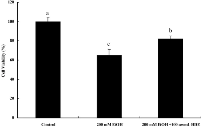
지구자 추출물의 알코올성 ROS 생성 및 산화 억제활성 DCF-DA는 세포막을 통해 확산되며 세포내 esterase에 의해 효소적으로 가수분해 되어 비형광성 DCF-H가 되며, 세포내 ROS가 존재할 경우 매우 빠르게 높은 형광을 띈 DCF로 산화된다(20). DCFH-DA를 이용하여 세포내 ROS 생성량을 측정함으로써 지구자 추출물의 알코올성 산화에 의한 ROS 생성 억제능을 평가하였고, 그 결과 Fig. 1에서와 같이 알코올에 의해 세포내 다량 생성된 ROS가 지구자 알코 올추출물(HDE)에 의해 억제됨을 확인할 수 있었다. 또한 3일간의 200 mM 알코올에 의해 유도된 알코올성 산화는 HDE에 의해 억제되었다(Fig. 2). 알코올로 야기되는 세포독 성은 ROS 생성 증가에 의한 것이라고 알려져 있으며, 이러 한 ROS 생성의 증가는 알코올로 유도되는 CYP2E1에 의한 간 손상의 주요 경로이다. 이상의 결과로부터, 지구자 추출
0 500 1000 1500 2000 2500 3000
Control 200 mM EtOH 200 mM EtOH +100 μg/mL HDE
Fluorescence Unit
c
a
b
Fig. 1. Effects of extract from Hovenia dulcis fruit on intra- cellular ROS level in ethanol treated HepG2/2E1 cells. Data express the mean±SD. Different letters above the bar indicate statistically significant differences by Duncan’s multiple range test (p<0.05).
0 20 40 60 80 100 120
Control 200 mM EtOH 200 mM EtOH +100 µg/mL HDE
Cell Viability (%)
a
c
b
Fig. 2. Effects of extract from Hovenia dulcis fruit on etha- nol-induced oxidative damage in ethanol treated HepG2/2E1 cells. Data express the mean±SD. Different letters above the bar indicate statistically significant differences by Duncan’s multiple range test (p<0.05).
물은 이러한 알코올 유도 CYP2E1에 의해 발생되는 ROS를 억제할 수 있을 것으로 사료된다.
체중증가량과 체중 당 간 무게의 비율
알코올과 HDE를 마우스 각 군에 1주일간 경구투여한 후 측정한 체중증가율, 간 무게 및 체중 당 간 무게의 비율은 Table 1과 같다. 체중증가량과 간 무게에서는 각 군 사이에 유의적 차이를 보이지 않았다. 그러나 체중 당 간 무게의 비율은 알코올 투여에 의해 통계적으로 유의하게 증가되었 다. 이는 알코올로 유도된 염증성 반응으로 간의 상대중량의 증가에 의한 결과로 생각된다.
혈청 중 ALT, AST 및 ALP 활성
알코올 투여에 따른 혈청 ALT, AST 및 ALP활성 상승에 대한 HDE의 억제효과는 Table 2와 같다. 혈청 amino- transferase 활성은 오랫동안 간 손상의 지표로서 유용하게 사용되어 왔다(21). 간세포 손상은 수송기능 및 막 투과성에 변화를 초래하여 결국 세포로부터 효소들을 혈액으로 방출 시킨다(22). 따라서 ALT 및 AST의 순환계로의 많은 방출 은 알코올에 의한 독성화 과정 동안에 간 조직 막에 심각한 손상을 의미한다. 본 연구에서 마우스에 알코올 투여는 혈청 ALT, AST 및 ALP 양의 급격한 상승을 초래하였는데, 이로 써 간 독성효과가 나타남을 알 수 있었다. 이러한 결과는 알코올 단독투여에 의한 혈청 효소들의 증가를 보고한 Ahn 등 연구 결과와 일치하였다(23). 이상의 결과로 알코올이 간 의 대사과정에 작용하여 대사 이상을 초래함으로써 간세포
Table 1. Changes in weight gain, liver weight, and liver in- dex in experimental mice1)
Group2) Weight gain (g)
Liver weight (g)
Liver index3) (%) NC
ET ET-HD
2.06±0.73a4) 1.89±0.67a 1.93±0.63a
1.56±0.14a 1.75±0.12a 1.48±0.21a
3.97±0.21b 4.38±0.10a 3.84±0.24b
1)Values are mean±SD of ten mice per each group.
2)The mice were given saline (NC), 5 g/kg body weight ethanol (ET), and 5 g/kg body weight ethanol plus 1 g/kg body weight ethanolic extract of Hovenia dulcis fruit (ET-HD).
3)Liver weight (g)/ body weight (g)×100.
4)Values with different letters in the same column are sig- nificantly different among groups by Duncan’s multiple range test (p<0.05).
Table 2. Analyses of hepatochemical parameters1)
Group2) ALT
(Karmen unit) AST
(Karmen unit) ALP (K-A unit) NC
ET ET-HD
39.07±2.09b3) 51.27±0.86a 37.19±1.68b
78.43±2.13c 116.28±1.56a 92.01±3.14b
21.98±0.64c 38.85±0.94a 26.29±0.82b
1)Values are mean±SD of ten mice per each group.
2)Groups are described at Table 1.
3)Values with different letters in the same column are sig- nificantly different among groups by Duncan’s multiple range test (p<0.05).
Table 3. Hepatic antioxidant enzyme activities1)
Group2) CAT
(U/mg protein)
SOD (U/mg protein)
GST (U/mg protein) NC
ET ET-HD
58.52±4.28a3) 37.22±5.55b 40.28±2.45b
28.96±0.75a 25.26±0.48b 27.55±0.21a
14.10±0.63a 9.80±0.52c 11.80±0.47b
1)Values are mean±SD of ten mice per each group.
2)Groups are described at Table 1.
3)Values with different letters in the same column are sig- nificantly different among groups by Duncan’s multiple range test (p<0.05).
손상이 증가되고 이러한 알코올성 간 손상은 지구자 추출물 투여에 의해서 보호활성을 나타내는 것으로 추정할 수 있다.
간 조직 중의 항산화 효소 활성
CAT는 cytochrome계를 보유하고 있는 모든 호기성 세포 에 널리 분포되어 있는 효소로서 특히 간에 가장 많이 함유 되어 있다. 이 효소는 과산화수소수를 물과 산소로 분해시키 는 역할을 담당하고 있다(24). 알코올 투여 시 CAT 활성은 급격히 감소되었다(Table 3). 이는 알코올에 의해 유도된 세 포내 라디칼에 의하여 효소활성이 억제되거나 또는 효소 활 성능 이상의 라디칼의 과생산에 기인되는 것으로 추정된다.
알코올성 간 손상에서 HDE 투여에 따른 CAT 활성 향상효 과는 나타나지 않았다. SOD는 반응성이 높으며 독성을 유발 하는 라디칼인 superoxide anion radical을 과산화수소수로 dismutation 시키는 역할을 담당하는 매우 효과적인 항산화 효소이다(25). 본 실험에서 마우스에 알코올 투여 시 SOD 활성은 유의적으로 감소되었다. 그러나 HDE를 함께 투여할 경우 SOD 효소활성은 통계적으로 유의하게 높아졌다 (Table 3). 간의 세포질 및 소포체에 존재하는 수용성 단백질 인 GST는 외인성 물질 및 그 활성 대사물들을 해독하는 역할을 한다(26). 또한, 과산화수소수를 비롯한 활성 대사물 들을 제거함으로써 산화적 손상과 세포막의 지질과산화로 부터 세포를 보호한다(27). 알코올 투여에 의해 GST 활성이 현저히 낮아졌으며 HDE 투여로 인해 GST 효소 활성이 통 계적으로 유의하게 증가하였다(Table 3). 이상의 결과들로 부터 마우스에 알코올 투여로 인한 간의 CAT, SOD 및 GST 활성의 유의적 감소는 알코올 섭취에 의한 자유라디칼의 급 격한 증가에 의한 것으로 생각되며, 지구자 추출물은 이러한 자유라디칼을 제거하는 항산화 효소들을 효과적으로 활성 화시킴으로써 알코올성 산화스트레스로부터 간 보호 효과 를 갖는 것으로 생각된다.
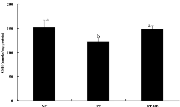
간 조직 중 GSH 함량
만성적 알코올 섭취는 간세포 손상을 일으킨다. GSH는 생명체에 존재하는 주요 비단백 thiol로서 인체의 산화적 손 상에 대한 방어체계에서 중요한 역할을 담당한다. 과산화물 을 환원시키는 다양한 효소과정에서 각종 효소들과 함께 세 포 내외적으로 주요한 역할을 하는 비효소계 항산화제인
Fig. 3. The levels of GSH in ethanol-intoxicated mice. Data express the mean±SD of ten mice per each group. Groups are described at Table 1. Different letters above the bar indicate stat- istically significant differences by Duncan’s multiple range test (p<0.05).
GSH는 redox 반응 및 해독작용을 통하여 정상 세포들의 구조 및 기능을 유지시켜준다(28). 본 연구에서 알코올군 (ET)은 대조군(NC)에 비하여 간의 GSH 함량이 약 30% 정 도 감소되었다(Fig. 3). 또한 HDE는 항산화 활성성분을 함 유하고 있음으로써 알코올 투여에 따른 간의 GSH 함량 감 소를 보호하였을 것으로 추측된다.
간조직 중 지질과산화물 함량
지질과산화는 활성산소종들에 의해 매개된 기작으로서 여러 동물 및 인체실험들을 통하여 다양한 종류의 간 손상을 일으키는 것으로 알려져 왔다(29). Malondialdehyde(MDA) 는 지질과산화 과정 중 생성되는 대표적 활성 알데히드로, 조직 내 알코올에 의해 유도된 지질과산화물의 함량은 MDA 양으로 측정될 수 있다(30). 대조군(NC)과 비교 시 알코올군(ET)에서는 간 MDA 함량이 유의적으로 증가하는 경향을 보였다(Fig. 4). 알코올과 함께 HDE를 투여할 때, 간 MDA 함량이 유의하게 감소함으로써 알코올에 의해 유 도된 지질과산화 과정이 지구자 추출물에 의해 효과적으로 억제됨을 추정할 수 있다.
0.0 0.5 1.0 1.5 2.0
NC ET ET-HD
MDA (nmoles/mg protein)
b
a
b
Fig. 4. The levels of MDA in ethanol-intoxicated mice. Data express the mean±SD of ten mice per each group. Groups are described at Table 1. Different letters above the bar indicate stat- istically significant differences by Duncan’s multiple range test (p<0.05).
0 50 100 150 200
NC ET ET-HD
GSH (nmoles/mg protein)
a
b
a
요 약
알코올에 의해 유도된 간 손상에 대한 지구자 추출물의 보호효과를 연구하였다. HepG2/2E1 세포에서 알코올로 유 도된 ROS 생성과 산화적 손상에 대한 지구자 추출물 보호효 과를 확인하였다. C57BL/6마우스를 대조군(NC), 알코올군 (ET), 알코올과 지구자 추출물 1 g/kg body weight 투여군 (ET-HD)으로 나누었다. 5 g/kg body weight의 알코올을 1주일간 ET와 ET-HD군에 투여하였다. 알코올 투여는 혈 청 alanine amintransferase(ALT), aspartate aminotrans- ferase(AST) 및 alkaline phosphatase(ALP)를 증가시키고, 지구자 추출물은 이러한 간 기능 지표효소의 증가를 억제시 켰다. 간조직의 항산화 효소 활성은 알코올 투여에 의해 감 소되었고, ET-HD군에서 SOD 및 GST 활성은 ET군과 비 교하여 통계적으로 유의하게 높아졌다. GSH 함량은 ET군 에서 NC군에 비하여 유의적으로 낮아졌고, ET-HD군에서 ET군과 비교하여 통계적으로 유의하게 높아졌으며, NC군 과 유사한 함량을 나타내어 간 보호 효과를 확인할 수 있었 다. 지질과산화물 함량은 ET-HD군과 NC군이 유사한 함량 을 나타냄으로써 알코올에 의해 유도된 지질과산화물 증가 에 의한 간손상으로부터 지구자 추출물의 보호 효과를 보여 주었다. 이상의 결과로부터, 지구자 추출물은 세포 및 동물 모델에서 알코올로 유도된 간 손상으로부터 항산화 방어 대 사의 증가와 지질과산화율의 감소에 의해 간세포 보호 활성 을 나타냄을 확인하였다. 이에 지구자 추출물은 알코올성 간 손상으로부터 보호 효과를 갖는 소재로 활용될 수 있을 것으로 사료된다.
문 헌
1. Maher JJ. 1997. Exploring alcohol’s effects on liver function.
Alcohol Health Research World 21: 5-12.
2. Neuman MG. 2003. Cytokines-central factors in alcoholic liver disease. Alcohol Res Health 27: 307-316.
3. Tuma DJ, Casey CA. 2003. Dangerous by-products of alco- hol breakdown-focus on adducts. Alcohol Res Health 27:
285-290.
4. Castillo T, Koop DR, Kamimura S, Triadafilopoulos G, Tsukamoto H. 1992. Role of cytochrome P-450 2E1 in etha- nol-, carbon tetrachloride and iron-dependent microsomal lipid peroxidation. Hepatology 16: 992-996.
5. García-Ruiz C, Morales A, Ballesta A, Rodés J, Kaplowitz N, Fernández-Checa JC. 1994. Effect of chronic ethanol feeding on glutathione and functional integrity of mi- tochondria in periportal and perivenous rat hepatocytes. J Clin Invest 94: 193-201.
6. 이시진. 1982. 본초강목. 인민위생출판사, 북경. p 1845-1846.
7. Kim TJ. 1996. Korean Resources Plants. Seoul National University Press, Seoul. p 72.
8. Yoshikawa M, Murakami T, Ueda T, Matsuda H, Yamahara J, Murakami N. 1996. Bioactive saponins and glycosides. Four methyl-migrated 16,17-seco-dammarane triterpene glycosides from Chinese natural medicine, hove-
niae semen seu fructus, the seeds and fruit of Hovenia dul- cis Thunb.: absolute stereostructures and inhibitory activ- ity on histamine release of hovenidulciosides A1, A2, B1, and B2. Chem Pharm Bull 44: 1736-1743.
9. Fang HL, Lin HY, Chan MC, Lin WL, Lin WC. 2007.
Treatment of chronic liver injuries in mice by oral admin- istration of ethanolic extract of the fruit of Hovenia dulcis.
Am J Chin Med 35: 693-703.
10. Ji Y, Li J, Yang P. 2001. Effects of fruits of Hovenia dulcis Thunb on acute alcohol toxicity in mice. Zhong Yao Cai 24: 126-128.
11. Carmichael J, DeGraff WG, Gazdar AF, Minna JD, Mitchell JB. 1987. Evaluation of a tetrazolium-based semiautomated colorimetric assay: assessment of chemosensitivity testing.
Cancer Res 47: 936-942.
12. Bradford MM. 1976. A rapid and sensitive method for the quantitation of microgram quantities of protein utilizing the principle of protein-dye binding. Anal Biochem 72: 248-25 13. Reitman S, Frankel SA. 1957. Colorimetric method for de- termination of serum glutamic oxalacetic and glutamic pyr- uvic transaminases. Am J Clin Pathol 28: 56-63.
14. Kind PRN, King EJ. 1954. Estimation of plasma phospha- tase by determination of hydrolysed phenol with amino antipyrine. J Clin Path 7: 322-326.
15. Aebi H. 1984. Catalase in vitro. Methods Enzymol 105:
121-126.
16. McCord JM, Fridovich I. 1969. Superoxide dismutase. An enzymic function for erythrocuprein (hemocuprein). J Biol Chem 244: 6049-6055.
17. Habig WH, Jakoby WB. 1981. Assays for differentiation of glutathione S-transferases. Methods Enzymol 77: 398-405.
18. Akerboom TP, Sies H. 1981. Assay of glutathione, gluta- thione disulfide, and glutathione mixed disulfides in bio- logical samples. Methods Enzymol 77: 373-382.
19. Draper HH, Hadley M. 1990. Malondialdehyde determi- nation as index of lipid peroxidation. Methods Enzymol 186:
421-431.
20. Lee YH, Ho JN, Dong MS, Park JH, Kim HK, Hong BS, Shin DH, Cho HY. 2005. Transfected HepG2 cells for evalu- ation of catechin effects on alcohol-induced CYP2E1 cyto- toxicity. J Microbiol Biotechnol 15: 1310-1316.
21. Molander DW, Wroblewsk F, La Due JS. 1955. Transami- nase compared with cholinesterase and alkaline phospha- tase an index of hepatocellular integrity. Clin Res Proc 3:
20-24.
22. Zimmerman HJ, Seeff LB. 1970. Enzymes in hepatic disease. In Dagnostic Enzymology. Goodley EL, ed. Lea and Febiger Publisher, Philadelphia. p 1-38.
23. Ahn YT, Bae JS, Kim YH, Lim KS, Huh CS. 2005. Effects of fermented milk intake on hepatic antioxidative systems in alcohol treated rats. Korean J Food Sci Technol 37:
631-635.
24. Baudrimont I, Ahouandjivo R, Creppy EE. 1997. Prevention of lipid peroxidation induced by ochratoxin A in vero cells in culture by several agents. Chem Biol Interact 104: 29-40.
25. Shim SI, Chung JW, Lee JM, Hwang KT, Sone J, Hong BS, Cho HY, Jun WJ. 2006. Hepatoprotective effects of black rice on superoxide anion radicals in HepG2 cell. Food Sci Biotechnol 15: 993-996.
26. Boyer TD, Bessey DA, Holcomb C, Saley N. 1984. Studies of the relationship between the catalytic activity and bind- ing of nonsubstrate ligands by the glutathione S-trans- ferases. Biochem J 217: 179-185.
27. Mari M, Cederbaum AI. 2001. Induction of catalase, alpha, and microsomal glutathione S-transferase in CYP2E1
overexpressing HepG2 cells and protection against short- term oxidative stress. Hepatology 33: 652-661.
28. Ahn TH, Yang YS, Lee JC, Moon CJ, Kim SH, Jun WJ, Park SC, Kim JC. 2007. Ameliorative effects of pycnogenol® on carbon tetrachloride-induced hepatic oxidative damage in rats. Phytother Res 21: 1015-1019.
29. Paradis V, Kollinger M, Fabre M, Holstege A, Poynard T,
Beddosa P. 1997. In situ detection of lipid peroxidation by-products in chronic liver diseases. Hepatology 26:
135-142.
30. Veca CE, Wilhelm J, Harms-Rihsdahl M. 1988. Interaction of lipid peroxidation product with DNA. A review. Mutat Res Rev Genet Toxicology 195: 137-149.
(2009년 1월 16일 접수; 2009년 1월 23일 채택)