비글견의 하악골에 식립된 임플랜트에 대한 공진주파수와 조직계측분석의 비교 연구
서울대학교 치과대학 치과보철학교실 김우영 장경수 김창회 김영수․ ․ ․
Comparison between Resonance Frequency and Histomorphometric Measurements of Mandibular Implants in Beagle Dogs
Woo-Young Kim, Kyung-Soo Jang, Chang-Whe Kim, Yung-Soo Kim
Department of Prosthodontics, College of Dentistry, Seoul National University.
The use of resonance frequency analysis (RFA) provides a possibility to clinically measure implant stability and osseointegration. The implant stability quotient (ISQ) value of RFA is well known that influenced by effective abutment length and stiffness of the implant in the surrounding tissues. Among these factors stiffness is not accurately defined histologically yet. And the purpose of this study was to find the histolgical relationship of RFA.
17 implants in 3 beagle dogs were used for this study. Among these implants 10 were survived for 7 months, 4 were survived for 3 months and 3 were immediate status after placement. Resonance frequency analyses were conducted and the dogs were sacrificed. Percentage of the bone to implant contact (BIC) in the interface, percentage of the mineralized bone (bone area) within the threads of the implant, and marginal bone level were measured under light microscopy. The correlation between resonance frequency and histomorphometric measurements were analysed and following results were obtained.
1. There was statistically significant correlation between ISQ value and BIC on healed implants. But ISQ value and BIC of all implants were not significantly correlated. (P<0.01)
2. Significant correlation between ISQ value and bone area was not found in this study.
3. There was statistically significant correlation between ISQ value and marginal bone level on all implants as well as on healed implants. (P<0.01)
Ⅰ 서. 론
임플랜트를 이용한 치과치료는 매우 안정적인 치 료로서 높은 성공률을 보인다고 보고되어 왔다 이. 러한 결과를 바탕으로 임플랜트 치료는 최근의 치 과진료에 있어서 빼놓을 수 없는 중요한 한 부분을 차지하게 되었다 또한 재료의 발전 및 다양한 술식. 의 개발로 임플랜트 치료의 영역이 점차 확대되고 있고 치료기간도 상당히 단축되고 있는 형편이, 다.1-9)
이러한 임플랜트 치료가 더욱 안정적이고 효율적 으로 이루어지기 위해서는 식립된 임플랜트에 대한 평가를 정확히 할 수 있어야 한다 골유착된 임플랜. 트를 평가할 수 있는 방법으로는 removal torque 조직계측등과 같은 파괴적인 방법과 타진 방
test, ,
사선사진, Periotest, 공진주파수 분석등과 같은 비파 괴적인 방법들이 있다 파괴적인 방법들이 골유착. 상태에 대한 정확한 정보를 알려 주지만 임상적으 로는 사용할 수 없기 때문에 비파괴적인 방법을 통 해 최대한 정확한 평가를 할 수 있어야 한다.
에 의해 소개된 공진주파수분석 방법은 Meredith
최근에 상품화된 제품이 공급되면서 주목을 받고
있다.10-13) 공진주파수분석의 결과는 ISQ값으로 표
현되는데 임플랜트의 안정성을 표시해 준다고 한 다. 이 ISQ값은 지대주의 유효 길이(effective 와 골내에 매식된 임플랜트의 견고 abutment length)
도(stiffness)에 의해 영향을 받는다고 알려져 있다. 그러나 이중 임플랜트의 견고도에 대한 조직학적인 의미는 아직 정확히 알려져 있지 않다.
본 연구에서는 동물실험을 통해 얻은 공진주파수 분석 결과와 조직계측치를 비교 분석함으로서 임플 랜트의 견고도가 의미하는 바에 대한 조직학적인 관련성을 알아 보고자 하였다.
Ⅱ 연구재료 및 방법. 실험동물 및 임플랜트
1.
비글견 마리에 식립된 임플랜트 중 실험동물의3 희생 직전에 실패하지 않고 남아있었던 17개의 임 플랜트를 대상으로 하였다. 임플랜트는 직경
길이 의
4.0mm, 10.0mm Kimplant(AVANA, Osstem, 가 사용 되었고 하악 견치 제 소구 Busan, Korea) , , 2 치 제 소구치 부위에 식립된 것이었다 구강 내에, 3 . 식립되어 있었던 기간은 개는 개월8 7 , 4개는 개월3 간 생존해 있었고, 3개는 희생직전에 식립된 것이 었다.
공진주파수 측정 2.
을 이용 Osstell(Integration Diagnostics AB, Sweden) 하여 공진주파수 측정을 하였다. Type F1, 높이 L5 의 변환기(transducer, Integration Diagnostics AB, 를 사용하였고 측정은 변환기의 위치를 근 Sweden) ,
심 원심 협측 설측의 방향으로 달리해 가면서 각, , , 4 각 측정하였다 측정결과는 공진주파수 그래프와. 공진주파수 값을 보정한 ISQ(implant stability 값으로 표시되는데 측정 결과의 공진주파 quotient) ,
비글견의 하악골에 식립된 임플랜트에 대한 공진주파수와 조직계측분석의 비교 연구
서울대학교 치과대학 치과보철학교실 김우영 장경수 김창회 김영수․ ․ ․
공진주파수의 그래프 결과를 보인 ISQ값은 사용하 지 않았다.
3. 조직계측
실험동물을 4% buffered paraformaldehyde로 관류 고정 한 후 희생하여 하악골을 적출한 뒤10% 포르 말린 용액에 추가 고정하였다.
조직을 70% 알코올에서 100%까지 농도를 증가 시키면서 탈수 시키고, glycolmethacrylate resin 독일 으로 침투 (Technovit 7200 VLC, Kulzer & Co, )
과정을 진행하였다 레진중합 후. Donath 방법14)에 따라 조직을 협설방향으로 절단 및 연마하여 최종 적으로 70 mμ 두께의 시편을 얻었다 이 시편을.
으로 염색하고 H&E(hematoxylin & eosin) 90%
을 이용하여 마운팅해서 조직슬라이드를 제 glycerol
작하였다.
조직계측은 컴퓨터에 연결된 광학현미경을 이용 하여 사진을 촬영하고 이를 Kappa ImageBase 프로 그램(Kappa opto-electronics GmbH, 독일 을 이용하) 여 분석하였다 변연골 높이는. 12.5배의 광학현미경 사진상에서 임플랜트의 platform에서부터 변연골이 임플랜트와 접촉하기 시작한 지점까지의 거리를 측 정하였다 골 임플랜트 접촉율은 임플랜트의 길이. - 에 대한 골조직과 임플랜트가 접촉하고 있는 길이 의 비율로서40배의 광학현미경 사진상에서 계측하 였다 골밀도는 각 임플랜트에 대해 대표적인 개. 3 의 나선을 선택하여 나선이 이루는 공간의 면적 중 에서 골조직이 차지하는 면적의 비율을 계측한 것 으로 100배의 광학현미경 사진상에서 계측하였다. 4. 통계분석
단순상관분석의 비모수적 방법인 Spearman 상관 계수로 변수들 간의 관련성을SPSS 10.0프로그램에 서 분석하였다.
Ⅲ 결. 과
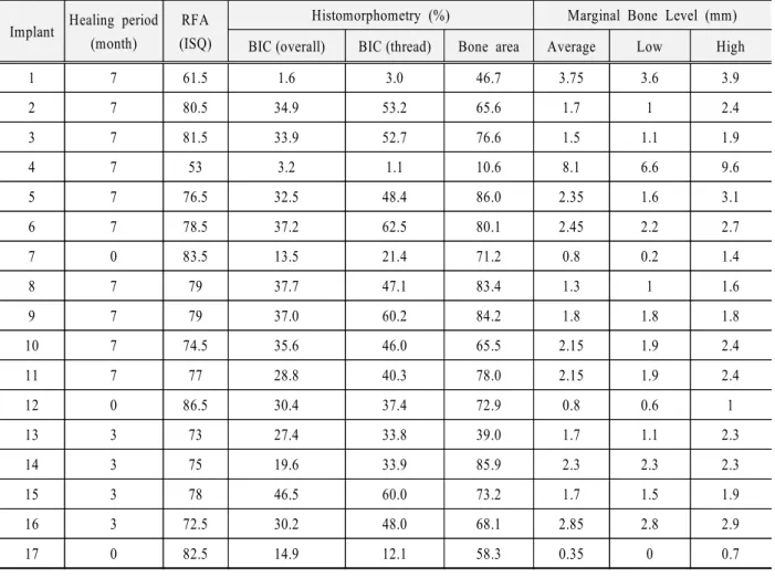
에 각각의 임플랜트에 대한 공진주파수분 Table 1
공진주파수분석 결과 소구치부위에 식립된 임플, 랜트는 협측이나 설측으로 변환기를 위치시킨 경우 다중 피크의 공진주파수 그래프를 보여ISQ값을 인 정할 수 없는 경우가 많았다 따라서 각 임플랜트에. 대한ISQ값은 근심과 원심 방향으로 변환기를 위치 시킨 경우의 ISQ값을 선택하여 그 평균을 구하였 다 최소. 53에서 최대 86.5까지의 값을 보였다.
조직계측분석 결과 중에서 골 임플랜트 접촉율은- 임플랜트 전체길이에 대한 것과 임플랜트의 나선 부분 만에 대한 접촉율을 각각 측정하였다 임플랜. 트 전체길이에 대한 골 임플랜트 접촉율은- 1.6%에 서 46.5%의 범위의 값을 보였고 임플랜트의 나선, 부분 만에 대한 골 임플랜트 접촉율은- 1.1%에서 의 범위의 값으로 전체 임플랜트에 대한 경우 62.5%
보다 전반적으로 높은 값을 보여 주었다 골밀도는. 최소10.6%에서 최대 86.0%까지의 값을 보였다. 변연골 높이는 현미경사진상에서 왼쪽과 오른쪽의 값을 각각 측정하여 둘 중에서 작은 값과 큰 값 두, 값의 평균을 구하여 표시하였다 임플랜트 식립 직. 후가 아닌 경우 변연골은 모두 임플랜트 platform 하방에서 접촉되었고, 1mm에서 9.6mm 범위의 값 을 보였다.
실험대상의 전체 임플랜트에 대한 공진주파수 분 석과 조직계측분석 및 변연골 위치와의 상관관계를 에 보여 주고 있다 공진주파수 분석과 골 임
Table 2 . -
플랜트 접촉율 골밀도간의 유의성 있는 관련성은, 관찰되지 않았고 공진주파수 분석과 변연골 높이, 와는 밀접한 관련이 있었다. (P<0.01)
식립된 직후의 임플랜트를 제외한 구강내에서 치 유과정을 겪은14개의 임플랜트에 대한 공진주파수 분석과 조직계측분석 및 변연골 위치와의 상관관계 를Table 3에 보여 주고 있다 공진주파수 분석과 골. 임플랜트 접촉율 변연골 높이간에 유의성 있는 관
- ,
련성이 관찰 되었다. (P<0.01)
Ⅳ 총괄 및 고안.
공진주파수 분석법은 임플랜트를 평가하는 비 파 괴적인 방법 중 최근에 가장 각광을 받고 있는 방법 중의 하나이다 측정의 재현성과 정확도가 신뢰할.
Table 1. Healing period, RFA, BIC, bone area and marginal bone level of implants used in this study.
Implant Healing period (month)
RFA (ISQ)
Histomorphometry (%) Marginal Bone Level (mm) BIC (overall) BIC (thread) Bone area Average Low High
1 7 61.5 1.6 3.0 46.7 3.75 3.6 3.9
2 7 80.5 34.9 53.2 65.6 1.7 1 2.4
3 7 81.5 33.9 52.7 76.6 1.5 1.1 1.9
4 7 53 3.2 1.1 10.6 8.1 6.6 9.6
5 7 76.5 32.5 48.4 86.0 2.35 1.6 3.1
6 7 78.5 37.2 62.5 80.1 2.45 2.2 2.7
7 0 83.5 13.5 21.4 71.2 0.8 0.2 1.4
8 7 79 37.7 47.1 83.4 1.3 1 1.6
9 7 79 37.0 60.2 84.2 1.8 1.8 1.8
10 7 74.5 35.6 46.0 65.5 2.15 1.9 2.4
11 7 77 28.8 40.3 78.0 2.15 1.9 2.4
12 0 86.5 30.4 37.4 72.9 0.8 0.6 1
13 3 73 27.4 33.8 39.0 1.7 1.1 2.3
14 3 75 19.6 33.9 85.9 2.3 2.3 2.3
15 3 78 46.5 60.0 73.2 1.7 1.5 1.9
16 3 72.5 30.2 48.0 68.1 2.85 2.8 2.9
17 0 82.5 14.9 12.1 58.3 0.35 0 0.7
Table 2. Spearman rank correlation coefficient between RFA and histomorphometric measurements on all implants.
BIC (overall) BIC (thread) Bone area
Marginal bone level
Average Low High
RFA 0.282 0.260 0.298 -0.848** -0.846** -0.812**
** : P < 0.01
Table 3. Spearman rank correlation coefficient between RFA and histomorphometric measurements on healed implants.
BIC (overall) BIC (thread) Bone area
Marginal bone loss
Average Low High
RFA .697** .755** .519 -.741** -.676** -.741**
** : P < 0.01
값으로 표현되는데 에서 까지의 숫자를
ISQ , 1 100
갖는다. Sennerby와 Meredith15)에 의하면, ISQ값이 이하이면 실패할 가능성이 매우 높고 이상이
45 , 65
면 immediate loading도 가능하다고 보고하였다. 임상적으로 ISQ값은 가지 변수의 영향을 받는2 다고 알려져 있다 하나는 지대주의 유효 길이. 로서 변연골 위로 노출된 (effective abutment length)
임플랜트와 지대주의 길이이다 그런데 지대주의. 길이는 변환기에 의해 보정되기 때문에 결국 임플 랜트 주위의 골 흡수 정도가 ISQ값에 영향을 준다 고 할 수 있다 또 한가지 변수는 식립된 임플랜트. 의 견고도(stiffness)이다 처음에 식립된 임플랜트는. 초기 고정이 견고도를 좌우하게 되고 시간이 지남, 에 따라 주변골의 치유와 재형성을 통해 이차적인 고정에 의해 견고도는 유지 또는 증가되게 된다.
임플랜트의 견고도를 조직학적으로 이해할 수 있는 공진주파수분석과 조직학적인 비교 연구는 그리 많 이 접할 수 있지는 않았다. Meredith 등12)은 토끼의 장골에 식립 후 개월 내지 개월 정도 치유된 임플1 5 랜트에 대해 공진주파수와 임플랜트 나선 주위 골 밀도를 비교하였는데 변환기, (transducer)를 수직방 향으로 위치시켰을 때의 공진주파수 값과 골밀도 간에 관련이 보였지만 대체로 측정치의 분포가 너 무 좁아 연관성을 파악하기에는 어려웠다고 보고한 바 있다. Paik16)은 우골을 사용한 비생체 실험에서 값은 골 임플랜트 접촉율과는 관련이 없었고
ISQ - ,
골밀도와는 연관성이 있다고 보고 하였다.
본 연구에 사용된 임플랜트는 식립직후의 것에서 부터 개월간 구강 내에 존재한 임플랜트까지 다양7 한 구성을 하고 있으며 임상적으로 기능하는 임플, 랜트만을 실험대상으로 선택 하였다 따라서 본 연. 구에서는 식립직후의 임플랜트 와 치유가 진행된 임플랜트를 모두 포함하고 있다.
식립직후의 임플랜트는 비록 골 임플랜트 접촉율- 은 낮았지만 ISQ값은 높게 나타났다 물론 변연골. 의 높이가 높고 골밀도가 낮지 않은 점이 식립직후, 임플랜트의 높은 ISQ값을 일부 설명할 수는 있지 만 이 변수만의 영향이라고 보기는 어렵다 왜냐하, . 면 변연골 높이가, 1mm 낮아질 때, ISQ값은 불량한 골질에서 2, 양호한 골질에서 3 정도 감소 한다고
트의ISQ값은 다른 요인도 복합적으로 작용된 결과 인 초기안정성을 표현해 준다고 볼 수 있다.
임플랜트가 치유되면서 ISQ값은 조직계측분석 결과 중 골 임플랜트 접촉율과 관련되어 있다는 결- 과를 본 실험에서 얻었다 이 결과는 치유가 진행된. 임플랜트에서 공명주파수가 골밀도와 관련이 있었 다는 Meredith 등의 결과와 약간의 차이는 있지만 공명주파수와 조직계측치 간의 관련성을 보여주고 있다 따라서 치유가 진행된 임플랜트의. ISQ값은 골개조 등의 결과로 얻어진 조직학적인 결과를 표 현해 준다고 볼 수 있었다.
공진주파수 측정은 일반적으로 변환기를 설측으 로 위치시켜서 치열궁에 수직방향으로 측정한다.
그러나 이 실험에서는 신뢰할 수 없는ISQ값 즉 다, 중공진(multiple resonance) 그래프를 보이는 경우가 이 방향에서 자주 나타나서 결국 치열궁에 평행한 방향으로 변환기를 위치시킨 경우의 값을 대표 ISQ 값으로 사용하였다 근원심 방향의 측정값이 협설. 측 방향의 값보다 약간 높게 나오지만17) 모든 임플 랜트에 대해 같은 방향의 측정값을 사용했으므로 결과의 해석에 영향을 주지는 않았다고 보았다.
본 실험에서는 시편의 수가 충분하지 않아 비모 수적인 통계 방법을 적용하여 상관관계를 분석하였 다 또한 식립직후의 임플랜트는 개에 불과하여. 3 공진주파수와 조직계측분석 간의 관계를 통계적으 로 분석하기는 어려웠다 따라서 향후 공진주파수. 값의 범위가 넓게 분포한 충분한 수의 시편을 대상 으로 한 연구가 진행된다면 공진주파수와 조직계, 측분석 사이의 관계를 더욱 명확히 이해할 수 있을 것이라 사료된다.
V. 결 론
마리의 비글견에 식립된 개의 임플랜트에 대
3 17
해 공명주파수분석과 조직계측분석을 시행하고 값과 골 임플랜트 접촉율 골밀도 변연골위치
ISQ - , ,
간의 상관관계를 조사한 결과 다음과 같은 결론을 얻었다.
치유가 진행된 임플랜트에서 값과 골 임플랜
1. ISQ -
트 접촉율간에 유의성있는 연관성이 관찰되었 고 식립직후의 임플랜트를 포함한 경우에는 연, 관성이 관찰되지 않았다. (P<0.01)
값과 골밀도 간에는 유의성있는 연관성이 관 2. ISQ
찰되지 않았다.
값과 변연골 위치 간에는 식립직후 임플랜트 3. ISQ
를 포함한 경우 및 치유된 임플랜트만을 조사한 경우 모두 유의성있는 연관성이 관찰 되었다.
(P<0.01)
참 고 문 헌
1. Ericsson I, Randow K, Glantz P-O, Lindhe J, Nilner K.
Clinical and radiographical features of submerged and non-submerged titanium implants. Clin Oral Impl Res 1994; 5:185-189.
2. Ericsson I, Randow K, Nilner K, Petersson A. Some clinical and radiographical features of submerged and non-submerged titanium implants. A 5-year follow-up study. Clin Oral Impl Res 1997; 8:422-426.
3. Becker W, Becker BE, Israelson H, et al. One-step surgical placement of Brånemark implants: a prospective clinical multicenter study. Int J Oral Maxillofac Implants 1997; 12:454-462.
4. Collaert B, deBruyn H. Comparison of Brånemark fixture integration and short-term survival using one-stage or two-stage surgery in completely and partially edentulous mandibles. Clin Oral Impl Res 1998; 9:131-135.
5. Schnitman PA, Whörle PS, Rubenstein JE. Immediate fixed interim prostheses supported by two-stage threaded implants: methodology and results. J Oral Implantol 1990; 16:96-105.
6. Schnitman PA, Whörle PS, Rubenstein JE, Silva JD, Want NH. Ten-year results for Br?emark implants loaded with fixed prostheses at fixture placement. Int J Oral Maxillofac Implants 1997; 12:495-503.
7. Henry PJ, Rosenberg I. Single-stage surgery for rehabilitation of the edentulous mandible. Preliminary results. Pract Periodont Aesthetic Dent 1994; 6:1-9.
8. Balshi TJ, Wolfinger GJ. Immediate loading of Brånemark implants in edentulous mandibles: a preliminary report. Implant Dent 1997; 6:83-88.
9. Randow K, Ericsson I, Nilner K, Petersson A, Glantz P-O. Immediate functional loading of Brånemark dental implants. An 18-month study. Clin Oral Impl Res 1999;
10:8-15.
10. Meredith N, Alleyene D, Cawley P. Qualitative determination of the stability of the implant-tissue interface using resonance frequency analysis. Clin Oral Implants Res 1996; 7:261-269
11. Meredith N. On the clinical measurement of implant stability and osseointegration. Göteborg, Sweden:
Department of Biomaterials/Handicap Research, University of Göteborg. Ph D Thesis 1997
12. Meredith N, Shagaldi F, Alleyne D, Sennerby L, Cawley P. The application of resonance frequency measurements to study the stability of titanium implants during healing in the rabbit tibia. Clin Oral Imp Res 1997;8:234-243 13. Friberg B, Sennerby L, Meredith N, Lekholm U. A comparison between cutting torque and resonance frequency measurements of maxillary implants. A 20-month clinical study. Int J Oral Maxillofac Surg 1999a;28:297-303.
14. Donath K., Breuner G. A method for the study of undecalcifed bones bones and teeth with attached soft tissues. J Oral Pathol 1982: 11: 318-326
15. Sennerby L, Meredith N. Resonance frequency analysis.
Current knowledge and clinical implications. Integration Diagnostics.
16. Paik HS. Quantitative determination of the primary stability of implant using resonance frequency analysis and histomorphometry. Ph D thesis(in press). 2003, Seoul National University, Korea.
17. Park CJ. In vitro comparative study between ISQ and Periostest values on the implant stability measurements according to the increased effective implant length. J Korean Academy of Prosthodontics 2001;39:625-635.