• 검색 결과가 없습니다.

Fusobacterium nucleatum GroEL signaling via Toll-like receptor 4 in human microvascular endothelial cells

N/A
N/A
Protected

Academic year: 2021

Share "Fusobacterium nucleatum GroEL signaling via Toll-like receptor 4 in human microvascular endothelial cells"

Copied!
7
0
0

로드 중.... (전체 텍스트 보기)

전체 글

(1)

*Correspondence to: Bong-Kyu Choi, Department of Oral Microbiology and Immunology, School of Dentistry, Seoul National University, 28 Yongon-Dong, Chongno-Gu, Seoul 110-749, Republic of Korea, TEL: 82-2-740-8640, Fax: 82-2-743-0311, E-mail: bongchoi@snu.ac.kr

Fusobacterium nucleatum GroEL signaling via Toll-like receptor 4 in human microvascular endothelial cells

Hae-Ri Lee

1

, and Bong-Kyu Choi

2,3

*

1

Korea Centers for Disease Centers for Disease Control and Prevention, Cheongwon-gun, Chungbuk, Korea

2

Department of Oral Microbiology and Immunology, School of Dentistry, Seoul National University, Seoul, Korea

3
Dental Research Institute, School of Dentistry, Seoul National University, Seoul, Korea

(received August 31, 2012 ; revised September 11, 2012 ; accepted September 11, 2012)

The GroEL heat-shock protein from Fusobacterium nuc- leatum, a periodontopathogen, activates risk factors for atherosclerosis in human microvascular endothelial cells (HMEC-1) and ApoE-/- mice. In this study, we analyzed the signaling pathways by which F. nucleatum GroEL induces the proinflammatory factors in HMEC-1 cells known to be risk factors associated with the development of atherosclerosis and identified the cellular receptor used by GroEL. The MAPK and NF-κB signaling pathways were found to be activated by GroEL to induce the expression of inter- leukin-8 (IL-8), monocyte chemoattractant protein 1 (MCP- 1), intercellular adhesion molecule 1 (ICAM-1), vascular cell adhesion molecule 1 (VCAM-1), E-selectin, and tissue factor (TF). These effects were inhibited by a TLR4 knockdown. Our results thus indicate that TLR4 is a key receptor that mediates the interaction of F. nucleatum GroEL with HMEC-1 cells and subsequently induces an inf- lammatory response via the MAPK and NF-κB pathways.

Key words: Fusobacterium nucleatum, GroEL, Periodon- titis, Signaling

Introduction

Periodontitis is a chronic infection initiated by pathogens in subgingival biofilm [1,2]. Accumulating data show that periodontitis is associated with infection-induced cardiovas- cular disease [3]. Bacterial GroEL is strongly immunogenic and is highly homologous with eukaryotic heat-shock pro- tein 60 (HSP60) [4]. It is regarded as the primary antigen that may cause an autoimmune response via molecular mimicry. Antibodies directed against bacterial GroEL cross- react with human HSP60 on endothelial cells, resulting in autoimmune responses that may lead to endothelial dys- function and the development of atherosclerosis. A positive relationship between periodontitis and the level of serum antibody against GroEL has been observed [5].

Fusobacterium nucleatum is one of the most frequently found organisms in the subgingival plaque of periodontitis patients. It plays a pivotal role in dental biofilm formation by bridging early-colonizing commensals and late-coloni- zing pathogenic bacteria [6]. F. nucleatum is invasive and has been found in human carotid endarterectomy specimens from patients undergoing surgical treatment of atheros- clerosis [7]. GroEL of F. nucleatum induced risk factors of atherosclerosis in human microvascular endothelial cells (HMEC-1) and ApoE

-/-

mice [8].

The objective of this study was to analyze the cellular

receptor and intracellular signaling pathways of F. nuclea-

130

(2)

tum GroEL in HMEC-1 cells to induce risk factors of atherosclerosis.

Materials and methods

Cell culture

HMEC-1 cells were cultured in MCDB131 medium (Gibco BRL, Paisely, UK) supplemented with 15% fetal bovine serum (FBS, Gibco BRL), 100 μg streptomycin (Gibco BRL), 100 U/ml of penicillin(Gibco BRL), 0.1 μg/ml of hydrocortisone (Sigma Chemical Co., St. Louis, MO, USA) and 0.1 μg/ml of epidermal growth factor (Invi- trogen Life Technology, Carlsbad, CA, USA). Cells were maintained in a humidified 5% CO2 atmosphere at 37°C.

Cloning and expression of F. nucleatum GroEL

F. nucleatum subsp. nucleatum (ATCC 25586) was grown in brain heart infusion broth supplemented with 5 μg/ml of hemin (Sigma Chemical Co.) and 0.2 μg/ml of menadione (Sigma Chemical Co.) under an anaerobic atmosphere (10% CO

2

, 5% H

2

, 85% N

2

). The groEL gene was amplified from genomic DNA of F. nucleatum by PCR and cloned in Escherichia coli M15 using the expression vector pQE-30 as described previously [8]. Recombinant F.

nucleatum GroEL was prepared and endotoxin deconta- mination was verified as described previously [6].

Treatment of HMEC-1 with GroEL and real time RT-PCR

HMEC-1 cells (2×10

6

cells/ml) were cultured in 6-well plates and then stimulated with GroEL (10 μg/ml) for various time points. Total RNA of GroEL-treated and non- treated HMEC-1 cells were isolated and cDNA was synthesized using 1 μg/ml of RNA followed by real time RT-PCR to detectmRNA expression levels of interleukin-8 (IL-8), monocyte chemoattractant protein 1 (MCP-1), inter- cellular adhesion molecule 1 (ICAM-1), vascular cell adhe- sion molecule 1 (VCAM-1), E-selectin, and tissue factor (TF) as described previously [8]. Glyceraldehyde dehydro- genase (GADPH) was used as a reference to normalize expression levels. The sequences of the primers for PCR were as follows: 5'-GTG AAG GTG CAG TTT TGC CA- 3' and 5'-TCT CCA CAA CCC TCT GCA C- 3' for IL-8;

5'-CAG CCA GAT GCA ATC AAT GC-3' and 5'-GTG

GTC CAT GGA ATC CTG AA-3' for MCP-1; 5'-CAT ATG CCA TGC AGC TAC AC-3' and 5'-AGT TGT ATG TCC TCA TGG TG-3' for ICAM-1; 5'-AAG CGG AGA CAG GAG ACA C-3' and 5'-TGG CAG GTA TTA TTA AGG AGG ATG-3' for VCAM-1; 5'-TGT GAG ATG CGA TGC TGT C-3' and 5'-AAC CTC TTC TGT CCA TTG TCC-3' for E-selectin; 5'-CAC CGA CGA GAT TGT GAA GGA T-3' and 5'-CCC TGC CGG GTA GGA GAA-3' for TF; 5'-GTG GTG GAC CTG ACC TGC-3' and 5'-TGA GCT TGA CAA AGT GGT CG-3' for GADPH.

Western blot analysis of intracellular signaling path- ways by GroEL in HMEC-1 cells

Antibodies against p38 MAPK, phospho-p38 MAPK (Thr180/Tyr182), phospho-SAPK/JNK (Thr183/Tyr185), phos- pho-p44/42 MAPK (Thr202/Tyr204), and IкBα were purc- hase from Cell Signaling Technology (Beverly, MA, USA).

The antibody against JNK1 (F-3), ERK1 (K-23), and β -actin were obtained from Santa Cruz (Santa Cruz, CA, USA). HMEC-1 cells (1×10

6

cells/well) were cultured in6-well plates and treated with GroEL (10 μg/ml) for various time points (0 to 60 min). The cells were washed with chilled PBS and then lysed in 100 µl of RIPA buffer (10 mM Tris-HC1, pH 7.5, 150 mM NaC1, 1% Triton X-100, 50 mM NaF, 1 mM EDTA, 5 µM Na

3

VO

4

, 1 mM PMSF). Lysates were clarified by centrifugation at 13,000

× g for 45 min at 4

o

C. Fifty µg of lysates were mixed with 5 × SDS-PAGE sample buffer and heated at 95

o

C for 5 min. The samples were separated by SDS-PAGE and transferred onto nitrocellulose membranes. For the detection of activated MAPKs and degraded IкBα, the membranes were blocked with 5% skim milk in PBS containing 0.2%

Tween and then incubated with specific antibodies at 4°C overnight. The membranes were developed using ECL system (Amersham Pharmacia Biotech, Buckingham, UK), and detected with LAS 1000 (Fujifilm, Tokyo, Japan).

Effect of MAPK and NF-κB inhibitors on GroEL-induced intracellular signaling

HMEC-1 cells were seeded onto 6-well plates at a

density of 1×10

6

cells/well and treated with NF-κB inhibitor

(5 μM of Bay 11-7082) or MAPK inhibitors (20 μM of

PD98059 for ERK, 20 μM of SP600125 for JNK, 10 μM

of SB202190 for p38) from Calbiochem (San Diego, CA,

USA) for 1 h before stimulation with GroEL (10 μg/ml),

(3)

E. coli LPS (0.5 μg/ml) or LTA (5 μg/ml) for 12 h. The cells were harvested and RNA isolation, cDNA synthesis, and real time RT-PCR were performed for detection of mRNA expression of IL-8, MCP-1, ICAM-1, VCAM-1, E-selectin, and TF as described above.

TLR4 RNA interference assay

HMEC-1 cells (1×10

5

cells/well) were cultured in 12-well culture plates in serum-and antibiotic-free medium.

Small interference RNA (10 μM siRNA, Invitrogen, TM, Carlsbad, CA, USA) specific for TLR4 or negative control siRNA was mixed with 0.5 μl of lipofectamin RNAiMAX (Invitrogen) in 100 μl of serum- and antibiotic-free medium for 20 min at RT. Then, the mixture was added to the HMEC-1 cells and incubated at 37

o

C (5% CO

2

atmosphere) for 4 h. The medium was replaced with fresh MCDB131 medium containing 15% FBS (Thermo Scien- tific HyClone, Waltham, MA, USA), 0.1 μg/ml of hydro- cortisone (Sigma Chemical Co.), and 0.1 μg/ml of epi- dermal growth factor (Invitrogen), and incubated for 48 h at 37°C. The transfected cells were washed and treated with GroEL (10 μg/ml), E. coli LPS (0.5 μg/ml) or the synthetic bacterial lipopeptide Pam3-Cys-Ser-Lys4 (Pam3 CSK4, 5 μg/ml) for 12 h. Then, mRNA expression levels ofIL-8, MCP-1, ICAM-1, VCAM-1, and E-selectin were determined by real time RT-PCR as described above.

TLR4 knockdown in siRNA-transfected cells were verified by RT-PCR and Western blot analysis. The sequences of siRNA specific for TLR4 were 5'-UAA GGU AGA GAG GUG GCU UAG GCU C-3' and 5'-GAG CCU AAG CCA CCU CUC UAC CUU A-3'.

Statistic analysis

An unpaired Student’s t-test was performed to compare the data between experimental groups. The data were consi- dered statistically significant at p-value of <0.05.

Results

Activation of NF-κB and MAPK signaling pathways by F. nucleatum GroEL

In order to investigate the signaling pathways of GroEL to induce proinflammatory mediators in HMEC-1 cells, HMEC-1 cells were treated with GroEL and examined for

activation of MAPK and NF-κB by Western blot analysis.

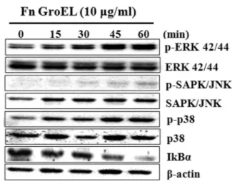
As shown in Fig. 1, phosphorylation of ERK and p38 was observed after 15 min of GroEL stimulation, and phosp- horylation of JNK was detected after 30 min stimulation.

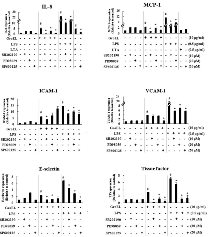
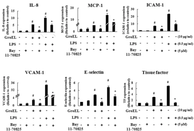
Additionally, IκBα degradation was detected after 15 min of GroEL stimulation. These results showed that F. nucleatum GroEL activates NF-κB and MAPK signaling pathways. We further tested whether the specific inhibitors of MAPKs and NF-κB inhibit the expression of chemokines, cell adhesion molecules, and TF induced by GroEL in HMEC-1 cells. As shown in Fig. 2 and 3, mRNA expres- sion of IL-8 and MCP-1 by GroEL was reduced by pret- reatment with MAPK and NF-κB inhibitors. In the case of adhesion molecules, ICAM-1 expression was reduced by the p38 and NF-κB inhibitors, but not by ERK and JNK inhibitors, whereas VCAM-1 expression was reduced by the JNK and NF-κB inhibitors, but not by p38 and ERK inhibitors. E-selectin expression was reduced by the p38, ERK, and NF-κB inhibitors, but not by JNK. TF expres- sion was reduced by pretreatment with p38, ERK, JNK, and NF-κB inhibitors. LPS-induced IL-8 mRNA expression in HMEC-1 cells was not regulated by JNK inhibitor as described previously [9]. In this study, we also observed that IL-8 mRNA expression induced by LPS was not affected by the JNK inhibitor. IL-8 mRNA expression

Fig 1. MAPK activation and IκBα degradation by F.

nucleatum GroEL in HMEC-1 cells. HMEC-1 cells (1 × 10

6

cells/well) were treated with GroEL (10 μg/ml) for various

times. The cell lysates were examined for phosphorylation of

ERK, JNK, and p38 and IκBα degradation by Western blotting

using antibodies against p38 MAPK, phospho-p38 MAPK

(Thr180/Tyr182), phospho-SAPK/JNK (Thr183/Tyr185), phospho-

p44/42 MAPK (Thr202/Tyr204), and IкBα.

(4)

Fig. 2. Effect of MAPK inhibitors on mRNA expression of inflammatory mediators and TF induced by GroEL in HMEC-1 cells.

HMEC-1 cells (1 × 10

6

cells/well) were pretreated with MAPK inhibitors (20 μM of PD98059 for ERK), 20 μM of SP600125 for

JNK), 10 μM of SB202190 for p38) for 1 h before stimulation with GroEL (10 μg/ml) for 12 h. The expression levels of IL-8,

MCP-1, ICAM-1, VCAM-1, E-selectin, and TF mRNA were determined by real time RT-PCR. The means and standard deviations

are shown.

#

p < 0.05, significant difference compared to the non-treated control and *p < 0.05, significant difference compared to the

stimulant-treated cells.

(5)

Fig. 3. Effect of the NF-κB inhibitor on mRNA expression of inflammatory mediators and TF induced by GroEL in HMEC-1 cells.

HMEC-1 cells (1 × 10

6

cells/well) were pretreated with the NF-κB inhibitor (5 μM of Bay 11-7082) for 1 h before stimulation with GroEL (10 μg/ml) for 12 h. The expression levels of IL-8, MCP-1, ICAM-1, VCAM-1, E-selectin, and TF mRNA were determined by real time RT-PCR. The means and standard deviations are shown.

#

p < 0.05, significant difference compared to the non-treated control and *p < 0.05, significant difference compared to the stimulant-treated cells.

induced by LTA used as a positive control was decreased by the JNK inhibitor.

Involvement of TLR4 in the expression of inflammatory mediators by GroEL in HMEC-1 cells

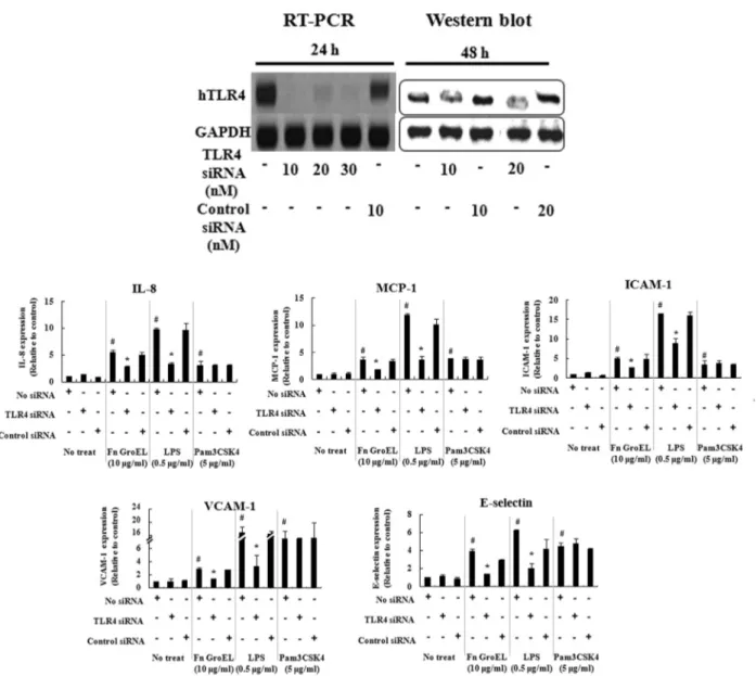
To characterize the host cell receptor for GroEL, we exa- mined the involvement of TLR4 in the expression of inflam- matory mediators by GroEL in HMEC-1 cells which were transfected with TLR4 siRNA. Knockdown of TLR4 in HMEC-1 cells was verified at the gene and protein level (Fig.

4A). As shown in Figure 4B, TLR4 knockdown resulted in a significant reduction of IL-8, MCP-1, ICAM-1, VCAM-1, and E-selectin mRNA expression induced by GroEL treat- ment for 12 h in HMEC-1 cells. These results indicate that TLR4 acts as a cellular receptor for induction of inflam- matory mediators by F. nucleatum GroEL in HMEC-1 cells.

Discussion

Our previous study showed that F. nulceatum GroEL

induced chmokines, cell adhesion molecules, and TF in

HMEC-1 cells, which are atherosclerotic risk factors [6]. In

this study, we have demonstrated that F. nucleatum GroEL

is recognized by TLR4 and its intracellular signaling was

induced via TLR4 to activate MAPK and NF-κB path-

ways. Human HSP60, the GroEL homolog, induced expres-

sion of pro-inflammatory cytokines such as TNF-α, IL-6

and IL-12 [10]. Bacterial GroEL also acts as a powerful

stimulator of host cells. GroEL of Propionibacterium acnes

induced IL-1α, TNF-α and GM-CSF production in kera-

tinocytes [11]. Chlamydial GroEL induced NF-κB activa-

tion and the expression of E-sectin, ICAM-1, VCAM-1,

and IL-6 in vascular endothelial cells from human umbili-

cal cord [10]. E. coli GroEL induced IL-6 secretion in

mice macrophages [12]. Identification of cellular receptors-

which recognize bacterial GroEL facilitates the unders-

tanding of the pathogenesis of the immunodominant anti-

gen. TLRs are the primary receptors to bind various patho-

(6)

Fig. 4. TLR4-dependent expression of chemokines and adhesion molecules by GroEL. HMEC-1 cells (1×10

5

cells/well) were transfected with siRNA specific for TLR4 or the control for 4 h. TLR4 knockdown was verified by RT-PCR and Western blot analysis (A). TLR4-transfected HMEC-1 cells were treated with GroEL (10 μg/ml), E. coli LPS (0.5 μg/ml), and Pam3CSK4 (5 μ g/ml) for 12 h and the expression levels of IL-8, MCP-1, ICAM-1, VCAM-1, and E-selectin mRNA were analyzed by real time RT-PCR (B). The means and standard deviations are shown.

#

p < 0.05, significant difference compared to the control siRNA-transfected cells and *p < 0.05, significant difference compared to non-treated cells.

gen-associated molecular patterns. Bacterial GroEL has been reported to activate TLRs such as TLR4 and/or TLR2 [10,13]. Among periodontopathogens, Porpyromonas gingi- valis GroEL, which was most intensively studied, has been shown to activate NF-κB via TLR2 and TLR4 in THP-1 cells [14]. In our study, we did not analyze whether F.

nucleatum GroEL interacts with TLR2. The involvement of TLR2 in F. nucleatum GroEL signaling needs to be analy- zed in other cell types, because HMEC-1 cells express very low level of TLR2 [15]. E. coli GroEL recognized class

B scavenger receptors (SR-B), such as CLA-1, CLA-2, and CD36, and mediated intracellular signaling in HeLa cells [10]. The SR-B recognition was TLR-independent.

As we used recombinant GroEL, there might be a con-

cern about endotoxin contamination in the recombinant pro-

tein, which may result in the recognition of TLR4. To

verify that the recombinant GroEL was not contaminated

with endotoxin, we tested the activity of F. nucleatum

GroEL using a Chinese hamster ovary/CD14 cell line trans-

fected with a plasmid expressing human TLR4 (CHO/

(7)

CD14/TLR4) in the presence or absence of polymyxin B.

The cell line has the gene encoding membrane CD25 with the human E-selectin promoter, which contains NF-κB binding sites [16]. Both GroEL and LPS increased CD25 expression. However, GroEL-induced CD25 expression was not affected by polymyxin B, whereas LPS-induced CD25 expression was inhibited by polymyxin B. The endotoxin activity of the recombinant GroEL was was about 1/75,000 of the same amount of E. coli LPS by Limulus amebocyte lysate assay. These results indicated that endotoxin conta- mination was minimal in the recombinant GroEL.

In conclusion, our study demonstrates that F. nuleatum GroEL induces its intracellular signaling in HMEC-1 cells via TLR4 to activate MAPK and NF-κB pathways, which induce proinflammatory mediators. Further analysis is required to reveal the involvement of other cellular receptors including TLR2 and SR-B. And identification of GroEL domains interacting with the specific receptors would reveal the detailed mechanisms underlying interac- tion between F. nuleatum GroEL and the cell.

Acknowledgements

This work was supported by the National Research Foundation of Korea (NRF) grant funded by the Korean government (MEST) (2009-0075889).

References

1. Bak EJ, Kim JH, Lee DE, Park BH, Ryu JH, Cha JH, Jeon RO, Yoo YJ. Inhibitory effect of SPA0355, a thiourea analogue, on inflammation and alveolar bone loss in rats with ligature-induced periodontitis. Int J Oral Biol. 2012;

37:63-68.

2. Choi HJ, Cho HK, Soh YJ. Protective Effect of HP08-0106 on Ligature-induced Periodontitis in Rats. Int J Oral Biol.

2011; 36:187-194.

3. Ayada K, Yokota K, Kobayashi K, Shoenfeld Y, Matsuura E, Oguma K. Chronic infections and atherosclerosis. Clin Rev Allergy Immunol. 2009;37:44–48.

4. Wick G, Perschinka H, Millonig G. Atherosclerosis as an autoimmune disease: an update. Trends Immunol. 2001;

22:665-669.

5. Tabeta K, Yamazaki K, Hotokezaka H, Yoshie H, Hara K.

Elevated humoral immune response to heat shock protein 60 (hsp60) family in periodontitis patients. Clin Exp Immunol. 2000;120:285-293.

6. Kolenbrander PE, Palmer RJ Jr, Periasamy S, Jakubovics, NS. Oral multispecies biofilm development and the key role of cell-cell distance. Nat Rev Microbiol. 2010;

8:471-480.

7. Ford PJ, Gemmell E, Chan A, Carter CL, Walker PJ, Bird PS, West MJ, Cullinan MP, Seymour GJ. Inflammation, heat shock proteins and periodontal pathogens in athe- rosclerosis: an immunohistologic study. Oral Microbiol Immunol. 2006; 21:206-211.

8. Lee HR, Jun HK, Kim HD, Lee SH, Choi BK. Fuso- bacterium nucleatum GroEL induces risk factors of atherosclerosis in human microvascular endothelial cells and ApoE

-/-

mice. Mol Oral Microbiol. 201227:109-123.

9. Wang W, Deng M, Liu X, Ai W, Tang Q, Hu J. TLR4 activation induces nontolerant inflammatory response in endothelial cells. Inflammation. 2011;34:509-518.

10. Kol A, Bourcier T, Lichtman AH, Libby P. Chlamydial and human heat shock protein 60s activate human vascular endothelium, smooth muscle cells, and macrophages. J Clin Invest. 1999103:571-577.

11. Graham GM, Farrar MD, Cruse-Sawyer JE, Holland KT, Ingham E. Proinflammatory cytokine production by human keratinocytes stimulated with Propionibacterium acnes and P. acnes GroEL. Br J Dermatol. 2004;150:421-428.

12. Baranova IN, Vishnyakova TG, Bocharov AV, Leela- havanichkul A, Kurlander R, Chen Z, Souza AC, Yuen PS, Star RA, Csako G, Patterson AP, Eggerman TL. Class B scavenger receptor types I and II and CD36 media- tebacterial recognition and proinflammatory signaling induced by Escherichia coli, lipopolysaccharide, and cytosolic chaperonin 60. J Immunol. 2012188:1371-1380.

13. Vabulas RM, Ahmad-Nejad P, da Costa C, Miethke T, Kirschning CJ, Häcker H, Wagner H. Endocytosed HSP60s use Toll-like receptor 2 (TLR2) and TLR4 to activate the toll/interleukin-1 receptor signaling pathway in innate immune cells. J Biol Chem. 2001;276:31332-31339.

14. Argueta JG, Shiota S, Yamaguchi N, Masuhiro Y, Hanazawa S. Induction of Porphyromonas gingivalis GroEL signaling via binding to Toll-like receptors 2 and 4.

Oral Microbiol Immunol. 2006;21:245-251.

15. Faure E, Equils O, Sieling PA, Thomas L, Zhang FX, Kirschning CJ, Polentarutti N, Muzio M, Arditi M.

Bacterial lipopolysaccharide activates NF-κB through toll-like receptor 4 (TLR-4) in cultured human dermal endothelial cells. Differential expression of TLR-4 and TLR-2 in endothelial cells. J Biol Chem. 2000275:

11058-11063.

16. Han SH, Kim JH, Martin M, Michalek SM, Nahm MH.

Pneumococcal lipoteichoic acid (LTA) is not as potent as staphylococcal LTA in stimulating Toll-like receptor 2.

Infect Immun. 200371:5541-5548.

수치

Fig 1. MAPK activation and IκBα  degradation by F.
Fig. 2. Effect of MAPK inhibitors on mRNA expression of inflammatory mediators and TF induced by GroEL in HMEC-1 cells
Fig. 3. Effect of the NF-κB inhibitor on mRNA expression of inflammatory mediators and TF induced by GroEL in HMEC-1 cells
Fig. 4. TLR4-dependent expression of chemokines and adhesion molecules by GroEL. HMEC-1 cells (1×10 5  cells/well) were  transfected with siRNA specific for TLR4 or the control for 4 h

참조

관련 문서

These results suggest that the bilobalide inhibits the cell proliferation and induces the apoptotic cell death in FaDu human pharyngeal squamous cell carcinoma via both

Taken together, these results suggested that latex containing the ficin inhibited the cell growth and induced apoptosis by caspase and Bcl-2 family signaling pathway in

Sequencing results of groEL gene of Anaplasma phagocytophilum detected in blood, kidney and spleen of wild rodents captured in Jeollanam-do area using a

These results indicated that KS28 could inhibit MAPK pathway signaling and that inhibition of the MAPK pathway could affect the suppression of

포에서 We s t e r nbl ot 을 통해 HJ ME가 i NOS의 단백질 발현을 농도의존적으로 저해 하는 것을 확인하였다( Fi g.6) .그러므로 환삼덩굴은 i NOS의 발현을

Conclusion I: Stem Cells Reprogramming of human chondrocytes via non-viral minicircle DNA delivery. Conversion of partially reprogrammed chondrocytes

7.3 Switching systems (Routers in Backbone) 7.4 Signaling systems... Dual-tone multi-frequency

Collectively, exogenous 8-OHdG decreased the expres- sion of NOX1/2/4, TLR2/4, and NF-κB by decreasing Rac1 activity in DOX-treated cells, which eventually led to