ISSN 0378-6471 (Print)⋅ISSN 2092-9374 (Online)
http://dx.doi.org/10.3341/jkos.2016.57.4.601
Original Article
녹내장 환자의 방수 내 사이토카인 농도 분석
Elevated Matrix Metalloproteinase in Aqueous Humor in Patients with Open-Angle Glaucoma
김효원⋅지동현⋅최진아
Hyo Won Kim, MD, Dong Hyun Jee, MD, Jin A Choi, MD, PhD
가톨릭대학교 의과대학 성빈센트병원 안과 및 시과학교실
Department of Ophthalmology and Visual Science, St. Vincent's Hospital, College of Medicine, The Catholic University of Korea, Suwon, Korea
Purpose: To compare the aqueous concentrations of cytokines in glaucomatous eyes with those of normal controls and to char- acterize the clinical factors associated with aqueous cytokine concentration.
Methods: In this cross-sectional study, aqueous humor samples were collected from 54 eyes of 54 patients (34 medically treated primary open angle glaucoma and 20 normal controls) during cataract surgery from January 2014 to January 2015. Glaucoma patients were divided into two groups: patients using prostaglandin analogue for more than 6 months (prostaglandin F2α ana- logue [PGA] user) and patients with no experience of PGA use (PGA non-user). The levels of cytokines (matrix metal- loproteinase [MMP]1, MMP9, MMP3, vascular endothelial growth factor, interleukin [IL]-1, IL-8, tumor necrosis factor [TNF]-α) in the aqueous of glaucoma and control subjects were quantified using a multiplex cytokine analysis.
Results: Aqueous humor collected from the glaucoma patients exhibited significantly increased concentrations of MMP1 (p = 0.002) and MMP9 (p = 0.026). Among glaucoma patients, PGA users showed significantly higher level of MMP 9 compared with PGA non-users (p = 0.003). In the univariate and multivariate linear regression analyses, PGA use (β = 0.351, p = 0.027) and ver- tical cup-to disc ratio (β = -0.401, p = 0.013) were the significant risk factors associated with the level of MMP9.
Conclusions: Primary open-angle glaucoma showed increased aqueous levels of MMP1 and MMP9. Especially, PGA use for longer than 6 months was associated with increased level of MMP9.
J Korean Ophthalmol Soc 2016;57(4):601-606
Keywords: Aqueous humor, Cytokines, Glaucoma, Matrix metalloproteinase, Prostaglandin
■Received: 2015. 10. 15. ■ Revised: 2015. 12. 18.
■Accepted: 2016. 2. 13.
■Address reprint requests to Jin A Choi, MD, PhD
Department of Ophthalmology, The Catholic University of Korea St. Vincent's Hospital #93 Jungbu-daero, Paldal-gu, Suwon 16247, Korea
Tel: 82-31-249-8310, Fax: 82-31-251-6225 E-mail: [email protected]
* This research was supported by the Catholic Medical Center Research Foundation made in the program year of 2014 (5-2014-B001-00184), 2015 (5-2014-B0001-00220) and the St.
Vincent’s Hospital, research institute of medical science (SVHR-2013-14).
ⓒ2016 The Korean Ophthalmological Society
This is an Open Access article distributed under the terms of the Creative Commons Attribution Non-Commercial License (http://creativecommons.org/licenses/by-nc/3.0/) which permits unrestricted non-commercial use, distribution, and reproduction in any medium, provided the original work is properly cited.
녹내장은 진행되는 녹내장성 시신경 병증 및 이에 상응 하는 시야 결손으로 정의되며 안압 상승이 현재까지 알려진 가장 중요한 위험 인자이다. 후방에서 생성된 방수(aqueous humor)는 섬유주세포(trabecular meshwork endothelial cell), 그리고 쉴렘관을 통하는 섬유주 쉴렘관 경로를 통하여 대 부분이 배출되는데, 이러한 배출로의 저항이 증가되었을 때 안압이 상승된다.1 개방각 녹내장에서 안압 상승의 원인 인자로는 변형된 스테로이드 대사, 아드레날린성 조절 장 애, 비정상적 면역학적 과정, 산화로 인한 손상 등의 가능성 이 제기되고 있지만, 방수 배출로의 저항 증가에 관계되는 정확한 원인에 대해서는 아직 뚜렷이 밝혀진 것이 없다.1
방수는 적절한 안압을 유지하는 기능 외에 안구 내 조직 의 생리적 기능과 대사적 항상성을 유지하는 중요한 기능 을 하며, 섬유주세포, 각막 내피세포, 홍채, 수정체 등과 직 접 접촉하고 있다. 따라서 방수의 특징을 통하여 간접적으 로 녹내장에 있어 섬유주세포 등 안압에 관계되는 전방 조 직의 특징을 밝히고자 하는 연구들이 있어 왔다.2-5 녹내장 안의 방수에서 염증성 사이토카인이 상승되어 있음이 보고 된 바 있다.4 또한 섬유주세포에서 생리적으로 extracellular matrix를 분해시켜 결과적으로 extracellular matrix의 순환 (turnover)에 관여하는 효소인 matrix metalloproteinase (MMP)의 농도가 녹내장 안에서 증가되어 있음이 보고된 바 있다.2,3,6 그러나 실제 녹내장 환자의 방수는 녹내장 자 체뿐 아니라, 환자의 나이, 성별, 녹내장의 정도, 그리고 녹 내장 환자에서 대부분 평생 유지해야 하는 안압 하강제 등 에 의해서도 영향을 받을 가능성이 많다. 그러나 녹내장 방 수에서 특징적으로 발현되는 사이토카인과 녹내장 환자의 임상적 특징의 관계에 대해서는 많은 보고가 이루어지지 않았다. 따라서 본 연구에서는 녹내장안과 정상안의 방수 를 분석하여 사이토카인의 발현 농도 차이를 알아보았다.
다음으로 녹내장안에서 어떠한 임상적 특징이 방수 사이토 카인 농도를 결정하는지 분석해 보고자 하였다.
대상과 방법
본 연구는 비교 분석적, 단면적 연구로 시행되었으며, 가 톨릭대학교 성빈센트병원 의학윤리심의위원회(IRB)의 승 인을 받았고(승인번호: VC14SISI0001) 헬싱키 선언을 따라 시행되었다. 각 환자에게 연구 시작 전 연구 절차 및 과정 에 대한 자세한 설명과 문서화된 동의서를 받은 후 연구를 진행하였다. 2014년 1월부터 2015년 1월까지 가톨릭대학 교 성빈센트병원 안과에 내원하여 백내장 수술을 받은 환 자 중 방수 수집에 동의한 개방각 녹내장(원발 개방각 녹내 장 및 정상안압 녹내장) 환자와 정상안을 대상으로 하였다.
모든 대상 환자에게 과거력을 청취하였고, 최대교정시력 검사, Goldmann 압평 안압계를 이용한 안압 검사, 굴절 검 사, 세극등 현미경 검사, 전방각 검사, 안구 길이, PentacamⓇ (Oculus, Wetzlar, Germany), 비접촉경면현미경을 이용한 각막 내피세포 검사가 시행되었다. 이 중 녹내장 환자들에게 는 안저 촬영 검사, Humphrey Visual Field Analyzer (Swedish Interactive Threshold Algorithm 24-2, Carl Zeiss Meditec, Dublin, CA, USA)를 이용한 표준자동시야검사 및 Stratus OCT (Carl Zeiss Meditec, Dublin, CA, USA)를 이용하여 시신경 단층촬영이 시행되었다. 녹내장은 시신경 검사상, 수직 시신경유두비의 비대칭, 테 얇아짐(rim thinning), 패
임(notching), 함몰(excavation), 망막신경섬유층 손상(retinal nerve fiber layer defect) 등의 녹내장성 시신경 변화를 보이 며 시야 검사에서 이에 상응하는 녹내장성 시야 결손을 보 이는 경우로 정의하였다. 정상안은 안압이 21 mmHg 이하 이면서, 안저 검사상 정상 시신경 모습(시신경유두테가 정 상이고, 시신경 유두 출혈, 시신경테 얇아짐 등이 없음)과 망막신경섬유층 결손이 없는 경우로 정의하였다.
연구 참여자는 세극등 현미경을 이용한 검사상 정상적인 전안부 소견을 보이고, 개방각을 가진 경우를 대상으로 하 였으며, 검사 결과에 영향을 미칠 수 있는 백내장 이외의 안내 질환(망막정맥폐쇄, 망막동맥폐쇄, 당뇨망막병증, 연 령관련황반변성 등)이나, 당뇨 등의 전신질환을 가진 경우 를 제외하였고, 이전의 안내 수술이나 레이저 치료를 시행 한 경우, 안구 내 염증이 있는 경우, 면역저하제 사용자 등 은 대상에서 제외하였다.
녹내장안은 수술 전 사용하던 약제의 종류, 기간, 개수를 기록 하였고, 약제의 종류에 따라 prostaglandin F2α analogue (PGA, Travoprost: Travatan, Alcon, Fort Worth, TX, USA; Latanoprost:
Xalatan, Pfizer, New York, NY, USA; Bimatoprost: Lumigan, Allergan Inc., Irvine, CA, USA; Tafloprost: Taflotan, Santen Inc., Osaka, Japan)를 6개월 이상 사용한 군(PGA 사용군) 과 PGA를 사용하지 않은 군(PGA 비사용군)으로 나누어 분석하였다. 6개월 이하로 PGA를 사용했던 환자는 대상에 서 제외하였다.4 모든 연구 참여자에게 고식적인 수정체 유 화술 및 인공수정체 삽입술을 시행하였다. 수술 시행 전에 전방 천자를 통하여 100 μL의 방수를 수집하였다. 방수 샘 플은 분석을 위하여 채취 2시간 이내에 –80oC에서 냉동 보 관되었다.
Luminex multiplex bead immunoassay (Human Cytokine LINCOplex kit; LINCO Research, St. Charles, MO, USA) 를 이용하여 희석되지 않은 방수 검체에 존재하는 MMP1, MMP9, MMP3, Vascular endothelial growth factor (VEGF), interleukin (IL)-1, IL-8, Tumor necrosis factor (TNF)-α의 농도를 측정하였다. 시료의 분석은 제조 회사의 검사 방침 을 준수하여 이전 연구에서 보고된 바와 같은 방법으로 시 행하였다.7
녹내장안 및 정상대조군 간, 그리고 녹내장안 중 PGA 사 용군과 PGA 비사용군 간의 임상적 특징 및 방수 내 사이 토카인의 농도에 대한 비교 분석을 시행하였다. 비교 분석 은 연속 변수에 대해서는 independent t-test로, 범주형 변수 에 대해서는 Pearson χ2을 통하여 검정하였다. 연속변수에 대한 결과는 평균 및 표준편차로 나타내었다. 다음으로 방 수 내 MMP9의 농도에 미치는 인자에 대하여 단변량 선형 회귀분석(univariate linear regression analysis)을 시행하였
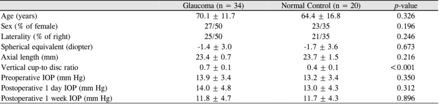
Table 1. Clinical characteristics of glaucoma patients and normal controls
Glaucoma (n = 34) Normal Control (n = 20) p-value
Age (years) 70.1 ± 11.7 64.4 ± 16.8 0.326
Sex (% of female) 27/50 23/35 0.196
Laterality (% of right) 25/50 21/35 0.246
Spherical equivalent (diopter) -1.4 ± 3.0 -1.7 ± 3.6 0.673
Axial length (mm) 23.4 ± 0.7 23.7 ± 1.5 0.216
Vertical cup-to disc ratio 0.7 ± 0.1 0.4 ± 0.1 <0.001
Preoperative IOP (mm Hg) 13.9 ± 3.4 13.2 ± 3.4 0.350
Postoperative 1 day IOP (mm Hg) 14.0 ± 4.8 13.0 ± 4.3 0.312
Postoperative 1 week IOP (mm Hg) 11.8 ± 4.7 11.7 ± 4.3 0.896
Values are presented as mean ± SD unless otherwise indicated.
IOP = intraocular pressure.
Figure 1. Comparisons of the aqueous concentrations of matrix metalloproteinase 1, 3, and 9 between glaucoma patients and normal
controls. All the comparisons were performed by independent t-test. MMP = matrix metalloproteinase.다. 단변량 분석에서 p<0.2인 인자를 다변량 분석에 포함시 켰으며, 후진제거법(backward elimination process)을 이용 하여 최종 다변량 선형 회귀 분석을 시행하였다. 통계학적 분석은 SPSS statistical software 18.0 (SPSS Inc., Chicago, IL, USA)을 사용하였고, p-value가 0.05 미만인 경우를 통 계학적으로 유의한 것으로 정의하였다.
결 과
개방각 녹내장 환자 34명의 34안, 정상안 20명의 20안이 포함되었다. 녹내장안 중 PGA 사용군은 21명 21안, PGA 비사용군은 13명 13안이었다. 녹내장안과 정상대조군의 평 균 나이는 각 70.1 ± 11.7세, 64.4 ± 16.8세로 두 군 간에 통계적으로 유의한 차이가 없었다(p=0.026, Table 1). 그 외 두 군 간에 성별, 우안의 비율, 구면수치 대응치, 안구 길이 역시 두 군 간의 유의한 차이는 없었다(p=0.196, 0.246, 0.673, 0.216, Table 1). 수직 시신경 함몰비는 녹내장안 0.7
± 0.1, 정상대조군 0.4 ± 0.1로 녹내장 군에서 유의하게 컸 다(p<0.001).
방수의 사이토카인 분석 결과, MMP1은 녹내장군에서 6.4 ± 7.0 pg/mL, 대조군에서 1.8 ± 2.9 pg/mL (glaucoma vs.
control, p=0.002), MMP3는 녹내장군에서 885.1 ± 943.7
pg/mL, 대조군에서 401.3 ± 719.2 pg/mL (p=0.090), 그리 고 MMP9은 녹내장군에서 69.4 ± 67.4 pg/mL, 대조군에서 34.8 ± 41.2 pg/mL로(p=0.026), 녹내장군에서 MMP1과 MMP9이 대조군에 비하여 유의하게 증가되어 있는 양상을 보였다(Fig. 1). 그 외 VEGF는 녹내장안에서 58.3 ± 40.6 pg/mL, 대조군에서 45.5 ± 33.5 pg/mL (p=0.285), IL-8은 녹내장안에서 6.12 ± 2.9 pg/mL, 대조군에서 18.57 ± 33.97 pg/mL (p=0.168), TNF-α는 녹내장안에서 0.08 ± 0.22 pg/mL, 대조군에서 0.03 ± 0.09 pg/mL (p=0.347)로 두 군 간에 유의한 차이가 없었으며, IL-1은 두 군 모두에서 검출 되지 않았다.
녹내장안을 PGA 사용군과 PGA 비사용군으로 나누어 분석했을 때, PGA 사용군에서 총 녹내장 약제 수가 유의하 게 많았으나(p=0.029), 사용하는 총 녹내장 약제 중 PGA를 제외한 타 약제를 사용하는 환자 비율(%)은 두 군 사이에 유의한 차이가 없었다(Table 2). 두 군 사이에 방수 사이토 카인 농도를 분석하였을 때 PGA 사용군에서 MMP9의 농 도가 90.68 ± 73.10 pg/mL로 PGA 비사용군 40.27 ± 47.16 pg/mL에 비하여 유의하게 증가되어 있는 양상을 보였다 (p=0.003, Table 2). 그 외 MMP1, MMP3, IL-8, TNF-alpha 등의 다른 사이토카인은 유의한 차이를 보이지 않았다 (Table 2). 녹내장 안에서 방수의 MMP9 농도와 관련된 인
Table 2. Comparison of clinical characteristics and aqueous cytokine concentrations between glaucoma patients using prostaglandin
analogues for longer than 6 months and patients with no experience of prostaglandin analoguesPGA user (n = 21) PGA non-user (n = 13) p-value Clinical characteristics
Age (years) 67.7 ± 13.6 74.2 ± 6.8 0.180
Average RNFL thickness (μm) 77.0 ± 22.5 80.53 ± 17.9 0.787
Axial length (mm) 23.3 ± 0.55 23.57 ± 0.97 0.181
Vertical cup-to disc ratio 0.73 ± 0.17 0.73 ± 0.10 0.775
Preoperative IOP (mm Hg) 13.8 ± 3.0 14.6 ± 5.3 0.649
Postoperative 1 day IOP (mm Hg) 12.5 ± 5.0 10.6 ± 4.1 0.699
Postoperative 1 week IOP (mm Hg) 12.5 ± 5.0 10.6 ± 4.1 0.243
Number of anti-glaucoma eyedrops (number) 1.5 ± 0.9 0.8 ± 0.6 0.029
Use of beta-blocker (%) 42.8 46.1 0.565
Use of carbonid anhydrase inhibitor (%) 28.6 30.7 0.594
Use of α-selective adrenergic drugs (%) 23.8 23.1 0.648
Use of miotic agents (%) 0.0 7.7 0.382
Duration of anti-glaucoma eyedrops (months) 18.2 ± 22.9 26.5 ± 34.1 0.960
Duration of PGA (months) 18.3 ± 22.8 0.0 ± 0.0 0.003
Aqueous cytokine concentration
MMP1 (pg/mL) 6.7 ± 6.2 5.9 ± 8.7 0.420
MMP9 (pg/mL) 90.7 ± 73.1 40.3 ± 47.2 0.003
VEGF (pg/mL) 67.0 ± 46.2 42.3 ± 27.4 0.275
MMP3 (pg/mL) 1251.5 ± 1078.0 396.6 ± 604.6 0.081
IL-1 (pg/mL) 0.0 ± 0.0 0.0 ± 0.0 N/A
IL-8 (pg/mL) 13.2 ± 9.5 6.9 ± 3.6 0.058
TNF-alpha (pg/mL) 0.0 ± 0.0 0.1 ± 0.1 0.588
Values are presented as mean ± SD unless otherwise indicated.
PGA = prostaglandina analogues; RNFL = retinal nerve fiber layer; IOP = intraocular pressure; MMP = matrix metalloproteinase; VEGF
= vascular endothelial growth factor; IL = interleukin; TNF = tumor necrosis factor; N/A = not applicable.
Table 3. Clinical factors affecting aqueous concentration of MMP9 in glaucoma patients
Parameters Univariate Multivariate
B SE β p-value B SE β p-value
Age -0.682 1.061 -0.113 0.525
Sex 15.21 24.75 0.108 0.543
PGA user 50.42 22.8 0.364 0.034 48.54 20.93 0.351 0.027
Axial length -5.92 16.06 -0.066 0.715
Preoperative IOP 0.179 3.64 0.009 0.961
Average RNFL thickness 0.533 0.667 0.152 0.431
Duration of PGA -0.047 0.621 -0.090 0.613
Number of anti-glaucoma eyedrops -5.818 13.68 -0.075 0.674
Vertical cup-to disc ratio -198.8 77.61 -0.412 0.015 -193.10 72.83 -0.401 0.013
MMP = matrix metalloproteinase; B = unstandardized coefficient; SE = spherical equivalent; β = standardized beta; PGA = prostaglandin analogue; IOP = intraocular pressure; RNFL = retinal nerve fiber layer.
자를 선형회귀분석을 통하여 알아보았을 때 단변량 및 다 변량 분석 모두에서 PGA 사용(β=0.351, p=0.027), 낮은 수 직 시신경함몰비(β=-0.401, p=0.013)가 MMP9 상승에 영향 을 미치는 독립적인 인자였다(Table 3).
고 찰
정상적으로 Matrix metalloproteinase는 섬유주세포에서 분비되며, 섬유주세포 사이에 존재하는 extracellular matrix의
분해를 촉진함으로써, extracellular matrix의 순환(turnover)에 핵심적인 역할을 하는 효소이다.8 녹내장 환자의 방수에서 정상인과 다른 MMP 농도를 나타냄이 이전 연구에서 보고 된 바 있다.2,6,9 Weinstein et al3은 개의 안구 조직을 조사한 연구에서 정상안에 비해 녹내장안의 방수 및 전방각 조직 에서 MMP2와 MMP9의 농도가 증가해 있다고 하였다. 그 러나 Pradhan et al5은 실제 환자의 방수에서 MMP2와 MMP9을 분석하였을 때, 유의한 차이가 없었고, 사용하는 프로스타글란딘 제제(prostaglandin F2α analogue)의 종류에
따라서도 유의한 차이가 없다고 하였다. 본 연구에서는 녹 내장안의 방수에 MMP1과 MMP9이 증가되어 있음을 확인 할 수 있었다(Fig. 1).
본 연구에서는 녹내장안에서 증가된 matrix metalloproteinase 가 녹내장의 병인에 관계하는지, 아니면 녹내장의 치료에 의한 이차적인 결과인지를 확인하기 위하여 녹내장의 임상 적 특징이 MMP의 농도를 결정하는지 알아보고자 하였다.
그 결과 MMP9의 경우 프로스타글란딘 제제를 6개월 이상 사용하였을 때 독립적으로 MMP9의 상승과 관련이 있음을 알 수 있었다(β=0.351, p=0.027, Table 3). 여기서 한 가지 흥미로운 점은 수직 시신경함몰비의 경우 오히려 작을수록, 즉 녹내장의 정도가 덜 심할수록 MMP9이 상승되는 방향 으로 나타났다는 점이다(β=-0.401, p=0.013). 이러한 점에 서 미루어 보았을 때, MMP9은 녹내장의 직접적인 병인 또 는 질환의 정도(severity)에 관계한다기보다 프로스타글란 딘 제제 사용에 의한 이차적인 결과일 가능성이 높다고 보 인다.
프로스타글란딘 제제는 특히 포도막공막유출로(uveoscleral outflow)에 영향을 줌으로써 안압을 하강시키는 가장 강력 한 단일 안압 하강제 중의 하나이다.10 프로스타글란딘 제 제는 안방수의 배출로뿐만 아니라 안구 전반에 걸쳐 영향 을 미친다고 보고되었다.11 이전 연구에서 프로스타글란딘 제제는 섬모체근육세포(ciliary smooth muscle cell)12 및 공
막13,14에 이르기까지 전반적으로 MMP의 농도를 상승시킨
다고 하였고, 이로 인하여 조직의 투과성이 증가되어, 프로 스타글란딘 제제에 의한 안압 하강의 기전에 일부 관여한 다고 하였다.13 이뿐 아니라 인간 섬유주세포 배양 후, lata- noprost로 처리하였을 때, 세포에서 MMP1, MMP3, MMP17 의 발현이 증가되었다.15 또한 최근에는 프로스타글란딘 제 제를 사용하는 녹내장 환자에서 눈물의 사이토카인 농도를 분석하였을 때 MMP1, MMP9의 증가와 함께, 길항작용을 하는 tissue inhibitor of metalloproteinase (TIMP)-1이 감소 되어 있음이 밝혀졌다.16
방수의 사이토카인은 녹내장 외의 다른 질환들의 병인과 도 밀접한 관계를 가진다. Jia et al17은 안축장의 길이에 따 라서 MMP2, TIMP-1, 2, 3가 증가한다고 보고하였다. 또한 당뇨 망막병증에서 MMP2, MMP9이 증가되어 있고,18,19 당 뇨의 신생혈관막에서 MMP9의 증가가 보고된 바 있다.20,21 이로 미루어 볼 때, 생리적인 MMP의 조절 메커니즘과는 별도로, 프로스타글란딘 제제의 장기 사용으로 인하여 2차 적으로 MMP1, 9의 농도가 증가하게 되며, 이로 인하여 MMP가 안구에 미칠 수 있는 영향에 대한 고려도 필요할 것으로 보인다.
프로스타글란딘은 염증성 반응의 매개 물질 중 하나로서
이론적으로 프로스타글란딘 제제의 사용이 안구의 염증을 유도할 가능성이 제시되어 왔다.22 MMP9 역시 다양한 염 증 반응을 조절하는 pro-inflammatory mediator로 알려져 있다.23 Chua et al4은 정상안과 녹내장안의 방수를 비교하 여 interferon-gamma, IL-10 등의 염증성 사이토카인이 개 방각 녹내장에서 증가되어 있다고 보고하였다. 그러나 본 연구에서는 IL-1, IL-8, TNF-α 및 VEGF 등의 염증성 사이 토카인들은 녹내장군과 정상군에서 유의한 차이가 발견되 지 않았으며, 프로스타글란딘 제제 사용 여부에 따라서도 유의한 차이가 발견되지 않았다(Table 2). 그러나 본 연구 는 백내장 수술 전에 채취한 방수에서 분석한 것으로, 다양 한 염증성 사이토카인에 대한 분석은 이루어지지 못하였다.
당뇨나 수술 등과 같이 전방 내 염증이 증가될 것이 예상되 는 스트레스 상황에서 MMP9이 전방 내 염증 반응에 미치 는 영향에 대해서는 추가 연구가 필요할 것으로 보인다.24,25 본 연구는 전향적 연구로 직접 사람의 방수를 대상으로 분석했다는 장점이 있다. 본 연구에서는 다변량 분석을 통 하여 프로스타글란딘 제제 사용 외에 방수 내 MMP 농도에 영향을 줄 수 있는 다른 인자를 보정하여 분석하였으나, 대 상안 수의 부족으로 인하여 프로스타글란딘 단일 제제만을 사용한 환자에 대한 분석이 별도로 시행되지 못했다는 점 이 연구의 제한점으로 사료된다. 결론적으로, 녹내장 환자 의 방수를 분석하였을 때, MMP1, MMP9이 상승되어 있었 고, 특히 6개월 이상의 프로스타글란딘 제제의 사용은 MMP9의 상승과 관련된 위험인자임을 알 수 있었다.
REFERENCES
1) Tamm ER. The trabecular meshwork outflow pathways: structural and functional aspects. Exp Eye Res 2009;88:648-55.
2) Markiewicz L, Pytel D, Mucha B, et al. Altered expression levels of MMP1, MMP9, MMP12, TIMP1, and IL-1β as a risk factor for the elevated IOP and optic nerve head damage in the primary open-angle glaucoma patients. Biomed Res Int 2015;2015:812503.
3) Weinstein WL, Dietrich UM, Sapienza JS, et al. Identification of ocular matrix metalloproteinases present within the aqueous hu- mor and iridocorneal drainage angle tissue of normal and glaucom- atous canine eyes. Vet Ophthalmol 2007;10 Suppl 1:108-16.
4) Chua J, Vania M, Cheung CM, et al. Expression profile of in- flammatory cytokines in aqueous from glaucomatous eyes. Mol Vis 2012;18:431-8.
5) Pradhan ZS, Dalvi RA, Lai T, et al. Prostaglandin agonist effect on matrix metalloproteinase aqueous levels in glaucoma patients. Can J Ophthalmol 2015;50:6-10.
6) Chintala SK, Wang N, Diskin S, et al. Matrix metalloproteinase ge- latinase B (MMP-9) is associated with leaking glaucoma filtering blebs. Exp Eye Res 2005;81:429-36.
7) duPont NC, Wang K, Wadhwa PD, et al. Validation and compar- ison of luminex multiplex cytokine analysis kits with ELISA: de-
= 국문초록 =
녹내장 환자의 방수 내 사이토카인 농도 분석
목적: 녹내장안과 정상안의 방수에서 사이토카인의 농도 차이를 알아보고, 녹내장안에서 어떠한 임상적 특징이 방수 내 사이토카인 농도를 결정하는지 분석해 보고자 하였다.
대상과 방법: 2014년 1월부터 2015년 1월까지 백내장 수술을 받은 환자 중 개방각 녹내장(원발 개방각 녹내장 및 정상안압 녹내장) 환자와 정상안을 대상으로 하였다. 녹내장안은 점안 프로스타글란딘 제제(prostaglandin F2α analogue, PGA)를 6개월 이상 사용한 군(PGA 사용군)과 PGA를 사용하지 않은 군(PGA 비사용군)으로 나누어 분석하였다.
결과: 녹내장 환자 34명의 34안, 정상인 20명의 20안이 포함되었고 녹내장안 중 PGA 사용군은 21안, PGA 비사용군은 13안이었다.
방수의 사이토카인 분석 결과, 녹내장안에서 MMP1과 MMP9이 정상 대조군에 비해 유의하게 증가된 양상을 보였다(p=0.002 and 0.026, MMP1 and MMP9). 녹내장안 중에서는 PGA 사용군에서 MMP9이 90.68 ± 73.10 pg/mL로 PGA 비사용군 40.27 ± 47.16 pg/mL에 비하여 유의하게 증가되어 있었다(p=0.003). 단변량 및 다변량 분석 모두에서 PGA 사용 여부(β=0.351, p=0.027) 및 낮은 수직 시신경함몰비(β=-0.401, p=0.013)가 MMP9 상승에 유의한 영향을 미쳤다.
결론: 녹내장 환자의 방수를 분석하였을 때, MMP1과 MMP9이 상승되어 있었고, 특히 6개월 이상 프로스타글란딘 제제의 사용은 MMP9의 상승에 영향을 미치는 인자임을 알 수 있었다.
<대한안과학회지 2016;57(4):601-606>
terminations of a panel of nine cytokines in clinical sample culture supernatants. J Reprod Immunol 2005;66:175-91.
8) Alexander JP, Samples JR, Van Buskirk EM, Acott TS. Expression of matrix metalloproteinases and inhibitor by human trabecular meshwork. Invest Ophthalmol Vis Sci 1991;32:172-80.
9) De Groef L, Van Hove I, Dekeyster E, et al. MMPs in the neuro- retina and optic nerve: modulators of glaucoma pathogenesis and repair? Invest Ophthalmol Vis Sci 2014;55:1953-64.
10) Nakakura S, Tabuchi H, Baba Y, et al. Comparison of the latano- prost 0.005%/timolol 0.5% + brinzolamide 1% versus dorzolamide 1%/timolol 0.5% + latanoprost 0.005%: a 12-week, randomized open-label trial. Clin Ophthalmol 2012;6:369-75.
11) Toris CB, Gabelt BT, Kaufman PL. Update on the mechanism of action of topical prostaglandins for intraocular pressure reduction.
Surv Ophthalmol 2008;53 Suppl1:S107-20.
12) Weinreb RN, Kashiwagi K, Kashiwagi F, et al. Prostaglandins in- crease matrix metalloproteinase release from human ciliary smooth muscle cells. Invest Ophthalmol Vis Sci 1997;38:2772-80.
13) Weinreb RN, Lindsey JD, Marchenko G, et al. Prostaglandin FP agonists alter metalloproteinase gene expression in sclera. Invest Ophthalmol Vis Sci 2004;45:4368-77.
14) Kim JW, Lindsey JD, Wang N, Weinreb RN. Increased human scler- al permeability with prostaglandin exposure. Invest Ophthalmol Vis Sci 2001;42:1514-21.
15) Oh DJ, Martin JL, Williams AJ, et al. Effect of latanoprost on the expression of matrix metalloproteinases and their tissue inhibitors in human trabecular meshwork cells. Invest Ophthalmol Vis Sci 2006;47:3887-95.
16) Lopilly Park HY, Kim JH, Lee KM, Park CK. Effect of prosta-
glandin analogues on tear proteomics and expression of cytokines and matrix metalloproteinases in the conjunctiva and cornea. Exp Eye Res 2012;94:13-21.
17) Jia Y, Hu DN, Zhu D, et al. MMP-2, MMP-3, TIMP-1, TIMP-2, and TIMP-3 protein levels in human aqueous humor: relationship with axial length. Invest Ophthalmol Vis Sci 2014;55:3922-8.
18) Mohammad G, Kowluru RA. Novel role of mitochondrial matrix metalloproteinase-2 in the development of diabetic retinopathy.
Invest Ophthalmol Vis Sci 2011;52:3832-41.
19) Mohammad G, Kowluru RA. Matrix metalloproteinase-2 in the de- velopment of diabetic retinopathy and mitochondrial dysfunction.
Lab Invest 2010;90:1365-72.
20) Hoffmann S, He S, Ehren M, et al. MMP-2 and MMP-9 secretion by rpe is stimulated by angiogenic molecules found in choroidal neovascular membranes. Retina 2006;26:454-61.
21) Das A, McGuire PG, Eriqat C, et al. Human diabetic neovascular membranes contain high levels of urokinase and metalloproteinase enzymes. Invest Ophthalmol Vis Sci 1999;40:809-13.
22) Hu J, Van den Steen PE, Sang QX, Opdenakker G. Matrix metal- loproteinase inhibitors as therapy for inflammatory and vascular diseases. Nat Rev Drug Discov 2007;6:480-98.
23) Kawasaki Y, Xu ZZ, Wang X, et al. Distinct roles of matrix metal- loproteases in the early- and late-phase development of neuro- pathic pain. Nat Med 2008;14:331-6.
24) Song YJ, Jung JH, Kim DG. Analysis of aqueous humor cytokines in diabetic retinopathy. J Korean Ophthalmol Soc 2014;55:1821-7.
25) Sin BH, Kim JY, Park JY, Park SP. Analysis of intraocular cyto- kines according to progression of diabetic retinopathy and macular edema in diabetic patients. J Korean Ophthalmol Soc 2013;54:618-26.