J Korean Ophthalmol Soc 2016;57(1):120-124 ISSN 0378-6471 (Print)⋅ISSN 2092-9374 (Online)
http://dx.doi.org/10.3341/jkos.2016.57.1.120
Case Report
10세 소년의 안와 골막하혈종에서 발생한 신생골 형성
Osteoneogenesis on Subperiosteal Orbital Hematoma in a 10-Year-Old Boy
손대용⋅우경인
Dae Yong Son, MD, Kyung In Woo, MD, PhD
성균관대학교 의과대학 삼성서울병원 안과학교실
Department of Ophthalmology, Samsung Medical Center, Sungkyunkwan University School of Medicine, Seoul, Korea
Purpose: We report the first case in Korea of rapid bone formation on a subperiosteal orbital hematoma after trauma.
Case summary: A 10-year-old boy who was in the intensive care unit after trauma showed proptosis and ocular movement limi- tation of the right eye associated with subperiosteal hematoma. On ocular examination, 3 mm of proptosis and limitation of right eye movement were observed; however, visual acuity was not decreased. At 1 month after the trauma, orbital computed tomog- raphy (CT) showed new bone formation at the margin of the hematoma border although the size of the hematoma decreased.
The patient underwent hematoma and bony tissue removal using anterior orbitotomy approach. A new bone was formed be- tween the orbital border and hematoma from the anterior orbital margin to the orbital apex. During pathological examination, wo- ven bone tissue with fibrotic tissue was observed in the hematoma wall. One year after surgery, the patient’s proptosis and limi- tation of ocular movement disappeared without any evidence of new bone formation.
Conclusions: Waiting for spontaneous absorption of orbital subperiosteal hematoma is usually recommended unless there is significant functional impairment. However, as in our case, new bone formation could occur during a short period of less than 1 month; imaging follow-up is necessary in patients having intensive care or showing delayed absorption of a hematoma.
J Korean Ophthalmol Soc 2016;57(1):120-124
Key Words: Orbital subperiosteal hematoma, Osteoneogenesis
■Received: 2015. 6. 12. ■ Revised: 2015. 7. 14.
■Accepted: 2015. 10. 16.
■Address reprint requests to Kyung In Woo, MD, PhD Department of Ophthalmology, Samsung Medical Center, #81 Irwon-ro, Gangnam-gu, Seoul 06351, Korea
Tel: 82-2-3410-3570, Fax: 82-2-3410-0029 E-mail: eyeminded@skku.edu
* This study was presented as a poster at the 110th Annual Meeting of the Korean Ophthalmological Society 2013.
ⓒ2016 The Korean Ophthalmological Society
This is an Open Access article distributed under the terms of the Creative Commons Attribution Non-Commercial License (http://creativecommons.org/licenses/by-nc/3.0/) which permits unrestricted non-commercial use, distribution, and reproduction in any medium, provided the original work is properly cited.
안와 혈종은 대부분 외상에 의해 발생하게 되는데, 위치 에 따라 안와내혈종과 골막하혈종으로 구분되며 안와내혈 종에 비해 골막하혈종의 빈도가 더 적다. 특히 소아에서 드 물게 관찰되는 안와내 외상성 골막하혈종은 주로 안와 상 부안와의 둔상과 연관이 있는 경우가 많다.
소아나 젊은 남성의 두개내 혈종에서 석회화가 일어난 경우는 보고된 바가 있으나,1-5 안와 골막하혈종에서 석회화 가 관찰되는 것은 매우 드문 소견으로 한 증례가 보고되어 있다.6 이 증례에서는 기존의 골막하혈종과 다른 임상적 영 상의학적 소견은 보이지 않았으며, 다만 수술 후 시행한 조 직검사에서 현미경적으로 발견된 골형성 현상만을 보고하 고 있다.6 저자들은 외상 후 집중치료를 받던 환자의 안와 내 골막하혈종에서 급격히 골조직을 형성하는 경우를 경험 하였기에 이를 영상의학적인 소견과 함께 보고하고자 한다.
증례보고
10세 남자 환아가 우안 안구돌출로 안과검진이 의뢰되었 다. 이전 안과 특이력이 없는 환아로 보행자 교통사고로 수
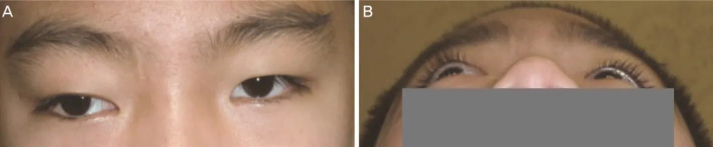
Figure 1. Clinical photographs of the patients eyelids. The photographs shows hypoglobus (A) and exophthalmos (B) of the right
eye.Figure 2. Orbit magnetic resonance imaging (MRI) taken 9 days after trauma. These pictures show subacute stage of hematoma.
T1-weighted axial images (A) and coronal images (B) after gadolinium enhancement with fat suppression reveal a soft tissue mass at the superior portion of right orbit (arrows). Mass shows a high signal intensity on T1-weighted sagittal image with enhancement (C, arrow) and low and high signal intensity on T2-weighted axial image (D, arrow).
상한 후 우안 안구돌출이 발생하였고, 수상 직후에는 우안 움직임이 거의 없을 정도로 심한 안구운동장애를 보였다.
눈 증상 외에도 외상으로 인한 경막하혈종, 광범위 축삭손 상, 폐좌상에 대한 치료를 위해 중환자실에서 2주간 집중치 료를 받았다.
외상 2주 후 시행한 안과검사에서 양안 교정시력은 1.0 이었으며, 안압은 우안 19 mmHg, 좌안 12 mmHg였다. 안구 돌출계검사에서 3 mm의 우안 안구돌출이 있었고, 우측 하 방으로 안구 편위를 보였다(Fig. 1). 환자의 눈꺼풀올림근기 능검사는 양안 11 mm로 측정되었고, 눈꺼풀각막반사간거리 1 (Marginal Reflex Distance 1, MRD1)은 양안 1.5 mm였 다. 안구운동검사에서 상전장애 -3, 외전장애 -1 정도의 우 안 안구운동제한이 있었으며, 중립위치에서 45PD 외사시, 8PD 우안 하사시 소견을 보였고 동반된 복시는 없었다. 상 대구심성동공장애는 없었으며 색각검사도 정상이었다.
외상 9일 후 시행한 안와 자기공명영상(magnetic resonance
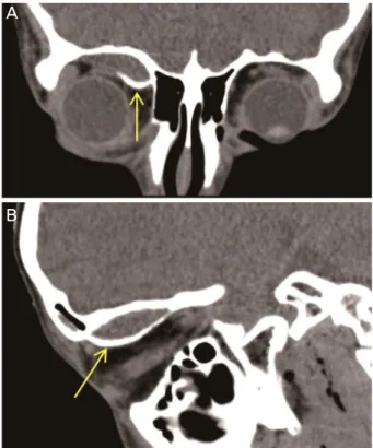
imaging, MRI)에서 우측 안와 상방에 연부조직 종괴가 관 찰되며 이는 T1 조영증강에서 고신호강도, T2 조영증강에서 저신호강도를 보이며, Gadolinium 조영제검사에서도 고신호강 도를 보여 아급성 단계의 혈종으로 보였고(Fig. 2), 외상 2주 후 시행한 안와 전산화단층촬영(computed tomography, CT)에서 미세한 부분적 석회화를 동반한 볼록한 모양의 종 괴가 관찰되었다(Fig. 3). 외상 4주 후 눈검사에서 호전을 보이지 않아 시행한 안와 CT에서 안와 상방의 종괴 아래 부 분에 경계가 분명한 석회화병변이 관찰되었다(Fig. 4).
전신마취하에 우측 속눈썹선위절개를 통한 앞쪽 안와절 개술을 시행하여 혈종 및 신생골제거술을 시행하였다. 골 막하부에 석회화된 혈종이 신생골을 형성하며 앞 안와연에 서 안와첨부쪽까지 단단하게 붙어 있는 양상이었다. 신생 골을 기존의 전두골과의 부착 부위에서 분리하였으며 단단 한 조직으로 되어 있는 혈종과 함께 모두 제거하였다. 병리 조직검사에서 혈종 내에 불규칙한 콜라겐 섬유와 골세포로
A B
A C D
B
Figure 3. Orbit CT taken at 2 weeks after trauma. Convex-shap-
ed mass lesion with fine spots of calcifications at the superior or- bit (arrow). CT = computed tomographyFigure 5. Histological examination. Calcification foci are denoted in the collagen fibril background (A: H&E stain, ×40). Immature
bony matrix lined with osteoblasts and osteoclasts was consistent with findings of woven bone (B: H&E stain, ×400).Figure 4. Orbit CT taken at 4 weeks after trauma. A convex le-
sion was slightly decreased, but it accompanies a clear-cut cal- cification line at the inferior margin, in the coronal (A, arrow) and sagittal (B, arrow) CT scans. CT = computed tomography.구성된 조직이 관찰되었으며, 미성숙 골기질과 동반된 골 세포, 파골세포가 관찰되었다(Fig. 5).
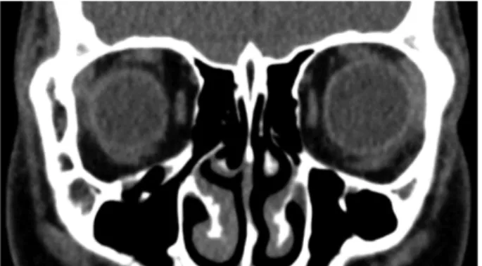
수술 1개월 후 시행한 안과검사에서 나안시력은 양안 1.0, 안구돌출계검사에서 안구돌출 소견은 없었고, 정면주 시 때 35PD 외사시 소견을 보여 우안 외직근 후전술 및 내 직근 절제술을 시행하여 외사시를 교정하였다. 안와 CT에 서 재발소견은 보이지 않았다(Fig. 6).
고 찰
안와 골막하출혈은 대부분 외상에 의해 발생하며 주로 안면부나 두부의 외상 후 바로 발생하지만 수년 후에 임상 양상이 발현하는 경우도 있다.7-10 울혈이 원인이 되는 경우 로는 지속적인 기침, Valsalva’s maneuver, 분만 중에 의해 증가된 복강압과 흉강압에 의해 중심정맥압 상승과 정맥울
혈이 일어나고 상대적으로 valve가 적은 안와내 정맥에 압 력이 전달되어 골막하혈종이 발생할 수 있다.11 비외상성으 로 발생한 경우는 백혈병, 혈우병, 괴혈병 등 전신질환으로 인해 출혈 경향이 있는 환자에서 발생하기도 하는데 혈종
A B
A
B
Figure 6. Orbit CT scan taken at 1month after surgery.
Hematoma and calcific wall was not observed at coronal CT scan. CT = computed tomography.
의 발생을 야기시킬 만한 특별한 외상이나 전신적인 질환 이 규명되지 않은 경우도 있다.12,13
골막하혈종은 골과 이를 둘러싼 골막 사이의 골막하 혈 관의 파열이 있거나 안와혈종의 직접적인 연장에 의해 발 생한다. 안와벽을 구성하는 전두골이 가장 크고 오목한 안 와면을 형성하며, 골막은 봉합선(suture line)을 따라 골에 단단히 붙어 있으나 안와 상벽의 경우 안와의 다른 부분과 는 달리 봉합부가 없고 다른 안와벽보다 골막이 골에 느슨 하게 붙어 있으므로 전두골아래가 안와내 골막하출혈의 주 요 호발 부위이다.14 이러한 골막하혈종은 대개 소아나 젊 은 연령에서 흔히 발생하며,8 호발하는 평균연령은 17세라 는 보고도 있다.9 본 연구에서도 10세 소아의 전두골 직하 방의 안와에서 발생하였다.
안와혈종에서 석회화가 일어나는 것은 매우 드문 현상이 며 기존에 발표된 증례는 1예가 있다. Sabet et al6이 발표한 증례에 따르면, 9세 남아가 운동 경기 중 우측 안면부를 수 상하였고, 수상 5일 후에 우측 안구 돌출과 수직 복시 증상 을 호소하여 병원에 왔다. 안와 CT에서 저명한 석회화 소견 은 관찰되지 않았으나 안와 상방의 혈종에 의해 외안근과 안 구가 하방으로 밀려있어, 앞측 안와절개술을 통한 혈종제거 술을 시행하였고, 조직검사에서 육아조직 내에 골기질과 함 께 뼈모세포가 동반되어 있었다.6 이 증례에서는 병리검사에 서 골형성이 발견되었을 뿐 기존의 골막하혈종과 다른 특이 한 임상소견을 보이지는 않았다. 이와 비교해 볼 때 저자들 의 증례에서는 외상 후 집중치료와 관련되어 안와를 영구적 으로 압박하는, 빠르게 진행하는 골형성이 있었고 CT 검사 에서 골형성면을 확인할 수 있다는 특이한 소견을 보였다.
두개내 혈종에서는 적지 않은 경우에 석회화가 발생하는 것으로 알려져 있다. 만성 경막하혈종에서 석회화 또는 골 화가 일어나는 비율은 0.8-10% 정도이며,1,15 초기 출혈이 발생한 시점부터 석회화가 일어나기까지 경막하혈종은 3개 월-3년 이상,3,5 경막외혈종은 17일-50년에 걸쳐 다양하게
나타날 수 있는 것으로 보고되고 있다.2 두개내 출혈이 있 을 때 출혈이 발생하는 위치에 따라 경막외(epidural), 경막 하(subdural), 지주막하(subarachnoid) 혈종으로 분류하며 발생 위치에 상관없이 혈종의 석회화가 관찰되었다.3-5,16-20
석회화의 기전은 역시 명확히 알려진 바는 없으나, 몇 가 지 가설이 제기되었다. Afra3에 따르면 혈관 내 혈전 생성 으로 인해 혈액순환이 원활하지 않은 경막하 공간에서 혈 종이 흡수되면서 세포 괴사, 결합조직의 유리질화 등을 초 래해 칼슘이 축적되는 것이라 하였다. Erdogan et al4에 따 르면 뼈나 경질막(dura mater)과 같은 혈관이 풍부한 조직 이 손상되면 염증, 복구, 재형성과 같은 일련의 조직반응이 촉발되는데, 소아에서 빠르게 골화가 일어나는 것은 급성 손상 이후에 발생하는 과도한 복구 반응이 일어나기 때문 이라 하였다. 또한 McLaurin and McLaurin19의 보고에 의 하면 양측성 만성 경막하혈종이 발생한 증례에서 일측성 석회화가 관찰되었는데, 이는 석회화 형성 기전에서 국소 적인 인자가 중요한 역할을 하는 것을 의미하며, 전신대사 요인 또한 영향을 미칠 수 있다고 하였다.
저자들의 증례 환자에서는 아마도 집중치료실에서 2주간 치료 받은 병력이 골형성의 선행요인으로 작용했을 가능성 이 높을 것으로 생각되었다. 골형성이 진행된 본 증례와 골 형성이 없는 일반적인 골막하혈종이 다른 감별점은 유발인 자의 하나로 중환자실 치료를 받았다는 점을 들 수 있고, 임상양상으로는 눈을 누르는 종괴 효과가 경과관찰 중에도 줄어들지 않고 계속되었다는 점을 들 수 있다. 영상의학적 소견으로는 CT에서 혈종 아랫면에서 골형성을 보였으며 수술 시 매우 단단한 뼈가 안와를 압박하고 있었던 것을 들 수 있겠다. 그러므로 골막하혈종이 잘 가라앉지 않거나 집 중치료를 받은 환자에서는 CT 영상을 이용한 경과관찰이 반드시 필요할 것으로 보인다.
안와의 골막하혈종에 대해서는 시력저하나 심각한 증상 이 없는 한 특별한 치료 없이 관찰하면서 보존적인 치료를 하는 것이 원칙이다.8,13,21 Wolter et al14은 안구나 시신경의 압박증상이 없는 경우에는 수술적 처치를 고려하기 전에 천 자흡인술을 시행해 보는 것이 좋다고 하였고, Markovits22 는 급성으로 발생한 안와출혈 환자에서 응급 천자배출 및 Hemovac을 연결하여 지속적인 흡인술로써 성공적인 치료 를 하였다고 보고하였다. 반면에 Bedrossian23은 급격한 시 력저하를 보이고 동공이 확대 고정되어 있으며 안저검사에 서 안동맥의 박동을 보이면 즉시 수술적인 안와 골막하 출 혈의 배출을 시행해야 한다고 하였고, Pope-Pegram and Hamill9, Kersten and Rice10는 천자 흡인보다 안와감압술의 경우 직접 출혈된 혈관을 지혈시킬 수 있고 재출혈 및 천자 흡인에 의한 추가적인 출혈의 예방과 동반된 안와골절을
= 국문초록 =
10세 소년의 안와 골막하혈종에서 발생한 신생골 형성
목적: 외상 후 안와에 발생한 골막하혈종에서 급격히 골형성이 일어난 증례 1예를 경험하였기에 이를 보고하고자 한다.
증례요약: 외상 후 집중치료를 받던 10세 소년에서 우안에 안구돌출 및 안구운동장애를 보이는 골막하혈종 소견이 있었다. 안과검사 에서 3 mm 안구돌출과 상측주시 때 우안 안구운동제한이 관찰되었으나 시력저하 소견은 없었다. 1개월 후 안와전산화단층촬영에서 골막하혈종 크기는 다소 감소하였으나 안와와의 경계면에서 신생골 형성 소견을 보였다. 앞측안와절개술을 시행하여 혈종을 제거하 고 골조직을 완전절제하였다. 혈종과 안와 사이의 경계에서 형성된 신생골이 앞쪽 안와연에서 안와첨까지 전두골에 단단하게 붙어 있었고, 조직검사에서 미성숙골조직이 관찰되었다. 수술 1년 후 경과관찰에서 우안 안구돌출 및 안구운동장애는 소실되고 새로운 골 형성은 보이지 않았다.
결론: 안와 골막하혈종은 대개 심한 신경이상을 초래하지 않으면 자연 흡수되도록 보존적인 치료를 하는 것이 권장되나 본 증례에서 와 같이 1개월 내의 짧은 시간에 골형성이 올 수도 있으므로, 집중치료를 받는 환자나 혈종의 흡수가 늦은 환자에 대해서는 주의 깊은 경과 관찰이 필요할 것으로 보인다.
<대한안과학회지 2016;57(1):120-124>
함께 정복할 수 있다는 장점으로 조기 수술적 처치를 선호 한다고 하였다.
저자들의 증례에서는 환자가 급격한 시력감소 및 시신경 압박의 징후는 보이지 않아 경과관찰하였으나, CT 검사상 에서 뚜렷하게 혈종 병변의 아래면에서 골형성을 보여, 안 구돌출과 안구 하방편위, 사시가 영구적으로 남을 가능성 이 있어 안와절개술을 통한 혈종 및 신생골제거술을 시행 하였다. 외상 후 집중치료를 받던 소아환자에서 발생한 안 와 골막하혈종에서 급격히 신생골이 형성되어 이를 수술을 통해 성공적으로 치료한 증례이기에 이를 보고하는 바이다.
REFERENCES
1) Hirakawa T, Tanaka A, Yoshinaga S, et al. Calcified chronic sub- dural hematoma with intracerebral rupture forming a subcortical hematoma. A case report. Surg Neurol 1989;32:51-5.
2) Chang JH, Choi JY, Chang JW, et al. Chronic epidural hematoma with rapid ossification. Childs Nerv Syst 2002;18:712-6.
3) Afra D. Ossification of subdural hematoma. Report of two cases. J Neurosurg 1961;18:393-7.
4) Erdogan B, Sen O, Bal N, et al. Rapidly calcifying and ossifying epidural hematoma. Pediatr Neurosurg 2003;39:208-11.
5) Niwa J, Nakamura T, Fujishige M, Hashi K. Removal of a large asymptomatic calcified chronic subdural hematoma. Surg Neurol 1988;30:135-9.
6) Sabet SJ, Tarbet KJ, Lemke BN, et al. Subperiosteal hematoma of the orbit with osteoneogenesis. Arch Ophthalmol 2001;119:301-3.
7) Wolter JR, Vanderveen GJ, Wacksman RL. Posttraumatic sub- galeal hematoma extending into the orbit as a cause of permanent blindness. J Pediatr Ophthalmol Strabismus 1978;15:151-3.
8) Seigel RS, Williams AG, Hutchison JW, et al. Subperiosteal hema- tomas of the orbit: angiographic and computed tomographic diagnosis.
Radiology 1982;143:711-4.
9) Pope-Pegram LD, Hamill MB. Post-traumatic subgaleal hema- toma with subperiosteal orbital extension. Surv Ophthalmol 1986;
30:258-62.
10) Kersten RC, Rice CD. Subperiosteal orbital hematoma: visual re- covery following delayed drainage. Ophthalmic Surg 1987;18:423-7.
11) Nakai K, Doi E, Kuriyama T, Tanaka Y. Spontaneous subperiosteal hematoma of the orbit. Surg Neurol 1983;20:100-2.
12) Law FW. Spontaneous orbital haemorrhage. Br J Ophthalmol 1971;55:556-8.
13) Carrion LT, Edwards WC, Perry LD. Spontaneous subperiosteal orbital hematoma. Ann Ophthalmol 1979;11:1754-7.
14) Wolter JR, Leenhouts JA, Coulthard SW. Clinical picture and man- agement of subperiosteal hematoma of the orbit. J Pediatr Ophthalmol 1976;13:136-8.
15) Ide M, Jimbo M, Yamamoto M, et al. Asymptomatic calcified chronic subdural hematoma-report of three cases. Neurol Med Chir (Tokyo) 1993;33:559-63.
16) Chen NF, Wang YC, Shen CC, et al. Calcification and ossification of chronic encapsulated intracerebral haematoma: case report. J Clin Neurosci 2004;11:527-30.
17) Loh JK, Howng SL. Huge calcified chronic subdural hematoma in the elderly-report of a case. Kaohsiung J Med Sci 1997;13:272-6.
18) Nagane M, Oyama H, Shibui S, et al. Ossified and calcified epi- dural hematoma incidentally found 40 years after head injury: case report. Surg Neurol 1994;42:65-9.
19) McLaurin RL, McLaurin KS. Calcified subdural hematomas in childhood. J Neurosurg 1966;24:648-55.
20) Kaplan M, Akgün B, Seçer HI. Ossified chronic subdural hema- toma with armored brain. Turk Neurosurg 2008;18:420-4.
21) Katz B, Carmody R. Subperiosteal orbital hematoma induced by the valsalva maneuver. Am J Ophthalmol 1985;100:617-8.
22) Markovits AS. Evacuation of orbital hematoma by continuous suction. Ann Ophthalmol 1977;9:1255-8.
23) Bedrossian RH. Emergency orbital decompression. Ophthalmic Surg 1985;16:293-5.