Cystic lymphangiomas involving the mesentery and the retroperitoneum are rare tumors. Most of the cystic lymphangiomas occur in the neck, and they generally have a single cavity with only only a small proportion of these tumor being multilocular. The cystic spaces are lined with a single layer of endothelium and there are small lymphoid aggregates in the cyst’s wall that help distinguish lymphangiomas from the simple cysts of the mesentery. We describe here an uncommon case of cys- tic lymphangioma presenting as a multilocular mass in- volving the mesentery and the retroperitoneum.
Case
A 20-year-old male patient was referred to the depart-
ment of general surgery for an operation on an intraab- dominal mass that was incidentally detected. He had back pain after minor trauma 4 weeks earlier, and mag- netic resonance imaging (MRI) demonstrated a huge cystic mass extending from the upper intraabdominal cavity to the pelvic cavity, and there was herniation of the nucleus pulposus at the L4-5 level (Fig. 1A). The pa- tient’s past medical history was unremarkable and he was asymptomatic. The physical examination revealed a blood pressure of 100/80 mm Hg, a pulse of 80 beats/min, respiration of 24 breaths/min, and a body temperature of 36.5℃. There was no palpable mass in the abdomen and the bowel sounds were hypoactive.
The laboratory data on admission, including the blood chemistry profile, coagulation studies and complete blood count revealed unremarkable findings. An admis- sion abdominal X-ray showed diffuse increased opacity that suggested a soft tissue mass in the abdomen. The transabdominal ultrasonography (US) revealed a multi- septated cystic mass without any definite wall (Fig. 1B).
The computed tomography (CT) scan showed a large cyst involving the mesentery and the retroperitoneum
J Korean Radiol Soc 2005;52:347-350
─ 347 ─
Cystic Lymphangioma Involving the Mesentery and the Retroperitoneum: A Case Report1
Dong Hun Kim, M.D.1, 2, Joo Nam Byun, M.D., Ji-Youn Jang, M.D.2
1Department of Radiology, Chosun University Hospital
2Department of Radiology, Soonchunhyang University Hospital Received January 7, 2005 ; Accepted April 14, 2005
Address reprint requests to : Dong Hun Kim, M.D., Department of Radiology, Chosun University Hospital, 588 Seoseok-dong, Dong-gu, Gwangju 501-717, Korea.
Tel. 82-62-220-3543 Fax. 82-62-228-9061 E-mail: kdhoon @ chosun.ac.kr
Cystic lymphangioma is uncommon angiomatous tumor that mainly occurs in the neck. Less than 1% of these tumors affect the mesentery, retroperitoneum and greater omentum. In particular, the cystic lymphangioma involving the mesentery and the retroperitoneum is a rare lesion. We report here on an uncommon case of cystic lym- phangioma that presented as a multilocular mass involving the mesentery and the retroperitoneum, and we also present a brief review of the relevant literature.
Index words :Mesentery, neoplasms Retroperitoneum, neoplasms Cysts, CT
Lymphangioma
(Figs. 1C, D). The possible clinical and radiological diag- noses at that time included mesenteric cystic lymphan- gioma, multicystic mesothelioma, other mesenteric cysts and lymphoma. Exploratory laparotomy revealed
a broad based cyst in the mesentery measuring 19×15
×6 cm; there were dense adhesions to the adjacent bowel loop, and the mass involved the retroperitoneum through the duodenal recess. The cyst’s wall was yel-
Dong Hun Kim, et al : Cystic Lymphangioma Involving the Mesentery and the Retroperitoneum
─ 348 ─
A B
C D
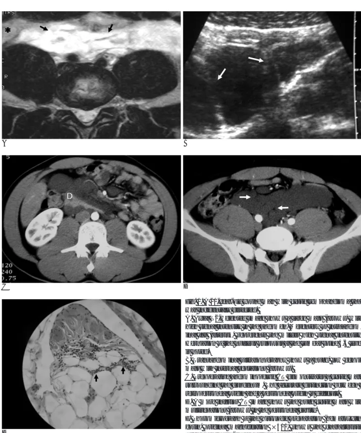
Fig. 1. A 20-year-old young man with cystic lymphangioma that was incidentally detected.
A. Axial T2-weighted image shows a large mass (arrows) with high signal intensity in the abdomen. Mesentery or intraabdom- inal fat (asterisk) represents the milder high signal intensity.
Herniation of the nucleus pulposus at the lumbar spine 4-5 level is noted.
B. Transabdominal ultrasonography shows a huge, low echoic mass with internal septations (arrows).
C. Preoperative abdominopelvic CT demonstrates a cystic mass surrounding the duodenum. The accurate distinction between a retroperitoneal origin and a peritoneal origin is difficult.
D. A more inferior CT image shows the huge cystic mass with multisepations (arrows) in the peritoneal cavity.
E. Photomicrograph of the histologic preparation (hematoxylin- eosin, original magnification ×400) shows the characteristic large endothelial-lined cystic spaces and stromal aggregates of lymphocytes (arrows).
E
lowish-gray and glistering; no enlarged lymph nodes were found. The rest of the abdomen was normal and any free intraabdominal fluid was not found. The mass was resected and upon sectioning, multicystic lobules with chylous whitish fluid were noted. Microscopically, the cyst’s wall showed an endothelial lining, smooth muscle fibers and fibrovascular adipose tissue.
Lymphocytic aggregates were seen throughout the cyst’s wall along with diffuse chronic inflammatory in- filtration being seen below the endothelial lining (Fig.
1E).
After surgery, the patient complained of diffuse ab- dominal discomfort and repetitive vomiting for 2 weeks.
Postoperative US and CT demonstrated a hematoma surrounding the duodenum and residual cystic mass.
Close observation was done and the patient made a re- covery without residual sequelae.
Discussion
Mesenteric cystic lymphangioma is an uncommon mesenteric mass. The gross and histological findings (Fig. 1E) in our case are similar to those findings report- ed in other series (1-5). However, when we clinically compare our case with the previous studies, our case demonstrated no symptoms, nor did it radiologically demonstrate any of the unusually involved sites like the mesentery and retroperitoneum.
Cystic lymphangiomas are usually located in the neck and axilla, and they rarely occur in the mediastinum, lungs, esophagus, diaphragm, duodenum, stomach, small and large bowels, spleen and liver. Less than 1%
of these tumors affect the mesentery, greater omentum and retroperitoneum. Lymphangioma involving the mesentery and retroperitoneum, such as our case, is rare. Meyer et al. have reported on a retroperitoneal lymphangioma in which the cystic tumor originated from the retroperitoneum with a broad attachment to the pancreas, and it had extended anteriorly into the root of the mesentery, thereby mimicking the radiologi- cal features of a mesenteric cyst (6). In our case, the tu- mor was clearly separable from the pancreas and the tu- mor was intraoperatively defined as a mesenteric mass growing posteriorly and taking up a retroperitoneal por- tion through the duodenal recess. Many other cystic tu- mors can involve the mesentery or retroperitoneum, and cystic mesothelioma, lymphangiosarcoma, myxoid degeneration of lymphangioma and also hemangioma must be included in the differential diagnosis.
Lymphangiomas are usually restricted to the mesentery, omentum, mesocolon and retroperitoneum. In contrast, benign cystic mesothelioma tends to have a pelvic loca- tion and there is involvement of the upper abdomen and retroperitoneum in some cases (7, 8).
Histologically, lymphangioma displays dilated lym- phatic vessels that are lined by flattened endothelium in between lobules of adipose, fibrous and lymphoid tis- sue. In contrast, the majority of mesenteric cysts often display a cuboidal or columnar epithelial lining that lacks smooth muscle cells or lymphatic elements.
The most common finding of mesenteric cystic lym- phangioma on plain abdominal radiographs is a soft-tis- sue mass with displacement of the bowel loops.
Transabdominal ultrasonography is a very sensitive imaging modality, and the mass generally appears on US as a sharply defined cystic or multicystic mass, and there are often internal septations such as was seen in our case (Fig. 1B). The fluid can be anechoic, or there are scattered internal echoes that represent infection or hemorrhage. CT and MRI can give important preopera- tive information regarding the anatomical location, the cyst size, organ involvement and the possible complica- tions. In addition, these two modalities can differentiate between chylous fluid, blood and pus.
The standard therapy for patients with mesenteric cystic lymphangioma is surgery. Total removal of the mass that invades the potentially respectable intraab- dominal structures such as the bowel, spleen or pan- creas is possible. However, adhesions to vital structures can sometimes make resection dangerous or even im- possible. Other treatment methods are palliative surgi- cal treatment or sclerosing agents. There are still high re- currence rates for the totally excised and incompletely resected mesenteric cystic lymphangiomas, and this in- dicates a continuing need to develop new and effective treatment options to supplement surgery (9).
In conclusion, cystic lymphangioma involving the mesentery and the retroperitoneum should be included in the differential diagnosis of cystic intraabdominal le- sions. Even when the patient is asymptomatic and this tumor is discovered incidentally, mesenteric cystic lym- phangioma must be treated surgically because of its po- tential to grow and invade vital structures, and this tu- mor can also develop life-threatening complications.
References
1. Losanoff JE, Richman BW, El-Sherif A, Rider KD, Jones JW.
J Korean Radiol Soc 2005;52:347-350
─ 349 ─
Mesenteric cystic lymphangioma. J Am Coll Surg 2003;196:598-603 2. Tsukada H, Takaori K, Ishiguro S, Tsuda T, Ota S, Yamamoto T.
Giant cystic lymphangioma of the small bowel mesentery: report of a case. Surg Today 2002;32:734-737
3. Konen O, Rathaus V, Dlugy E, Freud E, Kessler A, Shapiro M, et al. Childhood abdominal cystic lymphangioma. Pediatr Radiol 2002;32:88-94
4. Lopez-Gonzalez Garrido JD, Ramirez-Garrido F, Lopez-Gonzalez Garrido C, Marin Aznar JL, Valladares Mendias JC, Mingorance MA. Imaging diagnosis of mesenteric cystic lymphangioma: a case report in a newborn. Eur Radiol 1999;9:754
5. de Perrot M, Rostan O, Morel P, Le Coultre C. Abdominal lym-
phangioma in adults and children. Br J Surg 1998;85:395-397 6. Meyer T, Stohr G, Post S, Fayyazi A. Retroperitoneal lymphan-
gioma presenting as a mesenteric cyst. Eur J Radiol 1995;21:143- 144
7. Bui-Mansfield LT, Kim-Ahn G, O’Bryant LK. Multicystic mesothe- lioma of the peritoneum. AJR Am J Roentgenol 2002;178:402 8. Sheth S, Horton KM, Garland MR, Fishman EK. Mesenteric neo-
plasms: CT appearances of primary and secondary tumors and dif- ferential diagnosis. Radiographics 2003;3:457-473
9. Hancock BJ, St-Vil D, Luks FI, Di Lorenzo M, Blanchard H.
Complications of lymphangiomas in children. J Pediatr Surg 1992;
27:220-224
Dong Hun Kim, et al : Cystic Lymphangioma Involving the Mesentery and the Retroperitoneum
─ 350 ─
대한영상의학회지 2005;52:347-350
장간막과 후복막을 침범한 낭포성림프관종: 증례 보고1
1조선대학교병원 영상의학과
2순천향대학교병원 영상의학과 김동훈1,2・변주남・장지연2
낭포성 림프관종은 주로 목에서 발생하는 드문 혈관성 종양이다. 1% 미만에서 장간막, 후복막과 대망을 침범한다. 특 히 낭포성 림프관종이 장간막과 후복막을 함께 침범하는 경우는 드물다. 저자들은 장간막과 후복막을 침범한 낭포성 림프관종을 문헌고찰과 함께 보고하고자 한다.