Propofol protects against oxidative-stress-induced COS-7 cell apoptosis by inducing autophagy
Ji-Young Yoon
1, Chul-Woo Baek
1, Eun-Jung Kim
1, Bong-Soo Park
2, Su-Bin Yu
2, Ji-Uk Yoon
3, Eok-Nyun Kim
11Department of Dental Anesthesia and Pain Medicine, School of Dentistry, Pusan National University, Dental Research Institute, Yangsan, Republic of Korea
2Department of Oral Anatomy, School of Dentistry, Pusan National University, Yangsan, Republic of Korea
3Department of Anesthesia and Pain Medicine, School of Medicine, Pusan National University, Yangsan, Republic of Korea
Background: In oxidative stress, reactive oxygen species (ROS) production contributes to cellular dysfunction and initiates the apoptotic cascade. Autophagy is considered the mechanism that decreases ROS concentration and oxidative damage. Propofol shows antioxidant properties, but the mechanisms underlying the effect of propofol preconditioning (PPC) on oxidative injury remain unclear. Therefore, we investigated whether PPC protects against cell damage from hydrogen peroxide (H2O2)-induced oxidative stress and influences cellular autophagy.
Method: COS-7 cells were randomly divided into the following groups: control, cells were incubated in normoxia (5% CO2, 21% O2, and 74% N2) for 24 h without propofol; H2O2, cells were exposed to H2O2 (400 μM) for 2 h; PPC + H2O2, cells pretreated with propofol were exposed to H2O2; and 3-methyladenine (3-MA) + PPC + H2O2, cells pretreated with 3-MA (1 mM) for 1 h and propofol were exposed to H2O2. Cell viability was determined using 3-(4,5-dimethylthiazol-2-yl)-2,5-diphenyltetrazolium bromide thiazolyl blue (MTT) reduction.
Apoptosis was determined using Hoechst 33342 staining and fluorescence microscopy. The relationship between PPC and autophagy was detected using western blot analysis.
Results: Cell viability decreased more significantly in the H2O2 group than in the control group, but it was improved by PPC (100 μM). Pretreatment with propofol effectively decreased H2O2-induced COS-7 cell apoptosis.
However, pretreatment with 3-MA inhibited the protective effect of propofol during apoptosis. Western blot analysis showed that the level of autophagy-related proteins was higher in the PPC + H2O2 group than that in the H2O2 group.
Conclusion: PPC has a protective effect on H2O2-induced COS-7 cell apoptosis, which is mediated by autophagy activation.
Keywords: Autophagy; COS-7 Cells; Oxidative Stress; Propofol.
This is an Open Access article distributed under the terms of the Creative Commons Attribution Non-Commercial License (http://creativecommons.org/licenses/by-nc/3.0/) which permits unrestricted non-commercial use, distribution, and reproduction in any medium, provided the original work is properly cited.
Received: 2017. January. 24.•Revised: 2017. February. 24.•Accepted: 2017. March. 7.
Corresponding Author: Eok-Nyun Kim, Department of Dental Anesthesia and Pain Medicine, Pusan National University Dental Hospital, Geumo-ro 20, Yangsan, Gyeongnam, 626-787, Republic of Korea
Tel: +82-55-360-5370 Fax: 82-55-360-5369 E-mail: chemfrie@naver.com Copyrightⓒ 2017 Journal of Dental Anesthesia and Pain Medicine
INTRODUCTION
Oxidative stress occurs because of an imbalance between reactive oxygen species (ROS) production and the antioxidant defense system. ROS play a key role in oxidative stress and cause oxidative damage to lipids,
proteins, and DNA either directly or by altering signaling pathways [1]. ROS, including hydrogen peroxide (H2O2), superoxide radical, hydroxyl radical, and peroxynitrite, can be generated either exogenously or endogenously from several factors such as the mitochondrial respiratory chain and cytosolic enzyme systems [2,3]. In particular, in many clinical situations, such as surgery, acute
inflammatory processes, and anesthesia, ischemia/reper- fusion (I/R) injury can occur and induce ROS production, which contributes to cellular dysfunction and the initia- tion of the apoptotic/necrotic cascade [3,4].
Autophagy, which is the mechanism that decreases ROS concentration and oxidative damage to biomolecules and organelles, is activated by ROS and reactive nitrogen species in response to oxidative stress [5]. Autophagy is an intracellular catabolic mechanism for degrading and recycling unnecessary or dysfunctional cellular compo- nents in response to various conditions [6]. Through autophagy, cells maintain their energy levels and syn- thesize structures required for survival [5]. ROS have been reported to be crucial inducers of autophagy, but the unexpected energetic requirement that develops during oxidative stress, in turn, overburdens the mitochondria and impairs its function. Moreover, ROS is generated at high concentrations in such situations, thereby suppressing the antioxidant role of autophagy and accelerating apoptosis [7-9].
Propofol (2,6-diisopropylphenol) is a widely used intravenous sedative-hypnotic agent for the induction and maintenance of general anesthesia, as well as for sedation in intensive care units. Some studies have demonstrated the antioxidant properties of propofol both in vitro [10,11]
and in vivo [12]. Propofol has a phenolic hydroxyl group, which is a structural characteristic similar to that of α -tocopherol (vitamin E), a type of endogenous antioxi- dant; accordingly, propofol presents antioxidant pro- perties [13].
For protection against tissue damage from I/R injury, anesthetic preconditioning, which is defined as a brief exposure to anesthetic agents that protect the myocardium from the potentially fatal consequences of a subsequent prolonged period of myocardial I/R, has been increasingly used as a therapeutic strategy [14]. Propofol precondi- tioning (PPC) has been shown to protect the organs against I/R injury when propofol is administered before the ischemic period [15,16]. The mechanisms underlying the protective effects of PPC have been shown to be the activation of adenosine triphosphate-sensitive potassium
(KATP) channels, protein kinase C, protein tyrosine kinase, mitogen-activated protein kinase (MAPK), and protein kinase B (Akt) pathways [17-19]. Such mechanisms may be related to the antioxidant effects of propofol because ROS are produced during I/R, and therefore, oxidative stress is closely related to I/R injury. However, the mechanisms underlying the effect of PPC on oxidative injury remain unclear. In this study, we used COS-7 cells (African green monkey kidney fibroblast-like cell line) to investigate whether PPC protects against cell damage caused by H2O2-induced oxidative stress and influences cellular autophagy.
MATERIALS AND METHODS
1. Chemicals and antibodies
Propofol, H2O2, and Hoechst 33342 were purchased from Sigma Chemicals (St Louis, MO, USA). The reagents 3-(4,5-dimethylthiazol-2-yl)-2,5-diphenyltetrazolium bromide thiazolyl blue (MTT), acridine orange (AO), monodansylcadaverine (MDC), and 3-methyladenine (3-MA, class III phosphoinositide 3-kinase inhibitor) were ob- tained from Calbiochem (La Jolla, CA, USA). Antibodies used in the study were as follows: LC3-II (1:1,000) and Becline-1 (1:1,000) from Abcam, and p62 (1:1,000) and Atg5 (1:1,000) from Santa Cruz. Secondary antibodies against rabbit (1:5,000) and mouse (1:5,000) immunoglo- bulins were purchased from Bio-Rad (Richmond, CA, USA).
2. Cell cultures
The COS-7 cells were obtained from the American Type Culture Collection (Manassas, VA, USA). The COS-7 cells were grown in Dulbecco’s modified Eagle’s medium (DMEM, GIBCO) supplemented with 10%
inactivated fetal bovine serum (GIBCO) containing 500 μg/ml penicillin and 500 μg/ml streptomycin (GIBCO) and incubated at 37°C in a humidified atmosphere of 95%
air and 5% CO2. The medium was replaced every 3 days.
3. Treatment with propofol and H2O2
The propofol stock was prepared by dissolving it in dimethyl sulfoxide (DMSO) and was kept frozen at -4°C until use. The stock was diluted to necessary concentra- tions by using DMEM when needed. The cells were grown to about 80% confluence and treated with H2O2
(400 μM) for 2 h with or without propofol. Prior to the addition of H2O2, the cells were exposed to propofol (100 μM) for 2 h. The cells grown in a medium containing an equivalent amount of DMSO without propofol and H2O2 served as the controls. The cells were divided into the following four groups: control, cells were incubated in normoxia (5% CO2, 21% O2, and 74% N2) for 24 h without propofol; H2O2, cells were exposed to H2O2 for 2 h; PPC + H2O2, cells pretreated with propofol for 2 h were exposed to H2O2; 3-MA + PPC + H2O2, cells pretreated with 3-MA (1 mM) for 1 h were exposed to propofol for 2 h and H2O2. 3-MA was used to identify whether the effect of PPC was mediated by autophagy, because 3-MA suppresses autophagy by blocking auto- phagosome formation.
4. Determination of cell viability by using MTT reduction Cell viability was measured using the MTT assay, showing the mitochondrial activity of living cells. COS-7 cells (3 × 10⁴) were seeded in 96-well plates. After drug treatment as indicated, the cells were incubated with 100 μl MTT (final concentration 0.5 mg/ml) for 4 h at 37°C.
The reaction was terminated by adding 200 μl DMSO.
The cells were exposed to propofol at various concen- trations (0, 3, 30, 100, and 300 μM) for 2 h. To investigate the influence of H2O2 on COS-7 cell viability, the cells were treated with H2O2 at various concentrations (0, 50, 100, 200, and 400 μM) for 2 h. Cell viability was measured using an ELISA reader (Tecan, Männedorf, Switzerland) at 620 nm.
5. Fluorescence microscopy
After treatment with H2O2 (400 μM) for 2 h with or without propofol, the cells were stained with Hoechst
33342 (Molecular Probes, Eugene, OR, USA) and visualized under fluorescence microscopy to evaluate the morphological changes to the nuclei as a measure of apoptosis. To further evaluate the effect of propofol on the autophagy of COS-7 cells, we conducted auto- phagosome staining. Cells were grown on coverslips and treated with COS-7 cells. After 24 h, the cells were stained with 0.05 mM MDC, a selective fluorescent marker for autophagic vacuoles, at 37°C for 1 h. The cellular fluorescence changes were observed under a fluorescence microscope (Axioskop, Carl Zeiss, Germany). For further detection of the acidic cellular compartment, we used AO, which emits bright red fluorescence in acidic vesicles but green fluorescence in the cytoplasm and nucleus. The cells were stained with 1 μg/ml AO for 15 min and washed with phosphate-buffered saline (PBS). Autophagic vesicle organelle (AVO) formation was observed under a confocal microscope (LSM 700, Carl Zeiss).
6. Flow cytometry analysis
The cells were seeded in 60-mm dishes at 70% con- fluence and incubated overnight. The propofol-pretreated cells were incubated for different periods according to the conditions. The harvested cells were washed with PBS containing 1% bovine serum albumin (BSA) and cen- trifuged at 2500 rpm for 10 min. The cells were then resuspended in ice-cold 95% ethanol with 0.5% Tween 20 to a final concentration of 75% ethanol. After 24 h, the fixed cells were washed in 1% BSA-PBS solution, resuspended in 1 ml PBS containing 40 μg/ml RNase A (Sigma), incubated at 4°C for 30 min, and resuspended in 10 μg/ml propidium iodide solution (Sigma). The DNA contents were examined using a CYTOMICS FC500 flow cytometer (Beckman Coulter) and the data were analyzed using the MultiCycle software, which allowed simul- taneous estimation of cell-cycle parameters and apoptosis.
7. Western blot analysis
The cells (2 × 10⁶) were washed twice in ice-cold PBS, resuspended in 200 μl ice-cold solubilizing buffer (300 mM NaCl, 50 mM Tris-Cl [pH 7.6], 0.5% Triton X-100,
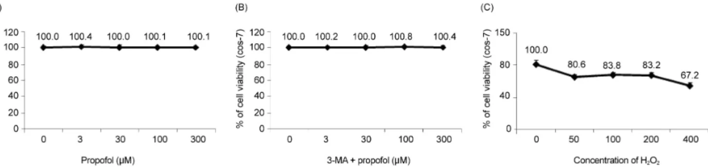
Fig. 1. The effect of H2O2 and propofol on COS-7 cell viability is assessed using the MTT assay. (A) Propofol at various concentrations shows no significant differences in cell viability. (B) Cell viability is maintained when the cells are pretreated with 3-MA before propofol administration. (C) The application of H2O2 to COS-7 cells at various concentrations (50-μM increments) results in a decrease in cell viability.
Fig. 2. Cell viability decreases significantly in the H2O2 group and is improved by propofol preconditioning (PPC) (100 μM). In the 3-MA + PPC + H2O2 group, cell viability is significantly lower than it is in the control group. *P < 0.05 compared with the control group.
2 mM phenylmethylsulfonyl fluoride, 2 μl/ml aprotinin, and 2 μl/ml leupeptin) and incubated at 4°C for 30 min.
The lysates were centrifuged at 14,000 rpm for 15 min at 4°C. Protein concentrations of the cell lysates were determined using the Bradford protein assay (Bio-Rad), and 25 μg of proteins were resolved by using 10% SDS/
PAGE. The gels were transferred to polyvinylidene fluoride membranes (Millipore, Billerica, MA, USA) and reacted with appropriate primary antibodies. Immuno- staining with secondary antibodies was detected using SuperSignal West Femto (Pierce, Rockford, IL, USA) enhanced chemiluminescence substrate and detected using AlphaImager HP (Alpha Innotech, Santa Clara, CA, USA).
8. Statistical analysis
The results were presented as the means ± standard deviation (SD). The study results were analyzed using analysis of variance followed by Tukey’s post-hoc test.
Simple pairwise comparisons were analyzed for signifi- cance by using Student’s t-test. Statistical significance was defined as P < 0.05.
RESULTS
1. Propofol improved the decrease in H2O2-induced COS-7 cell viability
We examined the effects of propofol and H2O2 injury on COS-7 cell viability by measuring MTT reduction.
Propofol treatment at various concentrations (0, 3, 30,
100, and 300 μM) produced no significant differences in COS-7 cell viability (Fig. 1A). No change in cell viability was observed when the cells were pretreated with 3-MA before propofol administration (Fig. 1B). However, the application of H2O2 to COS-7 cells at various concen- trations (0, 50, 100, 200, and 400 μM) for 24 h resulted in a decrease in cell viability (Fig. 1C). As shown in Fig.
2, compared to the control group, the H2O2 group showed a significant decrease in cell viability (P < 0.05), which was improved by PPC (100 μM). The 3-MA (1 mM) treatment 1 h before H2O2 exposure inhibited the protective effect of PPC and increased H2O2-induced COS-7 cell apoptosis (Fig. 2).
2. Propofol attenuated H2O2-induced COS-7 cell apoptosis
The effect of propofol on COS-7 cell apoptosis was
(A) (B)
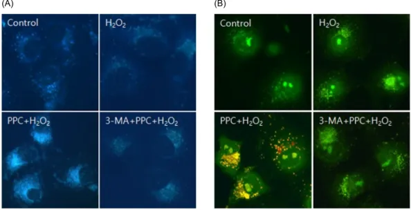
Fig. 4. (A) MDC staining of cytoplasmic vacuoles after propofol treatment in COS-7 cells. Compared to the control group, the propofol-pretreated group shows the accumulation of autophagosomes containing partially digested cytoplasmic contents. Propofol pretreatment dramatically increases the formation of autophagosomes, and the autophagy-pathway inhibitor 3-MA inhibits the propofol-mediated formation of autophagosomes. (B) AO staining of autophagosomes after propofol treatment in COS-7 cells. Autophagosomes with yellow or red vesicles during AO staining are detected in the PPC + H2O2 group, and 3-MA inhibits autophagosome formation.
(A) (B)
Fig. 3. (A) Hoechst staining: Morphological changes in COS-7 cells treated with H2O2, propofol (100 μM), and 3-MA. The cellular fluorescence changes are observed under a fluorescence microscope. Apoptotic bodies are observed in the H2O2 and 3-MA + PPC + H2O2 group cells. In contrast, apoptotic bodies markedly reduced in the PPC + H2O2 group cells. Control: no propofol treatment group; H2O2: H2O2 exposure group; PPC + H2O2: propofol pretreatment before exposure to H2O2 group; 3-MA + PPC + H2O2: pretreatment with 3-MA (1 mM) before 1 h and propofol treatment before exposure to H2O2 group. (B) The rate of apoptosis is assessed using a fluorescence-activated cell sorter.
examined using Hoechst 33342 staining. The cells were viewed under a fluorescence microscope (× 400) to evaluate the morphological changes in the nuclei (Fig.
3A). The majority of COS-7 cells in the control group showed normal morphology with round regular nuclei.
In contrast, apoptotic nuclei, characterized by bright blue chromatin that was highly condensed or fragmented, were observed in the H2O2 and 3-MA + PPC + H2O2 group cells. Pretreatment with propofol effectively decreased COS-7 cell apoptosis, as indicated by the restored cell
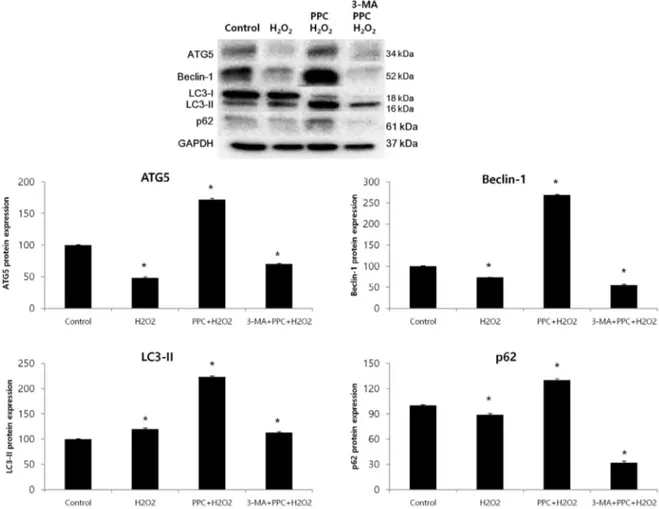
Fig. 5. Effects of propofol on autophagy markers in COS-7 cells are assessed using western blot analysis and densitometry. The levels of Atg5, Beclin-1, and p62 are significantly lower in the H2O2 group than in the control group as a result of the H2O2-induced apoptotic signaling; however, their levels increase in the PPC + H2O2 group. The level of LC3-II is significantly higher in the H2O2 group than in the control group, and is even higher in the PPC + H2O2 group. The levels of Atg5, Beclin-1, and LC3-II decrease when autophagy is suppressed by 3-MA. *P < 0.05 compared with the control group.
morphology. Upon determining apoptosis by using a fluorescence-activated cell sorter, we found that 10.5%
of the cells showed apoptosis in the H2O2 group, but in the PPC + H2O2 group, only 5.4% of the cells showed H2O2-induced apoptosis. These results suggested that PPC attenuated H2O2-induced COS-7 cell apoptosis. However, pretreatment with 3-MA inhibited the protective effect of propofol against apoptosis, and the percentage of apoptotic cells increased to 13% in the 3-MA + PPC + H2O2 group compared to the PPC + H2O2 group (Fig. 3B).
3. Propofol resulted in the induction of autophagy in COS-7 cells
Prominent accumulation of the autophagy-specific stain MDC was observed around the nuclei in the PPC group
COS-7 cells (Fig. 4A). We clarified the role of propofol- induced autophagy in COS-7 cells by investigating the consequences of treatment with 3-MA, a selective autophagy inhibitor. The inhibitory effects of 3-MA on AVO formation was confirmed by quantitatively measur- ing the red fluorescence ratio after AO staining. Yellow and red fluorescent spots appeared in the propofol- pretreated COS-7 cells, while the cells in the control and 3-MA + PPC + H2O2 groups showed mainly green cyto- plasmic fluorescence (Fig. 4B). We examined the activation of autophagy-related proteins in the H2O2- treated cells by using western blot analysis. The levels of Atg5, Beclin-1, and p62 decreased more significantly in the H2O2 group than in the control group after the H2O2-induced apoptotic signaling. The levels of Atg5,
Beclin-1, and p62 increased when autophagy was induced by propofol, and their levels decreased when autophagy was suppressed by 3-MA. The level of LC3-II increased more significantly in the H2O2 group than in the control group and increased even more in the PPC + H2O2 group.
However, pretreatment with 3-MA inhibited LC3-II expression (Fig. 5).
DISCUSSION
In this study, the protective effect of PPC against oxidative-stress-induced COS-7 cell apoptosis was pro- ven in an in vitro experimental model of H2O2-induced oxidative injury. We also demonstrated that the anti- oxidant effect of PPC was mediated by cellular autophagy activation by showing the formation of autophagosomes in COS-7 cells by using a fluorescence microscope and the expression of autophagy-related proteins by using western blot analysis.
Oxidative stress can induce toxic effects through the production of ROS that damage cardinal cellular com- ponents, such as proteins, lipids, and DNA, because of their highly reactive nature [20]. Navarro et al. [21]
reported that H2O2 was associated with cellular toxic effects, including the depletion of intracellular glutathione and ATP, an increase in NAD+ level, increase in free cytosolic Ca2+, and lipid peroxidation. Among the ROS, the hydroxyl radical can directly attack the DNA backbone by generating oxidative damages, such as oxidized bases, abasic sites, single-strand breaks, double- strand breaks, and DNA-protein crosslinks [22,23]. Via these processes, oxidative stress is involved in the development of cardiovascular, neurological, and oph- thalmological diseases and cancer, as well as in aging [1]. Therefore, we need to develop strategies for pre- venting oxidative injury.
The mediators associated with the cellular signaling pathway that respond to oxidative injury are the ex- tracellular signal-regulated kinase (ERK), c-Jun amino- terminal kinase (JNK), and p38 MAPK signaling cas-
cades; the phosphoinositide 3-kinase (PI3K)/Akt path- way; the nuclear factor (NF)-kB signaling system; p53 activation; and the heat shock response. In general, the ERK, PI3K/Akt, and NF-kB signaling pathways and the heat shock response produce a pro-survival effect during oxidative injury, whereas the activation of JNK, p38 MAPK, and p53 is more commonly involved in apoptosis [1]. Propofol exerts a protective effect against oxidative injury via these mediators. Wang et al. [24] demonstrated that propofol protects hepatocytes from H2O2-induced apoptosis, partly through the activation of the ERK pathway. In another study, they found that propofol suppressed p38 MAPK activation and attenuated oxidative-stress-induced apoptosis [25]. Previous reports have shown that heme oxygenase (HO) plays a critical role in the antioxidant pathway of propofol [26-28] and Acquaviva et al. [29] reported that the addition of a synthetic NF-kB inhibitor completely reversed propofol- mediated HO-1 expression, suggesting that propofol has a protective effect on oxidative injury via NF-kB acti- vation.
In our study, COS-7 cells were pretreated with the autophagy-pathway inhibitor 3-MA and propofol before they were exposed to H2O2-induced oxidative stress, and we investigated the changes in COS-7 cell apoptosis based on the assumption that autophagy might play an important role in the protective effect of propofol against oxidative stress. Hoechst 33342 staining revealed that the number of apoptotic bodies was higher in the 3-MA + PPC + H2O2 group cells than in the control and PPC + H2O2 group cells. This result suggests that autophagy has a cytoprotective function related to the protective mecha- nisms of PPC against oxidative injury. A recent study demonstrated that oxidative stress induced autophagy through molecular crosstalk between p62, an autophagy- related protein, and transcription factors related to cyto- protective genes in response to oxidative stress, and these had a protective effect against necrosis due to ATP depletion [30]. Other studies have shown that the suppression of autophagy by knockdown of Atg5 and Atg7, which are autophagy-related genes, greatly in-
creased H2O2-induced apoptosis; these studies also showed that autophagy played a cytoprotective role in H2O2-induced apoptosis [31,32]. In this study, PPC up- regulated the expression of autophagy-related proteins, such as Atg5, Beclin-1, and p62.
The molecular circuitry and signaling pathways re- gulating autophagy involve five key stages: (a) phago- phore formation or nucleation; (b) Atg5-Atg12 conjuga- tion, interaction with Atg16L, and multimerization at the phagophore; (c) LC3 processing and insertion into the extending phagophore membrane; (d) capture of random or selective targets for degradation; and (e) fusion of the autophagosome with the lysosome, followed by the proteolytic degradation of engulfed molecules by lysosomal proteases [33]. LC3 is expressed upon the induction of autophagy and is converted to LC3-II by LC3 processing. LC3 usually reveals two bands: LC3-I (18 kDa) and LC3-II (16 kDa). The amount of LC3-II is related to the number of autophagosomes and is used for analyzing autophagic activity [34]. The recruitment and integration of LC3-II into the growing phagophore, including LC3 processing, are dependent on Atg5-Atg12.
The Atg5-Atg12–dependent pathway of autophagy has been considered critical for survival during the starvation period in the first few days after birth [35,36]. However, a recent study discovered that the Atg5-Atg12–indepen- dent pathway was an alternative process for autophagy.
This pathway of autophagy was not associated with LC3 processing but appeared to specifically involve auto- phagosome formation from trans-Golgi and late endo- somes [37]. In our study, the expressions of Atg5, Beclin-1, and p62 decreased as a result of H2O2-induced oxidative stress, but LC3-II expression was higher in the treatment groups than in the control group. This finding suggests that LC3 processing is generated by an Atg5- independent pathway during H2O2-induced oxidative stress and that PPC activates the production of Atg5, thereby resulting in the increase of LC3-II expression through the Atg5-independent and Atg5-dependent path- ways in the PPC + H2O2 group. After all, the levels of autophagy-related proteins were higher in the PPC + H2O2
group than in the control group, suggesting that PPC enhanced autophagy. However, we did not investigate the roles of PI3K/Akt, p38 MAPK, and JNK, which are known to be involved in the autophagy signaling path- way. This is a limitation of our study.
In conclusion, this study suggests that PPC has a protective effect on H2O2-induced COS-7 cell apoptosis and this effect seemed to be mediated by autophagy.
Whether autophagy has a positive influence on oxidative injury and cell survival remains a controversial topic, and more research is needed to clarify the role of autophagy.
Nevertheless, our study provides evidence that propofol inhibits oxidative-stress-induced apoptosis through the induction of cellular protective autophagy.
NOTES: Thesis for the degree of Doctor of Philosophy.
There are no financial or other issues that might lead to conflict of interest.
REFERENCES
1. Finkel T, Holbrook NJ. Oxidants, oxidative stress and the biology of ageing. Nature 2000; 408: 239-47.
2. Geiszt M, Kopp JB, Varnai P, Leto TL. Identification of renox, an NAD(P)H oxidase in kidney. Proc Natl Acad Sci U S A 2000; 97: 8010-4.
3. Boveris A, Chance B. The mitochondrial generation of hydrogen peroxide. general properties and effect of hyper- baric oxygen. Biochem J 1973; 134: 707-16.
4. Chien CT, Lee PH, Chen CF, Ma MC, Lai MK, Hsu SM. De novo demonstration and co-localization of free- radical production and apoptosis formation in rat kidney subjected to ischemia/reperfusion. J Am Soc Nephrol 2001; 12: 973-82.
5. Filomeni G, De Zio D, Cecconi F. Oxidative stress and autophagy: The clash between damage and metabolic needs. Cell Death Differ 2015; 22: 377-88.
6. Kroemer G, Marino G, Levine B. Autophagy and the integrated stress response. Mol Cell 2010; 40: 280-93.
7. Hamacher-Brady A, Brady NR, Logue SE, Sayen MR, Jinno M, Kirshenbaum LA, et al. Response to myocardial ischemia/reperfusion injury involves Bnip3 and autophagy.
Cell Death Differ 2007; 14: 146-57.
8. Filomeni G, Desideri E, Cardaci S, Rotilio G, Ciriolo MR.
Under the ROS...thiol network is the principal suspect for autophagy commitment. Autophagy 2010; 6: 999-1005.
9. Murphy MP. How mitochondria produce reactive oxygen species. Biochem J 2009; 417: 1-13.
10. Tesauro M, Thompson WC, Moss J. Effect of staurosporine- induced apoptosis on endothelial nitric oxide synthase in transfected COS-7 cells and primary endothelial cells. Cell Death Differ 2006; 13: 597-606.
11. Xu JJ, Wang YL. Propofol attenuation of hydrogen peroxide-mediated oxidative stress and apoptosis in cultured cardiomyocytes involves haeme oxygenase-1. Eur J Anaesthesiol 2008; 25: 395-402.
12. Kobayashi K, Yoshino F, Takahashi SS, Todoki K, Maehata Y, Komatsu T, et al. Direct assessments of the antioxidant effects of propofol medium chain triglyceride/
long chain triglyceride on the brain of stroke-prone spontaneously hypertensive rats using electron spin re- sonance spectroscopy. Anesthesiology 2008; 109: 426-35.
13. Ansley DM, Lee J, Godin DV, Garnett ME, Qayumi AK.
Propofol enhances red cell antioxidant capacity in swine and humans. Can J Anaesth 1998; 45: 233-9.
14. Stadnicka A, Marinovic J, Ljubkovic M, Bienengraeber MW, Bosnjak ZJ. Volatile anesthetic-induced cardiac pre- conditioning. J Anesth 2007; 21: 212-9.
15. Liu KX, Rinne T, He W, Wang F, Xia Z. Propofol attenuates intestinal mucosa injury induced by intestinal ischemia-reperfusion in the rat. Can J Anaesth 2007; 54:
366-74.
16. Kamada N, Kanaya N, Hirata N, Kimura S, Namiki A.
Cardioprotective effects of propofol in isolated ischemia- reperfused guinea pig hearts: Role of KATP channels and GSK-3beta. Can J Anaesth 2008; 55: 595-605.
17. Zaugg M, Lucchinetti E, Uecker M, Pasch T, Schaub MC.
Anaesthetics and cardiac preconditioning. part I. signalling and cytoprotective mechanisms. Br J Anaesth 2003; 91:
551-65.
18. Das M, Das DK. Molecular mechanism of preconditioning.
IUBMB Life 2008; 60: 199-203.
19. Assad AR, Delou JM, Fonseca LM, Villela NR, Nascimento JH, Vercosa N, et al. The role of KATP channels on propofol preconditioning in a cellular model of renal ischemia-reperfusion. Anesth Analg 2009; 109: 1486-92.
20. Imlay JA, Linn S. DNA damage and oxygen radical toxicity.
Science 1988; 240: 1302-9.
21. Navarro A, Boveris A. The mitochondrial energy transduc- tion system and the aging process. Am J Physiol Cell Physiol 2007; 292: C670-86.
22. Cooke MS, Evans MD, Dizdaroglu M, Lunec J. Oxidative DNA damage: Mechanisms, mutation, and disease. FASEB J 2003; 17: 1195-214.
23. Cadet J, Delatour T, Douki T, Gasparutto D, Pouget JP, Ravanat JL, et al. Hydroxyl radicals and DNA base damage.
Mutat Res 1999; 424: 9-21.
24. Wang H, Xue Z, Wang Q, Feng X, Shen Z. Propofol protects hepatic L02 cells from hydrogen peroxide-induced apoptosis via activation of extracellular signal-regulated kinases pathway. Anesth Analg 2008; 107: 534-40.
25. Wu XJ, Zheng YJ, Cui YY, Zhu L, Lu Y, Chen HZ.
Propofol attenuates oxidative stress-induced PC12 cell injury via p38 MAP kinase dependent pathway. Acta Pharmacol Sin 2007; 28: 1123-8.
26. Li Volti G, Basile F, Murabito P, Galvano F, Di Giacomo C, Gazzolo D, et al. Antioxidant properties of anesthetics:
The biochemist, the surgeon and the anesthetist. Clin Ter 2008; 159: 463-9.
27. Li Volti G, Sorrenti V, Murabito P, Galvano F, Veroux M, Gullo A, et al. Pharmacological induction of heme oxygenase-1 inhibits iNOS and oxidative stress in renal ischemia-reperfusion injury. Transplant Proc 2007; 39:
2986-91.
28. Scapagnini G, Foresti R, Calabrese V, Giuffrida Stella AM, Green CJ, Motterlini R. Caffeic acid phenethyl ester and curcumin: A novel class of heme oxygenase-1 inducers.
Mol Pharmacol 2002; 61: 554-61.
29. Acquaviva R, Campisi A, Murabito P, Raciti G, Avola R, Mangiameli S, et al. Propofol attenuates peroxynitrite- mediated DNA damage and apoptosis in cultured astro-
cytes: An alternative protective mechanism. Anesthesiology 2004; 101: 1363-71.
30. Hayashi K, Dan K, Goto F, Tshuchihashi N, Nomura Y, Fujioka M, et al. The autophagy pathway maintained signaling crosstalk with the Keap1-Nrf2 system through p62 in auditory cells under oxidative stress. Cell Signal 2015; 27: 382-93.
31. Huang Q, Wu YT, Tan HL, Ong CN, Shen HM. A novel function of poly(ADP-ribose) polymerase-1 in modulation of autophagy and necrosis under oxidative stress. Cell Death Differ 2009; 16: 264-77.
32. Huang Q, Shen HM. To die or to live: The dual role of poly(ADP-ribose) polymerase-1 in autophagy and necrosis under oxidative stress and DNA damage.
Autophagy 2009; 5: 273-6.
33. Glick D, Barth S, Macleod KF. Autophagy: Cellular and
molecular mechanisms. J Pathol 2010; 221: 3-12.
34. Mori F, Tanji K, Odagiri S, Toyoshima Y, Yoshida M, Kakita A, et al. Autophagy-related proteins (p62, NBR1 and LC3) in intranuclear inclusions in neurodegenerative diseases. Neurosci Lett 2012; 522: 134-8.
35. Kuma A, Hatano M, Matsui M, Yamamoto A, Nakaya H, Yoshimori T, et al. The role of autophagy during the early neonatal starvation period. Nature 2004; 432: 1032-6.
36. Komatsu M, Waguri S, Ueno T, Iwata J, Murata S, Tanida I, et al. Impairment of starvation-induced and constitutive autophagy in Atg7-deficient mice. J Cell Biol 2005; 169:
425-34.
37. Nishida Y, Arakawa S, Fujitani K, Yamaguchi H, Mizuta T, Kanaseki T, et al. Discovery of Atg5/Atg7-independent alternative macroautophagy. Nature 2009; 461: 654-8.