Background and Purpose Middle East respiratory syndrome (MERS) has a high mortality rate and pandemic potential. However, the neurological manifestations of MERS have rarely been reported since it first emerged in 2012.
Methods We evaluated four patients with laboratory-confirmed MERS coronavirus (CoV) infections who showed neurological complications during MERS treatment. These 4 patients were from a cohort of 23 patients who were treated at a single designated hospital during the 2015 outbreak in the Republic of Korea. The clinical presentations, laboratory findings, and prognoses are described.
Results Four of the 23 admitted MERS patients reported neurological symptoms during or after MERS-CoV treatment. The potential diagnoses in these four cases included Bickerstaff’s encephalitis overlapping with Guillain-Barré syndrome, intensive-care-unit-acquired weakness, or other toxic or infectious neuropathies. Neurological complications did not appear concomi- tantly with respiratory symptoms, instead being delayed by 2–3 weeks.
Conclusions Neuromuscular complications are not rare during MERS treatment, and they may have previously been underdiagnosed. Understanding the neurological manifestations is important in an infectious disease such as MERS, because these symptoms are rarely evaluated thoroughly during treatment, and they may interfere with the prognosis or require treatment modification.
Key Words Guillain-Barré syndrome, Middle East respiratory syndrome, neurological complications, peripheral neuropathy.
Neurological Complications during Treatment of Middle East Respiratory Syndrome
INTRODUCTION
Since the first case of Middle East respiratory syndrome (MERS) was reported in Saudi Ara- bia in 2012, 1,826 laboratory-confirmed cases have been documented in 27 countries, and 35.5% of these patients have died from this novel virus.1,2 In May 2015, the Republic of Ko- rea suffered the largest outbreak of MERS outside of the Middle East, in which 186 patients were infected and 38 deaths were confirmed.3
Patients with MERS-coronavirus (CoV) infections typically exhibit fever, myalgia, cough, and dyspnea, which typically proceed to pneumonia. Although some infections are asymp- tomatic, many cases present with severe symptoms that can result in acute respiratory dis- tress syndrome (ARDS), septic shock, multiorgan failure, and death.4 MERS-CoV is in the Betacoronavirus genus, whose species are known to be potentially neuroinvasive.5 Howev- er, very little information is currently available on the neurological manifestations of MERS and their incidence rates. A retrospective study in Saudi Arabia found that 25.7% of MERS patients developed confusion and 8.6% experienced a seizure.6 Only four cases with central nervous system involvement (acute disseminated encephalomyelitis, stroke, and encephali- Jee-Eun Kima
Jae-Hyeok Heoa Hye-ok Kimb Sook-hee Songb Sang-Soon Parka Tai-Hwan Parka Jin-Young Ahna Min-Ky Kima Jae-Phil Choic
a Department of Neurology, Seoul Medical Center, Seoul, Korea
b Division of Pulmonology and Critical Care Medicine, Department of Internal Medicine, Seoul Medical Center, Seoul, Korea
c Division of Infectious Diseases, Department of Internal Medicine, Seoul Medical Center, Seoul, Korea
pISSN 1738-6586 / eISSN 2005-5013 / J Clin Neurol 2017;13(3):227-233 / https://doi.org/10.3988/jcn.2017.13.3.227
Received February 15, 2017 Revised May 16, 2017 Accepted May 18, 2017 Correspondence Jee-Eun Kim, MD, PhD Department of Neurology, Seoul Medical Center, 156 Sinnae-ro, Jungnang-gu, Seoul 02053, Korea Tel +82-2-2276-8637 Fax +82-2-2276-8509 E-mail [email protected] Jae-Phil Choi, MD
Division of Infectious Diseases, Department of Internal Medicine, Seoul Medical Center,
156 Sinnae-ro, Jungnang-gu, Seoul 02053, Korea Tel +82-2-2276-8663 Fax +82-2-2276-7820 E-mail [email protected]
cc This is an Open Access article distributed under the terms of the Creative Commons Attribution Non-Com- mercial License (http://creativecommons.org/licenses/by-nc/4.0) which permits unrestricted non-commercial use, distribution, and reproduction in any medium, provided the original work is properly cited.
JCN
Open Access ORIGINAL ARTICLENeurological Complications in MERS
JCN
tis) and one case with critical-illness polyneuropathy have been reported for patients with MERS.7,8
Here we describe four patients with neurological manifesta- tions that developed while they were being treated for MERS at a single hospital designated to treat MERS during the 2015 outbreak in the Republic of Korea.
METhODS
We retrospectively reviewed the clinical records and laborato- ry and radiological findings of all laboratory-confirmed MERS- CoV-infected patients who were admitted to Seoul Medical Center (Korea), which was one of the main designated isola- tion hospitals for the treatment of patients with severe MERS during the Korean MERS outbreak in May and June 2015.
Specifically, the medical records of the four patients who expe- rienced neurological complications were evaluated in detail by two experienced neurologists.
The presence of MERS-CoV infection was confirmed by applying real-time reverse-transcription polymerase chain reactions (RT-PCR) to specimens from the lower respiratory tract (collected sputum and endotracheal aspirates) accord- ing to the recommendations of the World Health Organiza- tion.9 Clinical stages of sepsis were defined according to the third International Consensus Definitions for Sepsis and Sep- tic Shock (Sepsis-3).10 We calculated the Pneumonia Severity Index and the Simplified Acute Physiology Score II to identi- fy the severity of pneumonia and the condition of each patient upon admission to the intensive care unit (ICU).11,12 This study was approved by the Institutional Review Board of Seoul Medi- cal Center (approval no. 2015-069).
RESUlTS
Demographic features of all admitted patients with MERS
Twenty-three laboratory-confirmed MERS-CoV-infected pa- tients were admitted during the study period. The virus trans- mission was associated with health-care facilities in all of the identified patients. The mean follow-up period was 49 days [interquartile range (IQR)=12–290 days]. Of the total 23 confirmed cases, 19 patients survived following treatment with a combination of antiviral agents and maximal support- ive care. A triple antiviral treatment regimen comprising sub- cutaneous pegylated interferon alpha-2a (180 µg per week for 2 weeks), high-dose oral ribavirin [2,000 mg loading dose, followed by 1,200 mg every 8 h (q8h) for 4 days and then 600 mg q8h for 4–6 days], and oral lopinavir/ritonavir (400 mg/100 mg q12h for 10 days) was administered to all patients regardless of disease severity, which was in accordance with
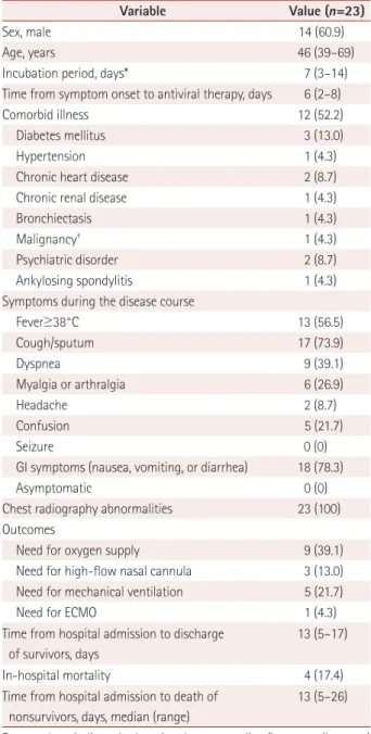
the interim recommendations generated during the early pe- riod of the Korean MERS epidemic.13 The demographic characteristics and clinical outcomes of all patients are pre- sented in Table 1. Four patients died due to early or late re- spiratory failure caused by the progression of the disease.
Table 1. Demographic features of patients with laboratory-con- firmed MERS coronavirus infection
Variable Value (n=23)
Sex, male 14 (60.9)
Age, years 46 (39–69)
Incubation period, days* 7 (3–14)
Time from symptom onset to antiviral therapy, days 6 (2–8)
Comorbid illness 12 (52.2)
Diabetes mellitus 3 (13.0)
Hypertension 1 (4.3)
Chronic heart disease 2 (8.7)
Chronic renal disease 1 (4.3)
Bronchiectasis 1 (4.3)
Malignancy† 1 (4.3)
Psychiatric disorder 2 (8.7)
Ankylosing spondylitis 1 (4.3)
Symptoms during the disease course
Fever≥38°C 13 (56.5)
Cough/sputum 17 (73.9)
Dyspnea 9 (39.1)
Myalgia or arthralgia 6 (26.9)
Headache 2 (8.7)
Confusion 5 (21.7)
Seizure 0 (0)
GI symptoms (nausea, vomiting, or diarrhea) 18 (78.3)
Asymptomatic 0 (0)
Chest radiography abnormalities 23 (100) Outcomes
Need for oxygen supply 9 (39.1)
Need for high-flow nasal cannula 3 (13.0) Need for mechanical ventilation 5 (21.7)
Need for ECMO 1 (4.3)
Time from hospital admission to discharge of survivors, days
13 (5–17)
In-hospital mortality 4 (17.4)
Time from hospital admission to death of nonsurvivors, days, median (range)
13 (5–26)
Except where indicated otherwise, data are median (interquartile range) or n (%) values.
*Five patients with an obscure incubation period were not included in the calculation, †One of the patients had a malignancy, hepatocellular carci- noma.
ECMO: extracorporeal membrane oxygenation, GI: gastrointestinal, MERS: Middle East respiratory syndrome.
Kim JE et al.
JCN
Characteristics of the patients with neurological complications
Four of the 23 included patients (2 men and 2 women) com- plained of neurological symptoms during or after MERS-CoV treatment, and all of these patients were referred to a neurolo- gist. Their median age was 46 years (IQR=27–46 years). The clinical presentations of these four patients and the thera- pies applied to them are summarized in Tables 2 and 3.
Patient 1
A 55-year-old man who may have been exposed to MERS- CoV in a hospital or through household contact between 5 and 18 days prior to his presentation at our hospital was ad- mitted because an RT-PCR of his sputum revealed positivity for MERS-CoV. He had a history of atrial fibrillation, diabetes mellitus, hypertension, chronic kidney disease, and hypo- thyroidism. At admission he denied any clinical symptoms, Table 2. Clinical presentations of patients with MERS who experienced neurological complications, and the therapies applied to them
Patient 1 Patient 2 Patient 3 Patient 4
Sex/age, years Male/55 Female/43 Male/46 Female/38
Incubation period, days 5–18 10 4 3
Initial symptoms Cough, dyspnea, and chest discomfort
Fever, myalgia, chills, cough, sputum, and headache
Fever, cough, dyspnea, and headache
Cough, sore throat, and fever
GI symptoms - Vomiting and nausea diarrhea -
Hospital course and treatment
Respiratory support Mechanical ventilation HFNC 5 L/min
nasal oxygen
2 L/min nasal oxygen
PSI 104 63 56 48
Sepsis severity* Septic shock Sepsis Sepsis Pneumonia
SAPS II 74 37 42 30
Antiviral regimen IFN, Rb, and LR IFN, Rb, and LR IFN, Rb, and LR IFN, Rb, and LR
Antibiotics used before onset of neurological symptoms
Ceftazidime, teicoplanin,
meropenem, and moxifloxacin - - -
IVIG treatment Yes No No No
Steroid treatment No No No No
*Sepsis is defined as life-threatening organ dysfunction from a dysregulated host reaction to an infection. Septic shock represents a subtype of sepsis that is accompanied by severe circulatory, cellular, and metabolic abnormalities that increase mortality.10
GI: gastrointestinal, HFNC: high-flow nasal cannula oxygen therapy, IFN: type 1 interferon, IVIG: intravenous immunoglobulin, LR: lopinavir/ritonavir, MERS: Middle East respiratory syndrome, PSI: Pneumonia Severity Index, Rb: ribavirin, SAPS II: Simplified Acute Physiology Score II.
Table 3. Neurological manifestations and laboratory findings in patients with laboratory-confirmed MERS
Patient 1 Patient 2 Patient 3 Patient 4
Neurological symptoms Hypersomnolence and weakness in all four limbs
Tingling/pain in both hands and below the knees, and
weakness in both legs
Tingling in distal parts
of both hands and feet Tingling in both hands
Days after MERS onset Unclear* 16 20 21
Neurological examination
Cranial nerves Ptosis and ophthalmoplegia Normal Normal Normal
Motor Weakness in all four limbs Proximal dominant weakness
in both legs Normal Normal
Sensory Normal Normal Hypesthesia in distal parts
of all four limbs Normal Deep tendon reflex Hyporeflexia in all four limbs Hyporeflexia in both legs Hyporeflexia in both legs Normal
Cerebellar function Limb ataxia Normal Normal Normal
Laboratory findings
CSF Normal NA NA NA
NCS Normal Normal Normal NA
Peak serum creatine kinase, U/L 45 48 99 NA
*Neurological symptoms were detected 24 days after the initial onset of respiratory symptoms.
CSF: cerebrospinal fluid, MERS: Middle East respiratory syndrome, NA: not available, NCS: nerve conduction study.
Neurological Complications in MERS
JCN
but chest radiography revealed ill-defined opacities in both of his lower lungs.
A triple antiviral regimen was initiated. He began to com- plain of dyspnea on hospital day (HD) 2, and his respiratory deterioration then progressed very rapidly. Antimicrobial therapy was added to his treatment regimen. He was intubat- ed, and a mechanical ventilator was added on HD 10. Acute respiratory failure was followed by ARDS, septic shock, and multiorgan dysfunction syndrome. His respiratory status be- gan to improve on HD 16, and he was taken off the mechani- cal ventilator on HD 28. The patient remained drowsy and ex- hibited bilateral ptosis up to 31 h after the administration of the sedative midazolam was stopped on HD 25. A neurological examination revealed complete external ophthalmoplegia and mild limb ataxia. Weakness was also suspected in all four limbs [Medical Research Council (MRC) grade 4]. Nystag- mus, sensory changes, and oropharyngeal or facial palsy were not observed. Deep tendon reflexes were decreased in all limbs.
The results of brain magnetic resonance imaging (MRI) and cerebrospinal fluid (CSF) studies were normal, including a negative CSF MERS-CoV RT-PCR assay. An electroenceph- alogram exhibited diffuse slow-wave activity. Nonspecific re- sults were obtained in laboratory studies performed at the onset of neurological symptoms, including in assays to deter- mine the glucose, thiamine, blood gas, ammonia, electrolyte, and creatinine levels. He was diagnosed with Bickerstaff’s en- cephalitis (BBE) overlapping with Guillain-Barré syndrome (GBS). IgM/IgG anti-GQ1b and IgM/IgG anti-GM1 antibody titers were analyzed on HD 39, and all were negative. The findings of nerve conduction studies performed on HD 46 were normal. His neurological complications began to im- prove on HD 30, and he had fully recovered by approximate- ly HD 60 (Fig. 1).
Patient 2
A 43-year-old woman who had been diagnosed with a labo- ratory-confirmed MERS-CoV infection was referred to our hospital. It was suspected that she had had contact with MERS- CoV-infected patients in another hospital 10 days before the onset of her symptoms. No underlying medical problems were present, but she initially presented with severe myalgia, chills, fever, cough, and headache. After 1 week (HD 2), she devel- oped gastrointestinal symptoms, including nausea, vomiting, and anorexia. On HD 3, she developed acute respiratory fail- ure, and the flow rate of her high-flow nasal cannula oxygen therapy was increased to 60 L/min with an inspiratory frac- tion of oxygen of 1.0. A chest radiograph displayed bilateral diffuse ground-glass opacities and dense consolidation.
A triple antiviral regimen was initiated on HD 1, with other treatments only including antiemetic, antitussive, and nonste- roidal anti-inflammatory drugs. Her clinical and radiological findings began to improve on HD 5, and oxygen therapy was stopped on HD 10. On the date of discharge (HD 10), she de- scribed a stinging pain in both hands and below the knees.
She felt that her legs were weakened, and she had difficulty walking for 1 km independently. A neurological examination revealed symmetric proximal dominant lower leg weakness, with her proximal and distal muscles at MRC grades 4– and 4, respectively. She experienced normal sensations to pinprick, temperature, vibration, and proprioception. Her deep tendon reflexes were mildly diminished in both legs. Laboratory find- ings did not indicate the presence of any underlying autoim- mune, infectious (except for a previous MERS infection), or nutritional diseases. Her creatine kinase level and the results of nerve conduction, electromyography, and evoked potential studies were normal. A CSF study was not performed. Her weakness began to improve 17 days after the original onset of the neurological symptoms, and had largely disappeared
Respiratory status starts to improve
Neurological symptoms fully recovered Paralysis
No weakness
TA(+) TA(+) TA(+) TA(+) TA(-) TA(-)
1 2 5 10 15 20 25 30 35 40 45 50 55 60 Intubation,
MV applied
IVIG infusion
Midazolam infusion Dyspnea,
pneumonia aggravation
Neurological symptoms start to improve Taken off MV Sedative drugs
stopped
Hypersomnolence, ophthalmoplegia, ataxia, and weakness in four limbs
Severity of weakness
Fig. 1. Timeline of clinical events and virological results in patient 1. The onset of neurological symptoms and their course during the sedative state were uncertain, and are indicated by the dotted lines. HD: hospital day, IVIG: intravenous immunoglobulin, MV: mechanical ventilator, TA: tracheal aspirate.
Time (HD)
Kim JE et al.
JCN
after 53 days. However, tingling and pain in the four distal limbs continued until the last follow-up (approximately 7 months after symptom onset). This patient was presumed to have ICU-acquired weakness or GBS.
Patient 3
A 46-year-old man with hypertension and a history of pul- monary tuberculosis was admitted due to infection with lab- oratory-confirmed MERS-CoV. He had previously experi- enced contact with patients with MERS in another hospital.
At 4 days after this exposure, he developed a fever, coughing, chest discomfort, dyspnea, and stool loss. A chest radiograph displayed ground-glass opacities in his left lung.
A triple antiviral regimen was initiated. No other drugs were administered that could cause peripheral neuropathy. Oxygen therapy was provided via a nasal prong and was increased to 5 L/min during admission. His pneumonia began to improve at 13 days after the onset of symptoms (HD 4), and the oxy- gen therapy was tapered off on HD 10. During this period he began to feel some tingling in the distal parts of his hands and feet. He denied experiencing any sensory symptoms pri- or to this admission. A neurological examination revealed normal mentation, cranial nerves, and muscle strength. He had bilateral decreases in pinprick and temperature sensa- tions up to the knees and elbows. His reflexes were decreased at both knees but were normal in both upper extremities. His other neurological findings were unremarkable. The results of nerve conduction studies and quantitative sensory tests were normal. He was assumed to have infectious or toxic poly- neuropathy, and his sensory symptoms gradually improved over 6 months.
Patient 4
A 38-year-old woman developed a cough, sore throat, and fe- ver 3 days after being exposed to patients with MERS in an- other hospital. A chest radiograph exhibited diffuse ground- glass opacities in both lungs.
A triple antiviral regimen was started. She reported tingling in both hands at approximately 3 weeks after the onset of her respiratory symptoms. She denied experiencing any sensory symptoms prior to her most recent admission. These sensory symptoms lasted for more than 4 months. A neurological ex- amination showed that her cranial nerves and muscle strength were normal. She experienced normal sensations in pinprick, temperature, vibration, and proprioception tests. Her reflexes were normal in all four extremities. A nerve conduction study was not performed because the patient was lost to fol- low-up. She was tentatively diagnosed with acute sensory neu- ropathy caused by a toxin or infection.
DISCUSSION
We have presented four patients with MERS-CoV infections who showed specific neurological manifestations during their treatment. These patients probably had BBE overlap- ping with GBS, ICU-acquired weakness, or acute sensory neu- ropathy that resulted from a toxin or infection. A particularly interesting finding was that almost one in five patients with MERS-CoV infections displayed neurological symptoms during or after the infection was confirmed. Although data were collected at a single institution, this represents a con- siderable proportion of infected patients, and suggests that such cases could have been underdiagnosed previously.
Patient 1 showed hypersomnolence, ophthalmoplegia, and weakness in all four limbs after a severe viral infection, and we therefore needed to differentiate between various condi- tions including GBS variant, Wernicke encephalopathy, pro- longed neuromuscular blockade, and critical-illness poly- neuropathy/myopathy. Because this patient had a preceding infection history, relative symmetric motor weakness, and a monophasic course with a clinical nadir within 4 weeks fol- lowed by a plateau, we considered GBS variant as a possible diagnosis.14 BBE, one of the GBS variants, is considered a sub- type of GQ1b antibody syndrome and is classically diagnosed based on its characteristic clinical features of progressive sym- metric external ophthalmoplegia, ataxia, and impaired con- sciousness.15 Because patient 1 had the classical triad of BBE accompanied by limb weakness, a diagnosis of BBE overlap- ping with GBS was suggested.14 We excluded many BBE- mimicking conditions in this patient based on his clinical histo- ry, brain MRI, and CSF findings. The presence of antiganglioside antibodies and albuminocytologic dissociation in the CSF supported the diagnosis of BBE as part of the GBS spectrum.
Nevertheless, a diagnosis of BBE is largely dependent on its clinical presentation. The absence of laboratory findings does not rule out a BBE diagnosis.16
Wernicke’s encephalopathy was an important condition to eliminate in this patient because it also frequently manifests with varying degrees of encephalopathy, ophthalmoplegia, and ataxia. However, we considered it unlikely that Wer- nicke’s encephalopathy was present in patient 1 for several reasons: 1) he showed none of the typical changes associated with Wernicke’s encephalopathy in brain MRI, which has particularly high sensitivity (97–100%) in patients without alcohol abuse,17 2) the ptosis and complete ophthalmoplegia that were observed in this patient are rare in Wernicke’s en- cephalopathy,18 and 3) thiamine deficiency was not confirmed in laboratory tests and the patient did not have dietary defi- ciencies or alcoholism. Critical-illness polyneuropathy/myop- athy appears frequently in ICU patients but was not thought
Neurological Complications in MERS
JCN
to be present in patient 1 because ophthalmoparesis and pto- sis are very rare in critical-illness polyneuropathy/myopa- thy.19 Prolonged neuromuscular blockade was another possi- ble consideration in this clinical setting, but a neuromuscular blocking agent was not administered in patient 1. The limb and ocular weakness had lasted for almost 2 months, and this duration would be an unusual response to the prolonged administration of a neuromuscular blocking agent. Neuro- logical signs typically completely recover within 1–2 weeks in patients with prolonged neuromuscular blockade.19
We hypothesized that patient 2 had ICU-acquired weak- ness or GBS. ICU-acquired weakness is diagnosed when a critically ill patient has limb weakness or ventilator depen- dency without heart or lung disease.20 GBS was another pos- sible diagnosis, since the patient experienced symmetric limb weakness following viral infection with a typical monophasic disease course.14 These two diseases are differentiated by the presence of albuminocytologic dissociation in the CSF, posi- tivity for antiganglioside antibodies, or demyelinating or ax- onal patterns in electrophysiology.18,19 Patient 2 was evaluated neurologically after discharge, when her neurological symp- toms had substantially improved; a lumbar puncture was there- fore not performed, and critical information from laboratory studies was also not available. However, we cautiously sup- ported a diagnosis of ICU-acquired weakness in patient 2, be- cause a paraparetic presentation (as observed in this patient) is extremely rare in GBS and because dysautonomia and cra- nial nerve palsy, which frequently accompany GBS, were not observed in this patient.18,21
Acute sensory neuropathy was the probable diagnosis in patients 3 and 4, which may have resulted from a toxin and/
or drug or viral infection. These two patients both received pegylated interferon alpha-2a, ribavirin, and lopinavir/rito- navir to treat MERS-CoV. Interferon alpha-2a rarely induc- es peripheral neuropathy,22 and several studies have found complications associated with sensory neuropathy, vasculitic neuropathy, Bell’s palsy, GBS, chronic inflammatory demye- linating polyneuropathy, and autonomic neuropathy.23,24 Rib- avirin is not associated with peripheral neuropathy,24 while lopinavir/ritonavir is another possible causative drug. How- ever, many people living with human immunodeficiency vi- rus have been treated with this protease inhibitor for many years, and the risk of peripheral neuropathy in patients receiv- ing lopinavir/ritonavir remains unclear.25
CoVs are a group of enveloped RNA viruses that include the Alphacoronavirus and Betacoronavirus [including MERS- CoV and severe acute respiratory syndrome (SARS) CoV]
genera.26 These viruses have neurotrophic and neuroinvasive characteristics, and CoV RNA has been detected in the cen- tral nervous systems of patients with various neurological
diseases.26,27 A recent in vitro study evaluated the human tissue tropism of MERS-CoV in diverse cell lines, and revealed that it has the ability to infect human neuronal cells (NT2).28 The neuropathological effects of MERS-CoV infections are sug- gested to result from immune-mediated processes, either di- rectly by viral invasion or via molecular changes that arise from systemic inflammatory response syndrome. The de- layed onset of neurological complications, the absence of the virus in the CSF, and the development of GBS-which is one of the prototypical immunological diseases observed in our patients-might support the immunological mechanisms of these phenomena. This topic requires further microbiologi- cal and pathological studies.
The structure of SARS-CoV is very similar to that of MERS- CoV, and these two viruses share many clinical characteris- tics.26 The neurological manifestations observed in patients with SARS were revealed during the Taiwan outbreak. Four patients who showed critical-illness polyneuropathy/myopa- thy have been described,29 while other groups have also re- ported central nervous involvement in ischemic stroke in pa- tients with SARS.30 Patients with MERS-CoV infections have a high probability of experiencing neurological complications, similar to other CoVs.
In conclusion, we encountered four neurological manifes- tations during MERS treatment. The most likely diagnoses were GBS (including its variant), BBE, as well as ICU-ac- quired weakness and toxins (including drugs) or virus-relat- ed sensory neuropathy. It was particularly interesting that the neurological complications did not appear concomitantly with respiratory symptoms, instead being delayed by 2–3 weeks. Understanding that neurological complications are not rare and may be delayed is important because it might be difficult for patients with MERS-CoV infections to contact a neurologist while they are in isolation and because thorough neurological evaluations of these patients are rarely performed.
Furthermore, some neurological complications interfere with the prognosis and require appropriate treatment, such as im- munoglobulin or plasmapheresis for GBS.
Conflicts of Interest
The authors have no financial conflicts of interest.
REFERENCES
1. who.int [internet]. Middle East Respiratory Syndrome Coronavirus Infections-Disease Outbreak News. [cited 2016 Nov 22]. Available from: http://www.who.int/emergencies/mers-cov/en.
2. Zaki AM, van Boheemen S, Bestebroer TM, Osterhaus AD, Fouchier RA. Isolation of a novel coronavirus from a man with pneumonia in Saudi Arabia. N Engl J Med 2012;367:1814-1820.
3. mers.go.kr [internet]. Summary of MERS Statistics in the Republic of Korea. [cited 2016 Nov 22]. Available from: https://www.mers.go.kr/
mers/html/jsp/main.jsp.
Kim JE et al.
JCN
4. WHO Mers-Cov Research Group. State of knowledge and data gaps of Middle East respiratory syndrome coronavirus (MERS-CoV) in hu- mans. PLoS Curr 2013;5:ecurrents.outbreaks.0bf719e352e7478f8ad85f a30127ddb8.
5. Desforges M, Le Coupanec A, Stodola JK, Meessen-Pinard M, Talbot PJ. Human coronaviruses: viral and cellular factors involved in neuro- invasiveness and neuropathogenesis. Virus Res 2014;194:145-158.
6. Saad M, Omrani AS, Baig K, Bahloul A, Elzein F, Matin MA, et al.
Clinical aspects and outcomes of 70 patients with Middle East respira- tory syndrome coronavirus infection: a single-center experience in Saudi Arabia. Int J Infect Dis 2014;29:301-306.
7. Arabi YM, Harthi A, Hussein J, Bouchama A, Johani S, Hajeer AH, et al. Severe neurologic syndrome associated with Middle East respirato- ry syndrome corona virus (MERS-CoV). Infection 2015;43:495-501.
8. Algahtani H, Subahi A, Shirah B. Neurological complications of Mid- dle East respiratory syndrome coronavirus: a report of two cases and review of the literature. Case Rep Neurol Med 2016;2016:3502683.
9. who.int [internet]. Technical Guidance on Laboratory: Middle East re- spiratory syndrome coronavirus (MERS-CoV). [cited 2016 Nov 22].
Available from: http://www.who.int/csr/disease/coronavirus_infec- tions/technical-guidance-laboratory/en.
10. Singer M, Deutschman CS, Seymour CW, Shankar-Hari M, Annane D, Bauer M, et al. The third International Consensus Definitions for Sep- sis and Septic Shock (Sepsis-3). JAMA 2016;315:801-810.
11. Fine MJ, Auble TE, Yealy DM, Hanusa BH, Weissfeld LA, Singer DE, et al. A prediction rule to identify low-risk patients with community- acquired pneumonia. N Engl J Med 1997;336:243-250.
12. Le Gall JR, Lemeshow S, Saulnier F. A new Simplified Acute Physiolo- gy Score (SAPS II) based on a European/North American multicenter study. JAMA 1993;270:2957-2963.
13. Chong YP, Song JY, Seo YB, Choi JP, Shin HS; Rapid Response Team.
Antiviral treatment guidelines for Middle East respiratory syndrome.
Infect Chemother 2015;47:212-222.
14. Wakerley BR, Uncini A, Yuki N; GBS Classification Group; GBS Clas- sification Group. Guillain-Barré and Miller Fisher syndromes--new diagnostic classification. Nat Rev Neurol 2014;10:537-544.
15. Mori M, Kuwabara S, Fukutake T, Yuki N, Hattori T. Clinical features and prognosis of Miller Fisher syndrome. Neurology 2001;56:1104- 1106.
16. Shahrizaila N, Yuki N. Bickerstaff brainstem encephalitis and Fisher syndrome: anti-GQ1b antibody syndrome. J Neurol Neurosurg Psychi-
atry 2013;84:576-583.
17. Galvin R, Bråthen G, Ivashynka A, Hillbom M, Tanasescu R, Leone MA; EFNS. EFNS guidelines for diagnosis, therapy and prevention of Wernicke encephalopathy. Eur J Neurol 2010;17:1408-1418.
18. Wakerley BR, Yuki N. Mimics and chameleons in Guillain-Barré and Miller Fisher syndromes. Pract Neurol 2015;15:90-99.
19. Dhand UK. Clinical approach to the weak patient in the intensive care unit. Respir Care 2006;51:1024-1040; discussion 1040-1041.
20. Latronico N, Bolton CF. Critical illness polyneuropathy and myopa- thy: a major cause of muscle weakness and paralysis. Lancet Neurol 2011;10:931-941.
21. van den Berg B, Fokke C, Drenthen J, van Doorn PA, Jacobs BC. Para- paretic Guillain-Barré syndrome. Neurology 2014;82:1984-1989.
22. Briani C, Chemello L, Zara G, Ermani M, Bernardinello E, Ruggero S, et al. Peripheral neurotoxicity of pegylated interferon alpha: a prospec- tive study in patients with HCV. Neurology 2006;67:781-785.
23. Meriggioli MN, Rowin J. Chronic inflammatory demyelinating poly- neuropathy after treatment with interferon-alpha. Muscle Nerve 2000;
23:433-435.
24. Khiani V, Kelly T, Shibli A, Jensen D, Mohanty SR. Acute inflammato- ry demyelinating polyneuropathy associated with pegylated interferon alpha 2a therapy for chronic hepatitis C virus infection. World J Gas- troenterol 2008;14:318-321.
25. Ellis RJ, Marquie-Beck J, Delaney P, Alexander T, Clifford DB, McAr- thur JC, et al. Human immunodeficiency virus protease inhibitors and risk for peripheral neuropathy. Ann Neurol 2008;64:566-572.
26. Zumla A, Hui DS, Perlman S. Middle East respiratory syndrome. Lan- cet 2015;386:995-1007.
27. Arbour N, Day R, Newcombe J, Talbot PJ. Neuroinvasion by human respiratory coronaviruses. J Virol 2000;74:8913-8921.
28. Chan JF, Chan KH, Choi GK, To KK, Tse H, Cai JP, et al. Differential cell line susceptibility to the emerging novel human betacoronavirus 2c EMC/2012: implications for disease pathogenesis and clinical mani- festation. J Infect Dis 2013;207:1743-1752.
29. Tsai LK, Hsieh ST, Chao CC, Chen YC, Lin YH, Chang SC, et al. Neu- romuscular disorders in severe acute respiratory syndrome. Arch Neu- rol 2004;61:1669-1673.
30. Umapathi T, Kor AC, Venketasubramanian N, Lim CC, Pang BC, Yeo TT, et al. Large artery ischaemic stroke in severe acute respiratory syn- drome (SARS). J Neurol 2004;251:1227-1231.