Ⅰ. 서 론
치과용 임프란트는 1960년대 Branemark등1)이 골유착의 가능성을 보고한 이래 1980년대부터 무치 악환자에서 임프란트의 사용이 임상술식으로 인정 받게 되었으며, 그 후, 발전을 거듭하여 현재는 완전 또는 부분 무치악 및 상실된 단일치아의 수복을 위 한 성공적인 치료법으로 이용되고 있다2-5). 임프란트 의 종류는 nonsubmerged type과 submerged type 이 있으며, submerged type의 경우 약 3 - 6개월의 치유기간후 임프란트 노출을 위한 2차 수술이 요구 된다6-9).
2차 수술을 위한 방법으로는 외과적 수술도를 이 용하는 방법10-16), Tissue punch를 이용하는 방법17), 전기 소작술에 의한 노출방법18), 레이저를 이용한 노
출방법19-23)등을 고려할 수 있으며 현재 일반적으로
시행되고 있는 외과적 수술도를 이용한 방법은 국소 마취하에서 치조정에 절개를 가하여 임프란트 덮개 나사를 노출시키는 방법으로 부착치은이 부족한 경 우 근단 변위 판막술, 결체조직 이식술, 유리치은 이 식술등 다양한 술식에 의해 부착치은을 확보한다.
레이저를 이용하는 방법은 외과적 수술도를 이용 한 방법과 비교시 마취가 거의 필요하지 않으며 지 혈효과24)에 의하여 수술시야가 향상되고, 인접 건강 한 조직의 손상을 최소화하며, 절개와 봉합이 불필 요하므로 시술시간이 단축되고 술후 부종 및 동통이 거의 없고25)출혈소견이 없어 신속하게 보철치료를 행할 수 있다. 또한 지대치 주위 연조직의 심미성을
증진시킬 수 있고 살균력에 의해 감염이 감소26)되는 등의 장점이 있다. 단, 부착치은이 충분한 경우와 술 자가 임프란트의 식립위치를 예측가능한 경우에 이 용할 수 있고 좁은 범위의 각화치은의 경우에는 사 용이 제한된다.
레이저는 1917년 Albert Einstein27)이 E=MC2의 방정식으로 그 기본원리를 밝힌 이래로 1958년 Townes28,29)에 의해 레이저의 전신인 Maser (Micro- wave Amplication by Stimulated emission of radiation)가 개발되었으며 1960년 S. Maiman30)은 Ruby를 사용하여 Laser(Light Amplication by Stimulated emission of radiation)의 발진에 성공하 였다. 레이저 연구 또한 활성화되어 여러종류의 고 체, 기체, 액체 및 반도체 레이저가 개발되어 우주항 공분야, 국방, 정보통신, 의학31-34), 치의학등으로 응 용분야가 확대되었다. 치의학 분야에서 레이저는 1964년 Goldman35)에 의해 처음 사용되었고 현재는
와동형성36-40), 근관 및 치근단의 치료41-44), 우식예방
45,46),치주치료47), 외과적 시술48)등에 이용되고 있으
며, 그 종류에 He-Ne, Argon, CO2, Er;YAG(Erbium yttrium Aluminum Garnet), Diode, Nd:YAG(neodymium:yttrium-aluminum-gar- net) 레이저등이 있다.
임프란트 주위연조직 시술에 레이저를 이용하는 것은 매우 흥미있는 연구분야로서, 레이저를 이용한 술식에는 1)임프란트의 2차 수술19-23), 2)임프란트 주위조직염증시 살균 또는 멸균효과20,22,26), 3)상피조 직의 근단이동 지연49), 4)비정상적인 임프란트 주위 대한치과보철학회지:Vol. 37, No. 2, 1999
레이저를 이용한 임프란트 이차수술시 온도변화에 관한 실험적 연구
대림성모병원 치과부 보철과 안현정∙김현철∙최병갑∙송언희∙김래경
연조직의 처치22,50)등을 생각할 수 있다. 레이저의 이 용은 시술범위가 다양하고 많은 장점이 있는 반면 레이저의 종류에 따라 임프란트 주위조직의 열손상 과 임프란트의 표면변화를 초래하여 그 사용이 제한 될 수 있다. 그러므로 본 연구는 Pulsed Nd;YAG, CO2, Er;YAG 레이저 각각을 임프란트에 적용시 주위조직의 온도변화와 임프란트의 표면변화에 대 한 관찰을 통하여 향후 임프란트에 대한 레이저의 임상적 적용을 위한 기초자료로 활용하고자 한다.
Ⅱ. 실험 재료 및 방법
직경 3.75mm, 길이 10mm의 pure titanium 임프
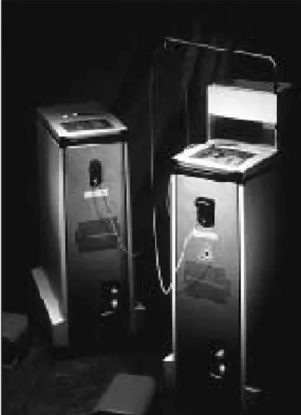
란트와 직경 4.0mm, 길이 10mm H-A coated 임프 란트 몸체의 상부와 말단부 각각에 thermocouple을 위치시켰으며(Fig. 1, 2), 2개의 thermocouple을 각각 thermometer에 연결하였다(Fig. 3).
이 때 레이저 에너지가 thermocouple에 직접 조사 되어 심한 온도상승이 초래되는 것을 방지하며 임프 란트 덮개나사에만 레이저 에너지가 조사될 수 있도 록 하기위해 실리콘 인상재를 이용하여 주형을 제작 하고 탄성 고무인상재를 이용하여 봉쇄하였다(Fig.
4, 5).
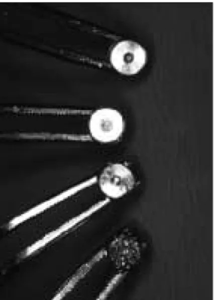
임프란트의 덮개나사 표면에 pulsed Nd-YAG laser (Sunlase, USA)(Fig. 6), continuous mode
& superpulse의 CO2laser(ILT, USA/ UMECO, Korea)(Fig 7, 8), Er-YAG laser (HOYA, Japan)(Fig. 9)를 30초동안 조사한후 10초, 20초, 30 초후 각각의 온도변화를 측정하였으며, 덮개나사의 표면변화를 관찰하였다(Fig. 10).
Fig. 2. H-A coated implant with two thermocouples Fig. 1. pure titanium implant with two thermocouples
Fig. 3. Thermometer used in this study Fig. 4. pulsed Nd:YAG laser irradiation
각 레이저는 연조직 시술시 임상적으로 사용가능 한 조건을 이용하였다.
1) pulsed Nd-YAG laser (Sunlase, U.S.A.) ; 2W, 20pps, contact mode
2) continuous mode의 CO2laser (ILT, U.S.A.)
; 주수 및 무주수상태, 2.5 - 3.5W noncontact mode
3) superpulse의 CO2laser (UMECO, Korea) ;
무주수상태, 3W, noncontact mode
4) Er-YAG laser ( HOYA, Japan) ; 주수상태, 10pps, 60mj, 80mj, 101mj, contact mode 무 주수상태, 10pps, 60mj, contact mode Fig. 5. Er:YAG laser irradiation Fig. 6. pulsed Nd:YAG laser (Sunlase/USA) used
in this study
Fig. 7. CO2laser (ILT/ USA) used in this study Fig. 8. CO2laser (UMECO/ Korea) used in this study
Ⅲ. 연구성적 1. 표면변화 (Fig.10)
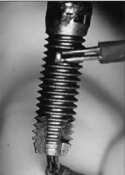
pulsed Nd-YAG laser를 이용한 경우 현저한 pit- ting과 darkening을 나타내었으며 CO2laser와 Er- YAG laser를 이용한 경우에는 특별한 변화를 보이지 않았다.
2. 온도변화
레이저 에너지가 임프란트에 조사되는 동안 ther- mocouple의 온도변화를 각 조건에서 10회씩 측정하 여 그 평균값을 기록하였다. 각각의 실험은 임프란 트에 연결된 thermometer가 실험전의 온도까지 내 려간 것을 확인한 후 시행되었다.
골유착된 임프란트의 경우 온도상승에 의한 손상 을 방지하기위해 정상체온 36.5℃를 기준시 47℃이 상의 온도상승을 나타내지 않아야 한다.
본 실험은 평균 초기온도 33℃에서 실시되었으므 로 43.5℃이하의 온도를 나타내어야 한다는 기준으 로 실시되었다.
1) Pure titanium 임프란트에서의 온도변화(Table 1, 2/ Fig. 11, 12)는 pulsed Nd:YAG laser의
경우 30초 동안 레이저를 조사한 후 10초 경과시 가 장 높은 온도상승으로 임프란트 몸체 상부에서 19.01℃, 말단부에서 11.78℃의 상당한 온도 변화를 나타내었다. 지속형의 CO2laser의 경우 무수하에서 레이저 조사후 20초 경과시 가장 높은온도 상승으로 임프란트 몸체 상부에서 4.74℃, 말단부에서 2.61℃
의 온도변화를 나타내었으며 펄스형의 CO2laser와 Er-YAG laser의 경우 무주수하에서 2.0℃이하 의 적은 온도변화를 나타내었고 주수의 조건에서는 온도감소를 나타내어 주수에 의해 레이저 에너지의 발열반응이 억제됨을 알 수 있었다.
2) 동일한 조건의 실험을 H-A coated 임프란트에 시행한 경우의 온도변화는 pure titanium 임프란트 에서와 비교시 더 적은 범위의 온도상승을 나타내었 다(Table 3, 4, Fig. 13, 14). pulsed Nd;YAG laser의 경우 레이저 조사후 20초 경과시 임프란트 몸체 상부에서 8.74℃, 말단부에서 3.78℃의 온도변 화를 나타내었으며, 지속형 CO2laser의 경우, 레이 저 조사후 10초 경과시 임프란트 몸체 상부에서 4.06℃, 말단부에서는 1.81℃의 온도변화를 나타내 었다. 펄스형 CO2laser와 Er-YAG laser의 경우 무 주수하에서 약 0.5℃이하의 적은 온도변화를 나타내 었으며 주수의 조건에서는 온도감소를 나타내었다.
Fig. 9. Er:YAG laser (HOYA/ Japan) used in this study
Fig. 10. Surface changes of implant cover screws
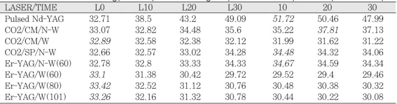
Table 1. Effect of lasers energy on temperature change in pure titanium implant at bottom thermocouple
Pulsed Nd-YAG 32.78 35.5 38.7 42.27 44.56 44.43 43.69
CO2/CM/N-W 32.69 32.43 32.77 34.45 35.01 35.3 35.13
CO2/CM/W 32.65 32.5 32.37 32.22 32.09 31.78 31.45
CO2/SP/N-W 32.44 32.87 33.04 34.32 34.44 34.04 33.68
Er-YAG/N-W(60) 33.11 33.07 33.28 33.73 34.17 34.32 34.23
Er-YAG/W(60) 33.42 33.04 32.32 31.8 31.42 31.24 31.1
Er-YAG/W(80) 33.44 33.28 32.82 32.38 32.02 31.84 31.74
Er-YAG/W(101) 33.54 33.42 32.88 32.38 31.98 31.74 31.58
CM ; continuous mode. SP ; superpulse.
W ; water infused state. N-W ; non-water infused state.
LASER/TIME L0 L10 L20 L30 10 20 30
Table 4. Effect of lasers energy on temperature change in H-A coated imlant at top thermocouple
Pulsed Nd-YAG 33.06 33.06 35.39 38.55 41.23 41.8 41.63
CO2/CM/N-W 32.15 32.7 35.31 37 37.21 36.31 35.43
CO2/CM/W 32.9 33.65 34.67 35.15 37.72 34.5 33.9
CO2/SP/N-W 32.3 32.3 32.5 32.6 32.7 32.8 32.8
Er-YAG/N-W(60) 33.18 33.1 33.09 33.24 33.44 33.61 33.59
Er-YAG/W(60) 33.52 33.82 31.82 31.04 30.72 30.56 30.58
Er-YAG/W(80) 33.32 32.58 31.2 30.5 30.14 30.02 30.06
Er-YAG/W(101) 33.54 33.14 32.06 31.58 31.24 31.24 31.4
LASER/TIME L0 L10 L20 L30 10 20 30
Table 3. Effect of lasers energy on temperature change in H-A coated implant at bottom thermocouple
Pulsed Nd-YAG 32.9 32.83 32.99 33.83 35.73 36.68 37.87
CO2/CM/N-W 32.09 31.99 32.45 33.33 33.9 33.61 33.59
CO2/CM/W 32.6 32.56 32.65 32.89 33.18 33.09 32.9
CO2/SP/N-W 32.22 32.25 32.37 32.5 32.58 32.72 32.72
Er-YAG/N-W(60) 33.06 32.97 33.04 33.2 33.21 33.3 33.33
Er-YAG/W(60) 33.38 33.38 33.34 33.24 33.08 32.9 32.78
Er-YAG/W(80) 32.96 32.94 32.86 32.7 32.5 32.24 31.36
Er-YAG/W(101) 33.2 33.24 33.22 33.1 32.94 32.78 32.64
LASER/TIME L0 L10 L20 L30 10 20 30
Table 2. Effect of lasers energy on temperature change in pure titanium implant at top thermocouple
Pulsed Nd-YAG 32.71 38.5 43.2 49.09 51.72 50.46 47.99
CO2/CM/N-W 33.07 32.82 34.48 35.6 35.22 37.81 37.13
CO2/CM/W 32.89 32.58 32.38 32.12 31.99 31.62 31.22
CO2/SP/N-W 32.66 32.57 33.02 34.28 34.48 34.32 34.06
Er-YAG/N-W(60) 32.78 32.8 33.33 34.33 34.67 34.59 34.34
Er-YAG/W(60) 33.1 31.38 30.42 29.72 29.52 29.4 29.46
Er-YAG/W(80) 33.42 32.52 31.12 30.76 30.48 30.38 30.32
Er-YAG/W(101) 33.26 32.16 31.32 30.78 30.44 30.22 30.08
LASER/TIME L0 L10 L20 L30 10 20 30
Fig. 1. Effect of lasers energy on temperature change in pure titanium implant at bottom thermocouple
Fig. 2. Effect of lasers energy on temperature change in pure titanium implant at top thermocouple
Fig. 3. Effect of lasers energy on temperature change in H-A coated implant at bottom thermocouple
Ⅳ. 총괄 및 고안
임프란트 2차 수술은 모든 임프란트시술의 장기적 인 성공에 중요한 요소의 하나이다. 효율적인 2차 수술을 위한 레이저의 응용은 술자에게 많은 이점을 부여하나 부적절한 레이저의 선택에 의한 치료는 임 프란트 주위 연조직 시술시 온도변화에 의한 열손상 및, 금속표면의 변화를 초래할 수 있으므로 주의하 여야 한다.
레이저와 생체조직간의 상호작용은 광자와 조직의 원자 내지는 분자의 작용으로 광선의 파장, 출력밀 도, 에너지 밀도, 촛점화와 비촛점화, 펄스기간, 에너 지 전달방법, 조직의 종류 등에 의해 결정된다. 레이 저 조사에 대한 조직의 반응은 각 레이저 파장의 흡 수 정도에 비례하며 광에너지가 유효한 에너지로 전 환되는 흡수, 광에너지가 일정하지 않은 방향으로 재방출되어 결국 더 넓은 표면에 흡수되므로 강도와 정확도가 낮은 분산된 열효과를 나타내는 산란, 광 에너지가 조직표면에서 거의 또는 전혀 흡수되지 않고 반사되어 조직에 아무런 영향도 주지않는 투 과, 그리고 광에너지가 어떠한 상호 작용도 없이 자 유롭게 조직을 통과하여 조직에 거의 또는 전혀 영 향을 주지않는 반사 등으로 다양하게 나타난다51,52).
또한, 레이저 조사에 대한 조직효과의 형태는 광화 학적 상호작용, 광열적 상호작용, 광기계적 상호작용 등이 있으며53), 첫째로, 광화학적 상호작용은 자외선 영역의 파장에서 나타나는 것으로 생화학적 반응을
유도하는 발색단에 의해 흡수되어 고에너지 상태로 흥분된후 산소방출에 의해 세포파괴를 유도하며 주 로 악성종양에 대한 치료와 충치 감별에 이용된다.
둘째로, 광열적 상호작용은 외과적 절제나 제거에 많이 이용되는 것으로 광에너지가 조직에 흡수되어 열에너지로 전환되어 전자적, 분자적 흥분을 유도하 고 열전도과정에 의해 조직을 분해하며 주변조직으 로 열이 확산되는 효과를 초래한다. 이와같은 기전 에 의한 조직반응은 온도상승에 따라 warming(37 - 60℃), welding(60 - 65℃), coagulation(65 - 90℃), protein denaturization(90 - 100℃), drying, vaporization(100℃), carbonization(300℃)등54) 으로 나타나며 주변조직의 혈류를 통한 냉각, 열전 도의 정도, 광선의 흡수와 산란의 정도, 레이저의 방 출양식 등에 의해 조직 손상의 정도가 좌우된다. 끝 으로, 광기계적 상호작용은 고에너지, 짧은 펄스의 레이저 에너지가 적은 면적의 조직을 빠르게 기화시 켜 프라즈마를 형성하며 빠른 기계적 충격에 수반되 는 고압발생으로 팽창을 통한 조직파괴를 나타낸다.
온도상승에 따른 골조직의 반응에 대하여 Green과 Matthews55)는 56℃의 경우 alkaline phosphatase 가 변성되어 비가역적인 변화가 초래된다고 하였으 며 Lundskog56)는 50℃에 30초동안 가열된 임프란트 주위에 골괴사의 조직화학적 증거를 보고하였다.
1984년 Ericksson과 Albrektsson은 44℃이하 온도 에서의 임프란트 시술은 주위 골손상없이 가능하며 44 - 47℃사이의 온도에서는 유해한 효과가 발생되 Fig. 4. Effect of lasers energy on temperature change in H-A coated imlant at top
thermocouple
기 시작하고 47℃ 에서 1분 또는 50℃에 순간적으로 노출되는 경우 골괴사를 야기하여 골유착이 파괴 되므로 수술동안의 온도조절이 중요하다고 강조하 였다57). 또한, 열손상의 중요성은 골괴사 보다는 골 의 분화가능성 여부에 있다고 하였다58).
Pulsed Nd-YAG laser (1.064um의 근적외선 파 장)는 광열적 작용에 의한 조직효과로 조직내의 수 분에 잘 흡수되지 않기 때문에 비접촉방식으로 사용 할 때 조직표면에서의 흡수가 적고 내부에 침투, 산 란되어 심한 조직괴사를 초래할 수 있다. 그러나, 접 촉방식으로 사용할 때에는 조직표면 흡수에 의해 약 200um의 조직소산효과가 기대된다59,60). 임프란트 2 차수술에 pulsed Nd:YAG laser의 사용은 Myer61,62) 가 처음 보고하였으며 Tadamasa63)는 비스듬히 조사 한 경우 정면으로 조사한 경우에 비해 심한 열손상 이 발생되지 않는다고 하였고 Bida64)는 금속면에서 약간의 pitting이 발생되나 마찬가지로 심한 열손상 이 발생되지는 않는다고 하였다. 그러나 Laurence65) 는 금속은 레이저열을 흡수하므로 표면구조가 변할 수 있어 사용하지 않는 것이 좋다고 하였으며, Block66)은 레이저를 임프란트 표면에 접촉 조사시 비 접촉 조사의 경우보다 훨씬 심한 손상이 나타남을 보고하였다. 본 연구에서도 이들의 결과와 유사하게 레이저에너지가 조사된 덮개나사부위에 심한 pit- ting, 탄화층형성이 초래됨과 동시에 심한 온도상승 을 관찰하였다.(레이저 조사후 10초 경과시 51.72℃
/ 온도 변화량: 19.01℃)
CO2laser(10.6um의 원적외선 파장)는 Patel67)에 의해 1964년에 개발되었으며 1979년68)에 처음으로 치과용으로 이용되었다. CO2레이저의 경우도 광열 적 작용에 의한 조직효과로 수분에 잘 흡수되므로 발생된 에너지의 95%가 치은조직에 신속히 흡수되 어 열에너지로 전환되며 목표조직의 세포내액과 세 포외액을 기화시켜 세포막을 파괴함으로써 조직이 제거되고 분화구양상의 결손이 형성된다. 조직은 열 전도가 낮으므로 열손상은 좁은범위에 국한되며69,70) 콜라겐 분자구조의 변화에 의해 0.5 - 1mm이내의 작은 혈관들이 폐쇄되어 조직제거에 더욱 효과적으 로 이용될 수 있다. 레이저에 대한 지속적인 연구에 의해 방출양식이 지속형에서 펄스형으로 변경되므 로써 충분한 조직효과를 낼 수 있는 만큼의 에너지 를 매우 짧은 지속 시간에 전달하게 하므로 보다 적
은 열손상으로 시술할 수 있다52,60). 그리고, CO2레이 저는 Titanium 덮개나사와 같은 금속 물질에는 반사 되므로 주위조직과 임프란트에 대한 열손상없이 효 과적인 시술이 가능하다는 이점을 가지고 있다71). Ganz52)는 continuous mode 2 - 4W, pulse mode 5 - 6W에서 안전하고 효과적인 시술이 가능하다고 하 였으나 온도상승효과와 노출시간을 최소화하기위해 on-and-off tachnique을 이용할 것을 추천하였다. 본 연구에서도 지속형에 비해 펄스형의 경우 더 적은 범위의 온도변화를 나타내었다(지속형;37.81℃/온 도 변화량:4.74℃, 펄스형:34.44℃/온도변화량:2
℃).
Er-YAG laser (2.94um의 중적외선 파장)는 광기 계적 작용에 의한 조직효과로 통상 치아를 포함한 경조직용으로 알려졌으나 수분의 광흡수 파장대 peak와 일치하고 생체조직에 대한 소산능력이 높으 며 작용부위는 조사부위의 매우 표층이고 주위조직 에 대한 영향은 거의 나타나지 않으므로 연조직 시 술도 기대할 수 있다. Er-YAG laser를 접촉방식으로 연조직 시술에 이용하는 경우 광에너지가 부분적으 로 열에너지로 전환되어 조직의 응고를 나타내며 CO2, pulsed Nd:YAG laser에서 탄화층을 형성하는 것과는 달리 절제된 치은면 직하방에 amyloid-like 치은 변성을 나타내고 레이저가 조사된 후의 조직면 이 비교적 매끈하다72). 본 연구에 이용된 일본 HOYA사의 제품은 불화물광섬유에 의한 도광이 가 능하여 접촉방식으로 임상에 응용하는 것이 더욱 용 이하게 되었으며 연조직 외과 수술72,73), scaling73), 와 동형성39)등에 이용될 수 있다.
Watanabe등73)은 치주질환 환자에서 치은절제술 을 위해 61.5mJ/pulse의 조건으로 평균 94.1초 동안 조사한 경우 수술도에 비해 절개속도가 늦으며 CO2 laser만큼 지혈능력이 우수하지 못하다는 단점을 가 지고 있으나 수술도를 이용한 절개시보다는 출혈이 적으며 조작성이 용이하고 치유경과가 통상의 경우 와 큰 차이가 없었다고 보고하였다. 임프란트 영역 에서의 Er-YAG laser의 문헌은 찾아볼 수 없었으나 본 연구의 결과로서 pulsed Nd;YAG, CO2laser와 비교시 가장 낮은 범위의 온도변화를 나타내었다(
34.67℃/온도변화량:1.99℃).
이상의 연구결과 각각의 레이저 종류에 따라 서로 다른 범위의 온도변화와 표면변화를 관찰할 수 있었
으나 많은 기계적인 인자들이 해석에 영향을 미칠 수 있으므로 이에 보완하여 임상적으로 적절한 레이 저의 표준화되고 의미있는 노출시간, 출력조건을 위해 생체실험을 통한 더 많은 연구가 필요하리라 사료된다.
Ⅴ. 결 론
본 기초실험과 문헌고찰을 통해 다음같은 결론을 얻을 수 있다.
1. 레이저를 이용한 임프란트 2차 수술시 골조직에 대한 레이저 조사로 골손상이 초래될 가능성이 있으므로 적절한 레이저를 선택, 사용하여야 한 다.
2. pulsed Nd-YAG 레이저의 경우 높은 온도상승(레 이저 조사후 10초 경과시 19.01℃)에 의한 주위 조직의 손상 가능성과 임프란트의 표면변화에 의 해 그 사용이 제한된다.
3. 무주수하의 10pps, 60mj, 및 주수하의 10pps, 60mj, 80mj, 101mj, 접촉방식의 Er-YAG 레이저 와 2-4W, continuous mode 및 0.05sec, 3W,.
superpulse의 CO2laser를 임프란트 노출을 위한 2차 수술 및 임프란트 주위 연조직 수술시 추천할 수 있다.
참 고 문 헌
1. Branemark, P. I., Brune, U., Adell, R., Hansson, B. O., Lindstrme J. and Ohlsson A., : Intraosseous anchorage of dental prostheses. : I. Experimental studies.
Scand. J Plasrt Reconstr Surg. 1969; 3: 81.
2. Adell R., Lekholm V., Rockler b. and Branemark P. I. : A 15-year study of osseointegrated implants in the treatment of the edentulous jaws. Int J Oral surg 1981;10: 387-416.
3. Adell R. et al. Marginal tissue reactions at osseointegrated titanium fixtures. Int J Oral Maxillofac Surg 1986; 15:39-52.
4. Branemark, P.I. : Osseointegration and its experimental background. J Prosthet Dent.,
1983; 50 : 399-410.
5. Albrektsson, T. and Jacobsson, M.: Bone- metal interface in osseointegration. J.
Prosthet. Dent., 1987; 57: 597-607.
6. Branemark, P.I., Hansson, b., Adell, R., Breine, U., Lindstrom, J., Hallen, O., Ohman, A.: Osseointegrated Implants in the Treatment of the edentulous Jaw : Experience from a 10-Year period. almqvist
& Miksell, Stockholm 1977.
7. Donath, K., Laab, M., Gunzl, H-J.: The histopathology of different foreign-body reactions in oral soft tissue and bone tis- sue. virchows arch. Abt. A 1992; 420 :131.
8. Donath, K. : Pathogenesis of bony pocket formation around dental implants. J. Dent.
Ass. J 1992 : 61.
9. Frost, H. M. : Bone Remodeling Dynamics.
Thomas, Springfield/III. : 1963.
10. Branemark, P.I., Albrektsson, T.:Titanium implants permanently penetrating human skin. Scand. J. Plast. Reconstr. Surg.
1982: 16;17.
11. Goldmann, H.M., Schuman,A., Ddemberg, G.: Die chirurgische Behandlung von Parodontalerkrankungen. Quintessenz, Berlin 1983.
12. Langer, B, Sullivan, D. : Osseointegration:
its impact on the interrelationship of periodontics and restorative dentistry.
Part II. Int J Periodont 1989:5:165.
13. Vence, M.G.: Die vestibulare verschiebung des palatinalen Lappens in der Implantologie.
Quintessenz 1992: 10:1569.
14. Simons, A.M., Darany, D.G., Giordano, J.R.:
The use of free gingival grafts in the treatment of periimplant soft tissue com- plications : Clinical report. Implant. Dent.
1993:2;27.
15. Stetler, K. J., Bissada, N. F.; Significance of the width of keratinized gingiva on the
periodontal status of teeth with submarginal restorations. J. Periodontol 1987: 58; 696.
16. Strub, J.R., Gaberthuel, T.W., Grunder, U.:
Die Rolle der attached gingiva fur die Gesunndheit der periimplantaren Gewebe be Kindernklinische Befunde. Int J Parodontol. 1991:11:305.
17. Beiber CL.; Implant surgery : Our expe- rience in performing Second stage surgery by a Punch approach:1997:23:56-58.
18. Gartner, A., behneke , N., Wagner, W.: Zur freilegung subgingivaler Implantatsysteme.
Z. Zahnarztl. Implantol. 1992.
19. Barak, S. and Horowitz, X.M. : Use of CO2 laser in titanium implant exposure. J.
Dent. Res. Israel Division Vol.72 No.4 1993.
20. Pick R.M, colvard mD. Current status of lasers in soft tissue dental surgery. J periodontol. 1993:64:589-602.
21. Bida DF. Current clinical applications of the dental laser- Part 2. Implant Soc 1991:
2(5):10-12.
22. Bida DF. Current clinical applications of the dental laser- Part 1. Implant Soc 1991:
2(4):7-11.
23. Bida DF. Current applications of the den- tal laser in implantology. Implant Soc 1991:2(2):8-10.
24. Hall RR. The healing of tissues incised by a carbon dioxide laser. Br J Surg 1971 : 58:
222-225.
25. Shuller DE. Use of the laser in the oral cav- ity. Otolaryngol clin North Am 1990:23:1- 5.
26. Laurence, j.W.: the ue of lasers in impal- tology: An overview. J. Oral Impalantology 335-339 Vol.18 No4. 1992.
27. Einstein A : Zur Quanten Theorie der Stralung. Phys Zeit 1917 ; 18 : 121.
28. Gordon JP, Zeigler HJ, Townes CH : THe Maser- new type of amplifier, frequency
standard and spectrometer. Physiol Rev 1955: 99:1264.
29. Schawlow AL, Townes CH: Infrared and optical red maser. Physiol Rev 1958:112:
1940.
30. Maiman,T.H. : Stimulated optical radia- tion in ruby. Nature 1960 :187: 493-494.
31. Carruth J, Mckenzie AL : Medical Lasers, science and Clinical Practice. Bristol and London, Adam Hilgar, Ltd., 1988.
32. Strong Ms, Jako GJ, Polanyi T, et al:
laser surgery in the aerodigestive tract. Am J Surg 1973: 126:529-533.
33. Haywood JK: The CO2laser. In Oswal VH, Kashima HK, Flood LM (eds): The CO2 Laser in Otolaryngology and Head and Neck Surgery. Boston, Wright, 1989.
34. Council on Scientific Affairs: Lasers in Medicine and surgery JAMA 1986:256:900- 907.
35. Goldman, L., Hornby, P., Meyer, R., and Goldman, B.: Impact of the laser on den- tal caries. Nature(London) :1964:25:417.
36. Kimura, H., Sohmura, T., Tsutsumi, N., and Okazaki, M. :Appication of laser on den- tal treatment. J.Osaka uni. Dent. Sch. 1993:
23:31-42.
37. Kumazaki M, Toyoda K: Removal of Hard Dental Tissue with Er:YAg laser; J Jap Soc Laser Dent 1995:6:16-24.
38. Kumazaki M et al : Exision of Dental Caries: J Jap soc LAser Dent 1992;3:23- 27.
39. Miserendino L, Pick R; Laser effect of dental hard tissue, Er-YAG laser effect on oral hard and soft tissues: Laser in den- tistry;Quintessence:1995:57-70:161-171.
40. Burkers e. et al: Wet versus dry enamel ablation by Er:YAG laser: J Prosthet Dent 1992:67:6:847-851.
41. Hardee MW, Miserendino L, Kos W.:
Evaluation of the antibacterial effects of
intracanal Nd:YAG laser irradiation. J Endodont 1990:16:194, abstract 20.
42. Khayat Aa, Stabholz Aa, Weeks DA, Torabinejad M.: Scanning electron micro- scopic study of resected teeth using Nd:YAG laser.
43. Stabholz A, Khayat A, Ravanshald SH, Torabinejad.: Effects of Nd:YAG laser on apical seal of teeth after apicoectomy and retrofill. J Endodont 1991:17:196, abstract 38.
44. Potts TV, petrou A.: Laser photopoly- merization of dental materials with poten- tial endodontic applications. J Endodont 1990:16:265-262.
45. Stewart I., Powell GL,Wright S.:
Hydroxyapatite attached by laser: A poten- tial sealant for pits and fissures. Operative Dent 1985:10:2-5.
46. Frentzen M, Sievers M, Koort HJ.: Laser application in pit and fissure sealing.
Innovation et technologie en biologie et med- icine. Deuxieme congres mondial Numero special Paris, 1990:11:numero special 1, 61-67.
47. Gold SI and Viladi MA : pulsed laser beam effects on gingiva J Clin periodontol 1994:21:391-396.
48. Pogrel MA. The carbon dioxide laser in soft tissue preprosthetic surgery. J Prosthet Dent 1989:61:203-208.
49. Rossman JA, Mcquade MJ, Turunen DE.
Retardation of epitherial migration in Monkeys using a dioxide laser : An animal study. J periodontol 1992:18:1-6.
50. Mason ML. Using the laser for implant maintenance. Dent Today 1992:11:74- 75.
51. Ganz CH. Evaluaton of the safety of the car- bon dioxide laser used in conjunction with root form implants: A pilot study. J Prosthet Dent 1994:71:27-30.
52. John E. Horton et al ; Lasers in Dentistry : Pulsed vs continuous wave lasers : 1994; 23-24.
53. Guy A. catone, Edward H. Jr.: Laser applications in oral and maxillofacial surgery. photobiology of lasers in oral and maxillofacial surgery :1997:29-44.
54. The American Academy of Periodontology.
Research in lasers in periodontics(position paper). Chicagos: The American Academy of periodontology: November 1992.
55. Matthews LS, Hirsch C. Temperatures measured in human cortical bone when drilling. J bone Joint Surg Am 1972:54:297- 308.
56. Lundskog, J.: Heat and Bone Tissue. An Experimental Investigation of the Thermal Properties of Bone Tissue and Threshold Levels for Thermal Injury. Thesis, University of Goteborg, 1972.
57. Ericksson R, Albrektsson T. The effect of heat on bone regeneration :An experi- mental study in the rabbit using the bione growth chamber. J Oral Maxillofac Surg 1984:42:705-711.
58. Ericksson A, Albrektsson T. Temperature threshold levels for heat-induced tissue injury:A vital microscopic study in the rabbit. J Prosthet Dent 1983:50:101-107.
59. Lundskog, J.: Heat and Bone Tissue. An Experimental Investigation of the Thermal Properties of Bone Tissue and Threshold Levels for Thermal Injury. Thesis, University of Goteborg, 1972.
60. White JM., Goodis HE., Rose CL., Use of the pulsed Nd:YAG laser for intraoral soft tissue surgery. Lasers in Surgery and Medicine 1991:11:455-461.
61. John E. Horton et al ; Lasers in Dentistry : Interaction with tissue:1994; 35-51.
62. Myer, T.D.: Emergence of lasers in dentistry.
J CA. Dent Assoc 1991:19;53-58.
63. Myer, T.D.: Lasers in dentistry. J Am Dent Assoc 1991:122:46-50.
64. Tadamasa, T. and Koukichi, M. : Scanning electron microscope finding of the lased implant surface. Program and Abstract handbook, The 4th International Congress on for Lasers in Dentistry, Singapore, 1994.
65. Bida D. current applications of the dental laser in impalntology. Implant Soc 1991;2(2):8-10.
66. Laurence, J.W.: The use of lasers in implantology:An overview. J Oral Implantology 1992:18:4: 335-339.
67. Block, C.M., Mayo, J.A. and Evans, G.H.
Effects of the Nd:YAG dental laser on plasma-sprayed and hydroxyapatite-coat- ed titanium dental implants : surface alteration and attempted sterilization. Int J Oral Maxilofac. Implants 1992:7:4:441- 449.
68. Pick R. Lasers in dentistry :Soft tissue pro- cedures. Dent Clin North Am 1993 : 337:281-296.
69. Strong MS, Vaughan CW. heal GB, Shapshag SM, jako GJ. Transoral man- agement of localized carcinoma of the oral cavity using the CO2laser. Laryngoscope 1979:89;897-905.
70. Research in lasers in periodontics. Chicago:
The American Academy of periodont-ology Position paper. May 1992:1-4.
71. Shlomo Barak et al : Thermal Changes in Endosseous Root-Form Implants as a Result CO2Laser Application: An In Vitro and Vivo Study : Int J Oral Maxilofac.
Implants1998:13:5:666-671.
72. Watanabe H., Ishikawa I. : Application of the Er:YAG laser for periodontal soft tis- sues-Animal and clinical study, in: pro- ceeding of the 4th Congress on laser in den- tistry. L. Hong-sai(ed.) Bologna : Monduzzi Editore, 1995:235-241.
73. Watanabe H., Ishikawa I., Suzuki M., Hasegawa K., : Clinical assessment of the Er:YAG laser for soft tissue surgery and Scaling:J Clinical Laser Medicine & Surgery 1996:14:2:67-75.
Submerged implants require secondary surgical uncovering of implants after healing period of 3 - 6 months. In surgical methods, there are surgical scalpel, tissue punch, electro-surgical, and laser-used uncovering, and so forth.
The objectives of this study are investigation and assessment of 1) thermal change in clinical application for uncovering of HA-coated implant and pure titanium implant irradiated by pulsed Nd-YAG, CO2, and Er-YAG laser. 2) surface change of cover screws after irradiation using laser energy.
The temperature of apex & side wall of implants were recorded at 10sec, 20sec, 30sec after 30sec irradiation to implant healing screw;
1) pulsed Nd-YAG laser; 2W, 20pps, contact mode
2) CO2laser; water-infused & non-water infused state, 2.5-3.5W, contibuous mode, noncon- tact mode
3) CO2laser; non-water infused state, 3W, superpulse, noncontact mode 4) Er-YAG laser; ① non-water infused state, 10pps, 60mj, contact mode
② water-infused state, 10pps, 60mj, 80mj, 101mj, contact mode
According to the results of this study, pulsed Nd-YAG laser is not indicated because of increased thermal change and pitting of metal surface of implant cover screw. By contrast, CO2laser & Er- YAG laser are presumed to indicate because of narrow range of thermal change & near abscence of thermal damage of metal surface.
Dental laser is thought to be much helpful to surgical procedure when it is used as optimal pow- er and time condition considering characteristics and indications of each laser. Further research is needed to verify that these techniques are safe and beneficial to implant success.
TEMPERATURE CHANGES OF IMPLANT SURFACE IN SECOND STAGE SURGERY WITH DETAL LASER : IN VITRO STUDY
*Hyun-Jeong Ahn, Hyoun-Chull Kim, Byeong-Gap Choi, Eon-Hee Song, Rae-Gyoung Kim Department of Prosthodontics, Dae Rim saint Mary’s Hospital
ABSTRACT
Key words : CO2laser, Dental implant, Er:YAG laser, Pulsed Nd:YAG laser, Temperature change