PGHN
Case Report
Clinical, Biochemical, and Genetic Characterization
of Glycogen Storage Type IX in a Child with Asymptomatic Hepatomegaly
Jung Ah Kim, Ja Hye Kim, Beom Hee Lee, Gu-Hwan Kim*, Yoon S. Shin
†, Han-Wook Yoo, and Kyung Mo Kim
Department of Pediatrics, *Medical Genetics Center, Asan Medical Center Children’s Hospital, University of Ulsan College of Medicine, Seoul, Korea, †University Children's Hospital and Molecular Genetics and Metabolism Laboratory, Munich, Germany
Glycogen storage disease type IX (GSD IX) is caused by a defect in phosphorylase b kinase (PhK) that results from mutations in the PHKA2, PHKB, and PHKG2 genes. Patients usually manifest recurrent ketotic hypoglycemia with growth delay, but some may present simple hepatomegaly. Although GSD IX is one of the most common causes of GSDs, its biochemical and genetic diagnosis has been problematic due to its rarity, phenotypic overlap with other types of GSDs, and genetic heterogeneities. In our report, a 22-month-old boy with GSD IX is described. No other manifestations were evident except for hepatomegaly. His growth and development also have been proceeding normally. Diagnosed was made by histologic examination, an enzyme assay, and genetic testing with known c.3210_3212del (p.Arg1070del) mutation in PHKA2 gene.
Key Words: Glycogen storage disease, Glycogen storage disease type IX, Phosphorylase b kinase 2, Phosphorylase kinase, Hepatomegaly
Received:October 9, 2014, Revised:November 3, 2014, Accepted:December 5, 2014
Corresponding author: Kyung Mo Kim, Department of Pediatrics, Asan Medical Center, University of Ulsan College of Medicine, 88 Olympic-ro 43-gil, Songpa-gu, Seoul 138-736, Korea. Tel: +82-2-3010-3380, Fax: +82-2-473-3725, E-mail: [email protected]
Copyright ⓒ 2015 by The Korean Society of Pediatric Gastroenterology, Hepatology and Nutrition
This is an openaccess article distributed under the terms of the Creative Commons Attribution NonCommercial License (http://creativecommons.org/licenses/by-nc/4.0/) which permits unrestricted noncommercial use, distribution, and reproduction in any medium, provided the original work is properly cited.
INTRODUCTION
Glycogen storage disease (GSD) is a group of dis- eases caused by inborn errors of glycogen metabo- lism that affect the liver and/or muscle. It is charac- terized by the accumulation of glycogen in various tissues and is caused by deficiencies of enzymes or transporter proteins involved in glycogen metabo-
lism [1]. GSD type IX (GSD IX) results from a defi- ciency of phosphorylase b kinase (PhK), which plays an essential role in regulating the breakdown of gly- cogen to glucose. Defects in PhK, corresponding to GSD IX, are responsible for 25% of all cases of GSD and occur with a frequency of 1 in 100,000 live births [2]. PhK activates glycogen phosphorylase, which catalyzes the glucosyl units from glycogen to release
Table 1.Laboratory Findings in a Korean Patient with Glyco- gen Storage Disease Type IXa
Laboratory finding
Age
22 months 35 months 3 years 9 months
AST (IU/L) 42 113 93
ALT (IU/L) 38 102 112
γ-GTP (IU/L) 13 19
Glucose (mg/dL) 81 66 77
Cholesterol (mg/dL) 141 110 142
TG (mg/dL) 169 111 93
Lactate (mg/dL) 1.1 1.9 0.5
PT (sec) 11.1 10.4
PTT (sec) 32.7
Albumin (mg/dL) 3.9 4.1 4.3
AST: aspartate aminotransferase, ALT: alanine transaminase, γ- GTP: gamma-glutamyl transpeptidase, TG: triglyceride, PT: pro- thrombin time, PTT: partial thromboplastin time.
glucose 1-phosphate [3]. PhK is a holoenzyme con- sisting of four subunits (α, β, γ, and δ subunits).
The α, β, and γ subunits are encoded by PHKA1 or PHKA2, PHKB, and PHKG2, respectively. PhK defi- ciency can be either hepatic or muscular. PHKA1 (Xq13.1) encodes the muscle form of the α subunit, whereas PHKA2 (Xp22.1) encodes the liver form.
PHKA1 mutations cause GSD IXd (OMIM 300559), a muscle form of GSD, whereas PHKA2 mutations cause a liver form of GSD IXa (OMIM 306000). GSD IXa is the most common GSD IX, accounting for about 75% of all GSD IX cases [4,5]. Both GSD IXa and GSD IXd are inherited in an X-linked recessive manner. Deficiencies of the other two PhK subunits, β and γ subunits, cause two recessive forms, GSD IXb (OMIM 261750), from PHKB (16q12.1) muta- tions [3], and GSD IXc (OMIM 613027), from PHKG2 (16p11.2) mutations, respectively [4,5]. GSD IXb in- cludes both liver and muscle PhK deficiencies, whereas GSD IXc is characterized by liver PhK deficiency.
Although GSD IX is a major subgroup of GSDs, its biochemical and genetic diagnosis has been prob- lematic due to its rarity, phenotypic overlap with other types of GSDs, and genetic heterogeneities. To date, only one case of GSD IX has been reported in the Korean population [6]. Here, we describe anoth- er Korean patient with GSD IXa confirmed by bio- chemical and genetic testing.
CASE REPORT
A 22-month-old boy was transferred our hospital due to persistent hepatomegaly for 5 months. Except for hepatomegaly and abdominal distension, he had no specific symptom.
On physical examination, his abdomen was dif- fusely distended and the liver was palpable at three finger breadths below the right costal margin. He had no jaundice or splenomegaly. His neurologic ex- ams were all normal. Mental state was alert, and muscle contracted normally full resistance. His height was 87 cm, a 0.24 standard deviation (SD) score of age- and sex-matched controls, and his body
weight was 14.5 kg (1.46 SD score). No history of hy- poglycemic episodes or hepatitis was present. There was no family history of liver disease.
On history taking, the patient was born by normal vaginal delivery after 40 weeks of gestation and his birth weight was 4,020 g. The prenatal and postnatal periods were uneventful and his developmental milestones had been normal. Mild abdominal dis- tension was noted at 12 months of age by his parents.
At 17 months of age, he visited a local clinic because of vomiting, diarrhea, and poor oral intake. On phys- ical examination, the abdomen was distended and the liver was palpable at three finger breaths below the right costal margin. Jaundice, lymphadenop- athy, or splenomegaly was not noted. His hemoglo- bin, white blood cell counts, and platelet counts were 12.8 g/dL (normal range, 10.5-14.0 g/dL), 14.65
×103/μL (6-15×10³/μL), and 572×103/μL (150-450
×103/μL), respectively. Serum aspartate amino- transferase (AST) was 53 IU/L (15-40 IU/L) and ala- nine aminotransferase (ALT) was 56 IU/L (10-40 IU/L). Serum total bilirubin was 0.5 mg/dL (0.2-1.2 mg/dL) and creatine kinase (CK) was 116 IU/L (32-294 IU/L). Serum ceruloplasmin was 36.1 mg/dL (15-44 mg/dL) and ferritin was 38.5 ng/mL (22-322 ng/mL).
In our hospital, his AST, ALT, and bilirubin levels
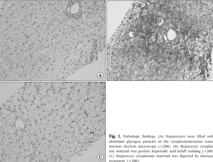
Fig. 1. Pathologic findings. (A) Hepatocytes were filled with abundant glycogen particles in the cytoplasmonroutine trans- mission electron microscopy (×200). (B) Hepatocyte cytoplas- mic material was positive byperiodic acid-Schiff staining (×200).
(C) Hepatocyte cytoplasmic material was digested by diastase- treatment (×200).
were 42 IU/L, 38 IU/L, and 0.7 mg/dL, respectively.
His serum cholesterol, uric acid, glucose, lactic acid, CK, and albumin levels were all normal (Table 1).
Serological tests revealed no evidence of infection such as hepatitis virus A, B, or C, Epstein-Barr virus, or cytomegalovirus. Plasma amino acid and urine or- ganic acid profiles were also unremarkable. On sono- graphic examination, his liver was enlarged with slightly increased parenchymal echogenicity but the intrahepatic and extrahepatic biliary ducts were not dilated. The gallbladder and spleen were normal. A liver biopsy was performed and, on histological ex- amination, the hepatocytes were filled with abun- dant glycogen particles in the cytoplasm with pe- ripheral displacement of organelles (Fig. 1).
With a high suspicion of GSD, genetic testing was performed to rule out GSD III (the AGL gene) and
GSD VI (the PYGL gene). Genomic DNA was isolated from peripheral leukocytes of the patient with a PureGene blood kit (Qiagen, Hilden, Germany). All exons and their intronic boundaries of AGL and PYGL were amplified and sequenced using a BigDye Terminator V3.1 Cycle Sequencing Ready Reaction kit (Applied Biosystems, Foster City, CA, USA). He had normal sequences in the coding regions of the AGL and PYGL genes. We then checked PhK activity in the red blood cells, which was 15 μmol/min/g Hb (normal range, 100-300 μmol/min/g Hb). With a suspicion of GSD IXa, the PHKA2 gene was tested.
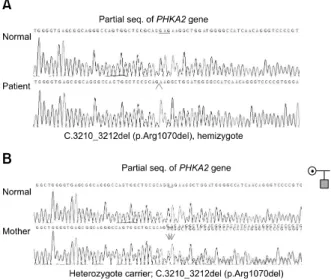
Thirty-three exons and their intronic boundaries were investigated, revealing a previously reported mutation, c.3210_3212del (p.Arg1070del) (Fig. 2A) [7]. His mother carried the mutation as a hetero- zygote (Fig. 2B). However, there was no family his-
Fig. 2. Partial genomic sequences of the PHAK2 gene. (A) The patient is a homozygotefor the c.3210_3212del (p.Arg1070del) mutation. (B) His mother is a heterozygote for this mutation.
tory of liver disease on his maternal side.
We educated his parents on preventing hypo- glycemia via frequent meals high in complex carbo- hydrates and protein, as well as cornstarch supple- ments. In particular, we advised that cornstarch should be fed to the patient before bed to maintain an adequate glucose level throughout the night. We recommended liver ultrasonography every 6months.
At the latest evaluation at 4.2 years of age, his height was 100 cm (−0.4 SD score) and his weight was 19.8 kg (1.32 SD score). He had not experienced any hy- poglycemic episodes. Hepatic dysfunction was un- changed except for elevated AST and ALT.
DISCUSSION
Our patient had no symptomatic presentation of liver-type GSD, such as hypoglycemia, delayed de- velopment, or growth retardation, and had only hep- atosplenomegaly with hepatic enzyme elevation.
GSD I is the most common liver type in Korea [8], but GSD I usually manifests in the neonatal period with severe and recurrent hypoglycemia. Patients with GSD III and GSD IV usually present mild fast- ing hypoglycemia, as in GSD IX. Most patients have both liver and muscle involvement in GSD III, which
shows hypotonia, weakness, wasting of skeletal muscle, and heart involvement. Osteopenia and fractures are associated with GSD III and GSD IV [9].
GSD IV typically presents in early infancy with hep- atosplenomegaly and growth failure. The disorder is rapidly progressive, leading to liver failure without transplantation [10,11]. Otherwise, there is no evi- dence of viral hepatitis and autoimmune hepatitis.
Liver and/or muscle cells are affected in GSD IX and when liver cells are affected in this disorder the signs and symptoms appear in early childhood [4,5]. Liver enlargement and slow growth are the initial fea- tures. The height of affected children is usually be- low the average of age- and sex-matched controls.
During prolonged fasting periods, hypoglycemia or increasing levels of ketones are one of the signs of be- ing affected, although ketotic hypoglycemia is not always noted in GSD IXa. In addition, patients may have delayed achievement of motor skills, and some have mild muscle weakness. These signs and symp- toms usually improve with age. However, some pa- tients may have a fibrosis in the liver tissue, which can in rare cases progress to irreversible liver cir- rhosis in childhood [12-14]. Therefore, it is very im- portant to make an early diagnosis of GSD IX and to undertake surveillance for hepatic dysfunction on a regular basis in these patients. Patients with mus- cle-type GSD IX may experience myopathy such as fatigue, cramps, and muscle pain, especially during exercise. Muscle-type GSD IX is a rare form, and most patients are diagnosed between 15 and 36 years of age [15]. Hypercholesterolemia and hyper trigly- ceridemia are common. CK levels are sometimes in- creased in muscle-type GSD IX. In myopathic var- iants, less frequent presentations include cardiac problems [10].
In the liver biopsy sample, we found glycogen par- ticles in hepatocytes, indicative of GSD. After GSD III (the AGL gene) and GSD VI (the PYGL gene) were ruled out by genetic testing, we evaluated PhK activ- ity to confirm a diagnosis of GSD IX and identified the c.3210_3212del (p.Arg1070del) mutation in the PHKA2 gene. This mutation was reported previously in a Finnish boy who also only had hepatomegaly
with no growth retardation or hypoglycemic epi- sodes [7]. As an in-frame deletion, the mutant pro- tein might have residual function, which could be evaluated by assessing the RNA and protein ex- pression of the mutant PHKA2.
Given the X-linked inheritance of GSD, we recom- mended genetic testing of the mother and sisters of our patient. His mother is a heterozygote for the same mutation of PHAK2. The testing has not yet been performed in his sisters. The screening of the carrier state of female members is important because some female carriers can manifest mild hep- atomegaly with hepatic dysfunction and growth re- tardation and the severity can differ according to the degree of the skewed inactivation of the X chromo- some. In addition, GSD IX can progress with age in some carriers [16]. Thus, regular surveillance of hep- atic function should be performed for female carriers with GSD IXa. Another reason for the importance of family screening is that the inheritance from a fe- male carrier to her son is 50% and the risks to future offspring should be carefully discussed.
Our current patient is the second reported Korean case of GSD IXa to date. Unlike the previous Korean case, however, our current patient had no specific symptoms such as ready fatigability or hypoglycemic events. Moreover, his growth was not significantly retarded and there was no familial history of liver disease. On the other hand, the first reported Korean case, a 5-year-old boy, presented with mild hypo- glycemic symptoms, easy fatigability, and hepato- megaly since the age of two [6].
There is no specific treatment for GSD IX, but com- plex carbohydrates and protein-rich foods can pre- vent hypoglycemia. Night-time cornstarch con- sumption can stave off a hypoglycemic attack during sleep. Patients should be monitored for hepatic dys- function on a regular basis. Though rare, some af- fected patients have fibrosis in the liver, which can progress to liver cirrhosis. In addition, echocardiog- raphy should be performed every 5 years to survey cardiac involvement in muscle-type GSD IX. Growth retardation is a common symptom of GSD IX but al- most all patients achieve complete catch-up in
growth.
In conclusion, our experience with the present case indicates the importance of screening for GSD IXa in male patients who are suspected of having GSD but manifest unusual presentations, such as those owe have here described. More Korean cases should be identified considering the prevalence of this GSD subgroup. Early identification and man- agement is important for patient prognosis.
REFERENCES
1. Lau CK, Hui J, Fong FN, To KF, Fok TF, Tang NL, et al. Novel mutations in PHKA2 gene in glycogen storage disease type IX patients from Hong Kong, China. Mol Genet Metab 2011;102:222-5.
2. Maichele AJ, Burwinkel B, Maire I, Søvik O, Kilimann MW. Mutations in the testis/liver isoform of the phos- phorylase kinase gamma subunit (PHKG2) cause auto- somal liver glycogenosis in the gsd rat and in humans.
Nat Genet 1996;14:337-40.
3. Newgard CB, Hwang PK, Fletterick RJ. The family of glycogen phosphorylases: structure and function. Crit Rev Biochem Mol Biol 1989;24:69-99.
4. Tsilianidis LA, Fiske LM, Siegel S, Lumpkin C, Hoyt K, Wasserstein M, et al. Aggressive therapy improves cir- rhosis in glycogen storage disease type IX. Mol Genet Metab 2013;109:179-82.
5. Bali DS, Goldstein JL, Fredrickson K, Rehder C, Boney A, Austin S, et al. Variability of disease spectrum in chil- dren with liver phosphorylase kinase deficiency caused by mutations in the PHKG2 gene. Mol Genet Metab 2014;111:309-13.
6. Park KJ, Park HD, Lee SY, Ki CS, Choe YH. A novel PHKA2 gross deletion mutation in a Korean patient with X-linked liver glycogenosis type I. Ann Clin Lab Sci 2011;41:197-200.
7. Crushell E, Treacy EP, Dawe J, Durkie M, Beauchamp NJ. Glycogen storage disease type III in the Irish population. J Inherit Metab Dis 2010;33 Suppl 3:S215- 8.
8. Choi J, Ko JM, Kim GH, Yoo HW. Clinical manifes- tation and effect of corn starch on height growth in Korean patients with glycogen storage disease type Ia.
J Korean Soc Pediatr Endocrinol 2007;12:35-40.
9. Beauchamp NJ, Taybert J, Champion MP, Layet V, Heinz-Erian P, Dalton A, et al. High frequency of mis- sense mutations in glycogen storage disease type VI. J Inherit Metab Dis 2007;30:722-34.
10. Moses SW, Parvari R. The variable presentations of gly- cogen storage disease type IV: a review of clinical, enzy- matic and molecular studies. Curr Mol Med 2002;2:177- 88.
11. Ban HR, Kim KM, Jang JY, Kim GH, You HW, Kim K, et al. Living donor liver transplantation in a Korean child with glycogen storage disease type IV and a GBE1 mutation. Gut Liver 2009;3:60-3.
12. Brushia RJ, Walsh DA. Phosphorylase kinase: the com- plexity of its regulation is reflected in the complexity of its structure. Front Biosci 1999;4:D618-41.
13. Rudolfová J, Slovácková R, Trbusek M, Pesková K, St'astná S, Kozák L. Identification of three novel muta- tions in the PHKA2 gene in Czech patients with X-
linked liver glycogenosis. J Inherit Metab Dis 2001;24:
85-7.
14. Hidaka F, Sawada H, Matsuyama M, Nunoi H. A novel mutation of the PHKA2 gene in a patient with X-linked liver glycogenosis type 1. Pediatr Int 2005;47:687-90.
15. Bak H, Cordato D, Carey WF, Milder D. Adult-onset ex- ercise intolerance due to phosphorylase b kinase deficiency. J Clin Neurosci 2001;8:286-7.
16. Morava E, Wortmann SB, van Essen HZ, Liebrand van Sambeek R, Wevers R, van Diggelen OP. Biochemical characteristics and increased tetraglucoside excretion in patients with phosphorylase kinase deficiency. J Inherit Metab Dis 2005;28:703-6.