ISSN 2234-3806 • eISSN 2234-3814
494 www.annlabmed.org http://dx.doi.org/10.3343/alm.2016.36.5.494 Ann Lab Med 2016;36:494-497
http://dx.doi.org/10.3343/alm.2016.36.5.494
Letter to the Editor
Diagnostic Hematology
Acute Myeloid Leukemia With MLL Rearrangement and CD4+/CD56+ Expression can be Misdiagnosed as Blastic Plasmacytoid Dendritic Cell Neoplasm: Two Case Reports
Ju-Mee Lee, M.D.1,2, In-Suk Kim, M.D.1,2, Jeong Nyeo Lee, M.D.3, Sang Hyuk Park, M.D.4 Hyung-Hoi Kim, M.D.4, Chulhun L. Chang, M.D.1,2, Eun Yup Lee, M.D.4, Hye Ran Kim, M.D.5, Seung Hwan Oh, M.D.5, and Sae Am Song, M.D.3
Department of Laboratory Medicine1, Pusan National University Yangsan Hospital, Yangsan; Research Institute for Convergence of Biomedical Science and Technology2, Pusan National University Yangsan Hospital, Yangsan; Department of Laboratory Medicine3, Haeundae Paik Hospital, Inje University College of Medicine, Busan; Department of Laboratory Medicine4, Pusan National University School of Medicine, Busan; Department of Laboratory Medicine5, Busan Paik Hospital, Inje University College of Medicine, Busan, Korea
Dear Editor,
Blastic plasmacytoid dendritic cell neoplasm (BPDCN), charac- terized by co-expression of CD4 and CD56 without any other lineage-specific markers, is an aggressive tumor type that shows a high frequency of skin involvement and nodal or marrow infil- tration with a propensity toward leukemic dissemination [1].This disease was formerly defined by the World Health Organization as blastic NK-cell lymphoma, and was later grouped with AML and related precursor neoplasms [2]. Here we report two AML cases with KMT2A (or MLL) rearrangements along with CD4+/
CD56+ expression, which had the potential to be misdiagnosed as BPDCN.
A 59-yr-old man presented with erythematous papules and vesicles on his whole body for one week. Complete blood counts showed anemia (Hb, 80 g/L), thrombocytopenia (62×109 plate- lets/L), and many circulating plasmoblast-like cells (65%). Bone
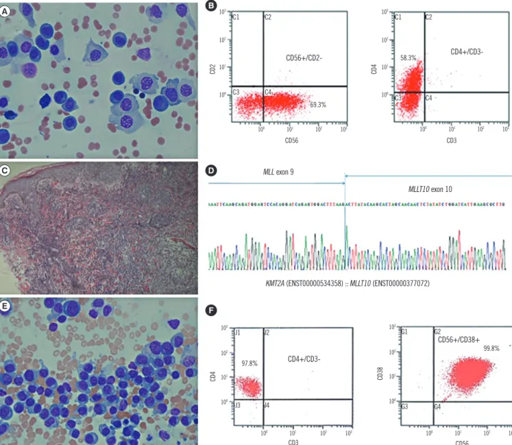
marrow examination showed hypercellular marrow (90%) mostly composed of plasmoblast-like cells with coarse nuclear chromatin, distinct nucleoli, and abundant basophilic cytoplasm (Fig. 1A). Flow cytometry revealed that the neoplastic cells were positive for CD4, CD33, CD38, CD56, CD117, and CD138 (Fig.
1B), and negative for other myeloid or lymphoid markers (Table 1). Skin lesions showed diffuse infiltration of medium- to large- sized agranular blastic cells with CD4 and CD56 co-expression (Fig. 1C). Bone marrow and skin were negative for CD123 im- munohistochemical stain. The chromosome study revealed 45,X,-Y[9]/46,XY[11] without 11q23 abnormalities. Multiplex, nested reverse transcription (RT)-PCR was performed for screening and detection of 28 chromosomal translocations us- ing the HemaVision kit (DNA Technology, Research Park, Aar- hus, Denmark), and the presence of MLL-MLLT10 rearrange- ment was demonstrated. It was confirmed by direct sequencing
Received: December 15, 2015 Revision received: April 27, 2016 Accepted: June 2, 2016 Corresponding author: In-Suk Kim
Department of Laboratory Medicine, Pusan National University Yangsan Hospital, 20 Geumo-ro, Mulgeum-eup, Yangsan 50612, Korea
Tel: +82-55-360-1878, Fax: +82-55-360-1880, E-mail: [email protected] Co-corresponding author: Jeong Nyeo Lee
Department of Laboratory Medicine, Haeundae Paik Hospital, Inje University, College of Medicine, 875 Haeun-daero, Haeundae-gu, Busan 48108, Korea
Tel: +82-51-797-3191, Fax: +82-51-797-3194, E-mail: [email protected]
© The Korean Society for Laboratory Medicine.
This is an Open Access article distributed under the terms of the Creative Commons Attribution Non-Commercial License (http://creativecommons.org/licenses/by-nc/4.0) which permits unrestricted non-commercial use, distribution, and reproduction in any medium, provided the original work is properly cited.
Lee J-M, et al.
CD4+/CD56+ AML with MLL rearrangement
http://dx.doi.org/10.3343/alm.2016.36.5.494 www.annlabmed.org 495
(Fig. 1D). Fluorescence in situ hybridization for MLL rearrange- ment revealed nuc ish(5´MLLx3,3´MLLx2)(5´MLL con 3´MLLx2) [170/200], suggesting the presence of an atypical MLL break- point. Although the lineage antigens expressed here were not specific to AML, he was diagnosed as having AML with MLL re- arrangement in bone marrow and skin.
A 30-yr-old woman presented with multiple lymphadenopathy for three weeks. Laboratory analysis revealed anemia (Hb, 81 g/
L), thrombocytopenia (82 ×109 platelets/L), and leukocytosis (16.77 ×10⁹ white blood cells/L) with circulating plasmacytoid cells (63%). Neither skin lesion nor monoclonal gammopathy was present. Bone marrow analysis showed hypercellular mar- row (100%) with leukemic cells containing abundant basophilic cytoplasm (Fig. 1E). Flow cytometry revealed that the neoplastic cells were positive for CD4, CD7, CD33, CD38, CD56, and CD64, (Fig. 1F), and were negative for other markers (Table 1).
Fig. 1. Morphological features, flow cytometric analysis, immunohistochemical stain, and genetic study of the two cases of CD4+/CD56+
AML. (A) Plasmoblast-like neoplastic cells in the first case (Wright Giemsa stain, 400×, Bone marrow); (B) Immunophenotyping features with CD56 and CD4 coexpression in the first case; (C) Skin biopsy showing diffuse infiltration of medium- to large-sized agranular blastic cells into the dermis in the first case (Hematoxylin and Eosin stain, 100×, Skin lesion); (D) The MLL-MLLT10 rearrangement confirmed by direct sequencing in the first case; (E) Plasmacytoid cells in the second case (Wright Giemsa stain; ×400, Bone marrow); and (F) The im- munophenotyping features with CD4 and CD56 coexpression in the second case.
B
D
F A
C
E
CD56+/CD2-
CD4+/CD3-
CD4+/CD3-
CD56+/CD38+
CD56
CD3
CD3
CD56
CD2CD4 CD4 CD38
C1
J1
C1
G1 C3
J3
C3
G3 C2
J2
C2
G2 C4
J4
C4
G4 69.3%
97.8%
103
103
103
103 102
102
102
102 101
101
101
101 100
100
100
100 100
100
100
100 101
101
101
101 102
102
102
102 103
103
103
103 58.3%
99.8%
MLL exon 9
MLLT10 exon 10
KMT2A (ENST00000534358) :: MLLT10 (ENST00000377072)
Lee J-M, et al.
CD4+/CD56+ AML with MLL rearrangement
496 www.annlabmed.org http://dx.doi.org/10.3343/alm.2016.36.5.494 The cytochemical stain for non-specific esterase in neoplastic
cells was negative. Immunohistochemical stains of bone mar- row biopsy specimens showed that the neoplastic cells were negative for CD123, CD138, kappa, and lambda. Lymph node biopsy analysis demonstrated that the neoplastic cells co-ex- pressing CD4 and CD56 were negative for CD123. Cytogenetic analysis revealed 46,XX,t(9;11)(p22;q23),t(9;21)(q12;p11.2) [20]. The MLL-MLLT3 rearrangement was detected by RT-PCR.
Although adequate lineage-specific markers were not observed, she was diagnosed as having an AML with MLL rearrangement in bone marrow and lymph nodes.
So far, three cases of CD4+/CD56+ hematologic malignancies with MLL rearrangements have been reported as rare cases of BPDCN with MLL rearrangement (Table 1). The leukemic cells reported in the literature expressed the myeloid and monocytic
markers CD33, CD117, CD11c, CD15, or CD64, which were not specific enough to identify a specific lineage [3-5]. These three cases did not express highly specific plasmacytoid dendritic cell-associated antigens, such as CD123, TCL1, CD2AP, or CD303 (BDCA2); therefore, additional immunohistochemical studies were necessary at their diagnosis. Variable expression of CD4 or CD56 in adult AML cases with MLL rearrangement has been reported [6], and BPDCN is often confused with mono- cytic leukemias. Based on the limited information on immuno- phenotypes of these cases, MLL-related monocytic leukemia would be a reasonable diagnosis (Table 1).
This study emphasizes that the diagnosis of BPDCN should only be considered after a full investigation of plasmacytoid den- dritic cell markers, ensuring there is no expression of myeloid or monocytic markers on the blasts [7]. Since hematologic neo- Table 1. The clinicopathologic characteristics of CD4+/CD56+ hematologic malignancies carrying the MLL rearrangement
Case 1 Case 2 Case 3 Case 4 Case 5
Reference [3] [4] [5] First case in this study Second case in this study
Diagnosis CD4+/CD56+
hematodermic malignancy
BPDCN with MLL-ENL rearrangement
BPDCN with MLL rearrangement
AML with MLL rearrangement
AML with MLL rearrangement
Age/gender 50/F 45/M 8/F 59/M 30/F
Race China Japan Korea Korea Korea
Chief complaint Not described Multiple disseminated skin
nodules Mild fatigue and petechiae
on extremities Multiple skin rash Multiple lymphadenopathy
Skin lesion Yes Yes No Yes No
Circulating tumor cells No Yes Yes Yes Yes
Bone marrow involvement Yes Yes Yes Yes Yes
Flow cytometric positivity CD4, CD45, CD56 CD4, CD11c, CD33, CD45RA, CD56, CD68, CD117, CD123, HLA-DR
CD4, CD15, CD33, CD56, CD64, CD117, HLA-DR, TdT
CD4, CD33, CD38, CD45, CD56, CD117, CD138
CD4, CD7, CD33, CD38, CD45, CD56, CD64,
HLA-DR Flow cytometric negativity CD2, CD3, CD5, CD7, CD8,
CD10, CD13, CD14, CD19, CD20, CD22, CD23, CD34,
HLA-DR, MPO
CD3, cCD3, CD5, CD7, CD8, CD10, CD11b, CD13, CD19, CD20, CD34, cMPO
CD2, CD3, cCD3, CD5, CD7, CD10, CD13, CD14, CD16, CD19, CD20, cCD22,
CD34, cMPO
CD2, CD3, CD5, CD7, CD10, CD13, CD14, CD19,
CD20, CD34, cMPO, TdT
CD3, CD5, CD8, CD10, CD11b, CD13, CD14, CD19,
CD20, CD34, cCD79a, CD117, CD138, cMPO, kappa, lambda, TdT
Karyotype 46, XX,t(4;9;11)
(q12;p22;q23) [18]/46, XX [10]
49, XY, +add(1)(p13), +8, +8,t(11;19)(q23;p13.3)
[20]
48,XX,+8,t(11;19) (q23;p13.3),+19[20]
45,X,-Y[9]/46,XY[11] 46,XX,t(9;11) (p22;q23),t(9;21)
(q12;p11.2)[20]
FISH, MLL gene rearrangement
Detected Detected Detected Detected atypical MLL
breakpoint
Detected
RT-PCR for MLL MLL-MLLT3 MLL-ELN MLL-MLLT1 MLL-MLLT10 MLL-MLLT3
Treatment CHOP combination
chemotherapy and allogeneic bone marrow
transplantation
After failure of CHOP chemotherapy, acute
leukemia-type chemotherapy was done
Induction chemotherapy (daunorubicin, vincristine,
prednisolone, and cytarabin)
Induction chemotherapy (daunorubicin and
cytarabin)
Induction chemotherapy (daunorubicin and cytarabin) and allogeneic stem cell transplantation Abbreviations: BPDCN, blastic plasmacytoid dendritic cell neoplasm; RT-PCR, reverse transcription-PCR; cMPO, cytoplasmic myeloperoxidase; TdT, termi- nal deoxynucleotidyl transferase; CHOP, cyclophosphamide, vincristine, epirubicin, and prednisolone.
Lee J-M, et al.
CD4+/CD56+ AML with MLL rearrangement
http://dx.doi.org/10.3343/alm.2016.36.5.494 www.annlabmed.org 497
plasms such as BPDCN, AML, extranodal NK/T cell lymphoma, nasal type, and mature T cell lymphomas with or without skin involvement may express CD56 with or without CD4, extensive immunohistochemical and genetic analyses are necessary be- fore definitively diagnosing BPDCN or AML [8, 9].
In conclusion, CD4+/CD56+ hematologic malignancies can be suspected to be BPDCN, and clinicians should conduct a full analysis including flow cytometry for adequate myeloid/
monocytic markers, immunohistochemical stain for highly spe- cific plasmacytoid dendritic cell-associated antigens, cytoge- netic, and genetic studies to make an exact diagnosis and de- termine effective treatment.
Authors’ Disclosures of Potential Conflicts of Interest
No potential conflicts of interest relevant to this article were re- ported.
Acknowledgments
This work was supported by the yearly clinical research grant from Pusan National University Yangsan Hospital.
REFERENCES
1. Garnache-Ottou F, Feuillard J, Saas P. Plasmacytoid dendritic cell leu- kaemia/lymphoma: towards a well defined entity? Br J Haematol 2007;
136:539-48.
2. Swerdlow SH, Campo E, et al. WHO classification of tumours of haema- topoietic and lymphoid tissues. France: IARC Press, 2008:145-7.
3. Leung R, Chow EE, Au WY, Chow C, Kwong YL, Lin SY, et al. CD4+/
CD56+ hematologic malignancy with rearranged MLL gene. Hum Pathol 2006;37:247-9.
4. Toya T, Nishimoto N, Koya J, Nakagawa M, Nakamura F, Kandabashi K, et al. The first case of blastic plasmacytoid dendritic cell neoplasm with MLL-ENL rearrangement. Leuk Res 2012;36:117-8.
5. Yang N, Huh J, Chung WS, Cho MS, Ryu KH, Chung HS. KMT2A (MLL)-MLLT1 rearrangement in blastic plasmacytoid dendritic cell neo- plasm. Cancer Genet 2015;208:464-7.
6. Muñoz L, Nomdedéu JF, Villamor N, Guardia R, Colomer D, Ribera JM, et al. Acute myeloid leukemia with MLL rearrangements: clinicobiologi- cal features, prognostic impact and value of flow cytometry in the de- tection of residual leukemic cells. Leukemia 2003;17:76-82.
7. Rush PS, Bennett DD, Yang DT. Hematopathology HP 15-5. ASCP case reports 2015;HP 15-5:1-20.
8. Bekkenk MW, Jansen PM, Meijer CJ, Willemze R. CD56+ hematological neoplasms presenting in the skin: a retrospective analysis of 23 new cases and 130 cases from the literature. Ann Oncol 2004;15:1097-108.
9. Herling M and Jones D. CD4+/CD56+ hematodermic tumor: the fea- tures of an evolving entity and its relationship to dendritic cells. Am J Clin Pathol 2007;127:687-700.