Recent treatment patterns and survival outcomes in pancreatic cancer according to clinical stage
based on single-center large-cohort data
Doo-ho Lee, Jin-Young Jang, Jae Seung Kang, Jae Ri Kim, Youngmin Han, Eunjung Kim, Wooil Kwon, and Sun-Whe Kim
Department of Surgery and Cancer Research Institute, Seoul National University College of Medicine, Seoul, Korea
Backgrounds/Aims: We performed a retrospective, single-center cohort study to evaluate the impact of various treat- ment modalities and recent changes in treatment modalities, including the increased application of chemotherapy, on survival in patients with pancreatic cancer. Methods: All patients with pancreatic cancer who were diagnosed and treat- ed at Seoul National University Hospital between January 2007 and December 2014 were registered in a prospective clinical database and included in this retrospective study. All patients’ radiologic imaging diagnoses were re-reviewed according to the National Cancer Center Network guidelines. The patients were divided into four groups according to their clinical stage, and each clinical stage group was further divided into four groups according to treatment modality.
Results: Overall, 475 (28.9%) patients had resectable pancreatic cancer, 129 (7.8%) patients borderline resectable pancreatic cancer, 384 (23.3%) patients locally advanced pancreatic cancer, and 658 (40.0%) patients metastatic pan- creatic cancer. Among the patients with borderline resectable pancreatic cancer, the median survival was significantly longer in the neoadjuvant therapy (NAT)+surgery groups (24 months) than the surgery without NAT (16 months) group (p=0.049). A multivariate survival analysis revealed that compared with the surgery group, the 5-year mortality risk was decreased by 35% in the NAT+surgery group (24 vs. 20 months, p=0.045). Conclusions: This retrospective cohort study showed that the rates of resectable and surgically treatable pancreatic cancer were 29.1% and 32.2%, which are higher than those reported previously, and aggressive NAT for select advanced-stage patients could lead to better survival outcomes. (Ann Hepatobiliary Pancreat Surg 2018;22:386-396)
Key Words: Neoadjuvant therapy; Survival rate; Pancreas neoplasms
Received: May 18, 2018; Revised: September 27, 2018; Accepted: September 27, 2018 Corresponding author: Jin-Young Jang
Department of Surgery and Cancer Research Institute, Seoul National University Hospital, Seoul National University College of Medicine, 101 Daehak-ro, Jongno-gu, Seoul 03080, Korea
Tel: +82-2-2072-2194, Fax: +82-2-741-2194, E-mail: [email protected]
Copyright Ⓒ 2018 by The Korean Association of Hepato-Biliary-Pancreatic Surgery
This is an Open Access article distributed under the terms of the Creative Commons Attribution Non-Commercial License (http://creativecommons.org/
licenses/by-nc/4.0) which permits unrestricted non-commercial use, distribution, and reproduction in any medium, provided the original work is properly cited.
Annals of Hepato-Biliary-Pancreatic Surgery ∙ pISSN: 2508-5778ㆍeISSN: 2508-5859
INTRODUCTION
Despite improvements in perioperative outcomes, pan- creatic cancer has a poor prognosis, with a 5-year survival rate of only 8%-10% among patients.1,2 Most patients are diagnosed in the advanced stages, and effective systemic therapies are lacking. According to the annual report of cancer statistics in Korea, the 5-year survival rate of over- all pancreatic cancer patients was 10% in 2014, with the higher rate due to increased early diagnoses following the introduction of a routine cancer screening program for all citizens conducted by the National Health Insurance, and increased detection of less aggressive pancreatic malign-
ancies.3 In addition, effective systemic treatments includ- ing neoadjuvant therapies (NATs) have been introduced in an attempt to overcome the limitations of prior treat- ments and to improve patient outcomes, especially in pa- tients with borderline resectable, locally advanced, and even metastatic pancreatic cancer; therefore, the outcomes of pancreatic cancer patients have been improved by addi- tional surgical resection after systemic treatment.4-6 However, because the definitions of borderline resectable and locally advanced pancreatic cancer differ among in- stitutions, it is impossible to compare survival rates ac- cording to clinical stage in pancreatic cancer patients. In addition, the data published by most institutions do not
Table 1. The National Comprehensive Cancer Network definitions for the CT-based staging of pancreatic cancer Resectable
Tumor-artery relationship: no radiographic evidence of arterial abutment (celiac, SMA, or hepatic artery)
Tumor-vein relationship: tumor-induced narrowing, if present, is ≤50% of the circumference of the SMV, PV, or SMV-PV confluence
Borderline resectable
Artery: tumor abutment (≤180° of the circumference) of SMA or celiac artery. Tumor abutment or short segment encasement (>180°) of the common hepatic artery. Tumor abutment >180° without involvement of the aorta and amenable to celiac re- section (HA-GDA not involved)
Vein: Tumor induced narrowing of >50 % of SMV, PV, or SMV-PV, or short segment occlusion of SMV, PV, SMV-PV with suitable PV (above) and SMV (below) to allow for safe vascular reconstruction.
Locally advanced
Artery: tumor encasement (>180° of the circumference) of SMA or celiac artery
Vein (SM-PV confluence): occlusion of SMV, PV, or SMV-PV without suitable vessels above and below the tumor to allow for reconstruction (no distal or proximal target for vascular reconstruction)
Extrapancreatic findings: no evidence of peritoneal, hepatic, extra-abdominal metastases Metastatic
Evidence of peritoneal or distant organ metastases
SMA, superior mesenteric artery; SMV, superior mesenteric vein; PV, portal vein; SMV-PV, superior mesenteric-portal vein;
HA-GDA, hepatic artery-gastroduodenal artery
include patients with metastatic or locally advanced pan- creatic cancer.4,7-9 SEER (Surveillance, Epidemiology, and End Results program, USA) data cover a large number of patients but are limited by the high variation among the involved hospitals and a lack of accurate clinical staging.10 Furthermore, as there are no recent data on clin- ical stage based on accurate imaging criteria in overall pa- tients, it is difficult to grasp the actual status of pancreatic cancer treatment in Korea. To date, very few studies have identified trends in the treatment of pancreatic cancers, in- cluding locally advanced and metastatic cancers, in a sin- gle-center study.
Therefore, the objective of this study was to categorize all patients according to clinical stage using uniform imaging diagnostic criteria and to evaluate the impact of treatment modality on the survival of pancreatic cancer patients included in a large-scale prospective database at Seoul National University Hospital.
MATERIALS AND METHODS
Database
We conducted a retrospective cohort study using pro- spectively recorded information of patients registered in a clinical oncology database at Seoul National University Hospital between January 1, 2007 and December 31, 2014. The study was approved by the Institutional Review Board at Seoul National University (1706-159-863).
Patients and groups
We included patients who were diagnosed with stage I-IV pancreatic cancer between January 1, 2007 and December 31, 2014 and aged ≥19 years at diagnosis. The diagnoses were made using computed tomography with a pancreas-specific protocol and/or magnetic resonance imaging, positron emission tomography, endoscopic ultra- sonography, with or without cytological or histological confirmation of conventional ductal adenocarcinoma of the pancreas. Patients were excluded if surgery was per- formed at another hospital or the diagnosis was made out- side the index period. The National Comprehensive Cancer Network 2017 guidelines were used for pancreatic cancer staging, based on computed tomography performed at the time of diagnosis (Table 1). The patients were first categorized into four groups according to the clinical stage, as follows: resectable (RPC), borderline resectable (BRPC), locally advanced (LAPC), or metastatic pancre- atic cancer (MPC).4 Patients in each stage group were fur- ther categorized according to the treatment modality, as follows: NAT followed by surgery (NAT+surgery group), surgery±adjuvant chemoradiotherapy (surgery group), che- motherapy alone (CTx group), and palliative care (palliative care group). NAT was defined as the first course of chemotherapy, radiation, or concurrent chemo- radiotherapy (CCRT) prior to surgery.
Data extraction
The primary outcome was survival time, which was de- fined as the time from the date of diagnosis to the date of death or the last follow-up. The patients’ character- istics, date of diagnosis, type of surgery, tumor location, tumor size, serum level of carbohydrate antigen (CA) 19-9 (a tumor marker; normal range <37 U/ml), and patho- logic reports were extracted from the medical records.11 We re-reviewed the images of computed tomography and magnetic resonance examinations performed at the time of diagnosis. The patients were categorized according to clinical stage (according to the National Cancer Center Network 2017 guidelines) and again by treatment modality. The tumor response was defined according to the Response Evaluation Criteria in Solid Tumors using computed tomography performed after NAT, and was categorized as complete response (disappearance of the target lesion), partial response (≥30% decrease in tumor diameter), stable disease (neither sufficient shrinkage to qualify as partial response nor sufficient increase to qual- ify as progressive disease), or progressive disease (≥20%
increase in diameter).12
The TNM residual tumor classification was recorded in patients who underwent pancreatectomy with curative intent.13 We used the tumor regression grading system of the College of American Pathologists, which comprises the following four grades according to the extent of re- sidual carcinoma in post-NAT pancreatectomy specimens:
grade 0, no viable residual tumor (pathologic complete re- sponse); grade 1, marked response (minimal residual can- cer with single cells or small groups of cancer cells);
grade 2, moderate response (residual cancer outgrown by fibrosis); and grade 3, poor or no response (extensive re- sidual cancer).14
Neoadjuvant chemotherapy and/or radiotherapy
Neoadjuvant chemotherapy regimens included gemcita- bine, conventional 5-fluouracil (5-FU), or FOLFIRINOX.
Gemcitabine chemotherapy consisted of 400 mg/m2 body surface area (BSA) intravenous gemcitabine administered weekly for 6 weeks. Three-dimensional conformal radio- therapy consisted of a total dose of 45 Gy (1.8 Gy daily fraction, 5 fractions per week for 5 weeks) with a boost dose of 9 Gy (1.8 Gy daily fraction, 5 fractions). 5-FU
based CCRT consisted of 20-Gy dose to the tumor given in 10 daily fractions over a 2-week period plus an intra- venous bolus of 5-FU (500 mg/m2 of BSA on each of the first 3 days of radiotherapy and again after a planned break of 2 weeks). FOLPIRINOX consisted of oxaliplatin at a dose of 85 mg/m2 followed by leucovorin at a dose of 400 mg/m2 both administered as a 2-hour intravenous infusion with the addition of 180 mg/m2 irinotecan after 30 minutes given over 90 minutes as an intravenous infusion. This treatment was followed by 5-FU at a dose of 400 mg/m2 administered as an intravenous bolus fol- lowed by a continuous infusion of 2,400 mg/m2 for a 46-hour period (one cycle) every 2 weeks. The choice of chemotherapy and/or radiotherapy was determined by each patient’s general performance and ease of access to the hospital. Dose were reduced depending on the pa- tient’s status or when adverse events were noted.
Adjuvant chemotherapy and/or radiotherapy Adjuvant chemotherapy and/or radiotherapy was rec- ommended to patients after operation. Adjuvant treatment regimen included gemcitabine, conventional 5-FU, or FOLFIRINOX. Similar to neoadjuvant treatment, the regi- men was determined considering each patient’s general performance and ease of access to the hospital.
Statistical analysis
Patient demographics, treatment modalities, and tumor characteristics were compared among the four clinical stages and four treatment groups using analysis of var- iance or t tests for continuous variables and 2 tests for categorical variables. Kaplan-Meier analyses were con- ducted to compare survival rates among treatment groups according to clinical stage, and log rank tests were used to compare differences in survival. The median survival time was estimated from the Kaplan-Meier curves. Cox proportional hazards models were used to evaluate risk factors affecting survival according to clinical stage. All analyses were performed using PASW software, version 18.0 (SPSS Inc., Chicago, IL, USA). Differences were considered significant at p-value <0.05.
Table 2. Baseline characteristics of patients with pancreatic cancer stratified by clinical stage Variable
RPC (n=475, 28.9%)
BRPC (n=129,
7.8%)
LAPC (n=384,
23.3%)
MPC (n=658,
40.0%)
All patients (n=1646)
p-value RPC vs
BRPC
RPC vs LAPC
RPC vs MPC Age, years 64.9±10.8 63.1±10.3 64.8±9.9 63.0±11.0 64.0±10.7 0.368 0.998 0.020
Sex 0.990 0.560 0.984
Male 288 (60.6) 77 (59.7) 213 (55.5) 412 (62.6) 990 (60.1) Female 187 (39.4) 52 (40.3) 171 (44.5) 246 (37.4) 656 (39.9)
Treatment 0.184 <0.001 <0.001
Surgery 434 (91.4) 45 (34.9) 16 (4.2) 0 (0.0) 495 (30.1)
NAT+surgery 0 (0.0) 28 (21.7) 4 (1.0) 3 (0.5) 35 (2.1)
CTx 17 (3.6) 43 (33.3) 285 (74.2) 449 (68.2) 794 (48.2)
Palliative care 24 (5.1) 13 (10.1) 79 (20.6) 206 (31.3) 322 (19.6)
Tumor location 0.090 <0.001 <0.001
Head 329 (69.3) 102 (79.1) 195 (50.8) 215 (32.7) 841 (51.1) Body or tail 144 (30.3) 27 (20.9) 184 (47.9) 438 (66.6) 793 (48.2)
Diffuse 2 (0.4) 0 (0.0) 5 (1.3) 5 (0.8) 12 (0.7)
Tumor size, mm 26.5±10.0 32.0±9.1 38.9±14.0 44.0±17.6 36.8±16.1 <0.001 <0.001 <0.001
Initial CA 19-9 0.984 0.139 0.338
Normal range 124 (26.1) 30 (23.3) 93 (25.5) 117 (19.1) 362 (22.9) Elevated 351 (73.9) 99 (76.7) 273 (74.5) 495 (80.9) 1218 (77.1) Values are expressed as n (%) or means±standard deviation
RPC, resectable pancreatic cancer; BRPC, borderline resectable pancreatic cancer; LAPC, locally advanced pancreatic cancer;
MPC, metastatic pancreatic cancer; NAT, neoadjuvant therapy; CTx, chemotherapy; CA 19-9, carbohydrate antigen 19-9
RESULTS
Patient characteristics
Overall, 1,646 patients were included in this retro- spective study. The patients were divided into four groups according to their clinical stage: RPC (n=475, 28.9%), BRPC (n=129, 7.8%), LAPC (n=384, 23.3%), and MPC (n=658, 40.0%). There were significant differences among the four groups in terms of clinical stage, tumor location, tumor size, and serum CA 19-9 level, but not sex (Table 2). Patients in the RPC group was older than those in the MPC group (64.9 years vs. 63.0 years, respectively, p=0.020). The proportions of males and females were similar in all four groups (approximately 60% males).
Pancreatic head cancer was dominant in the RPC group compared to the LAPC and MPC groups (Both p<0.001).
The rate of pancreatic body or tail cancer was greater in the LAPC (47.9%) and MPC (66.6%) than in the RPC (30.5%) and BRPC (20.0%) groups. Tumor size was greater in the BRPC (32.0 mm), LAPC (38.9 mm) and MPC (44.0 mm) groups than in the RPC (26.5 mm) group (All p<0.001).
Treatment groups
The patients in each clinical stage group were further classified according to treatment modality: surgery (n=495, 30.1%), NAT+surgery (n=35, 2.1%), CTx (n=794, 48.2%), and palliative care (n=322, 19.6%).
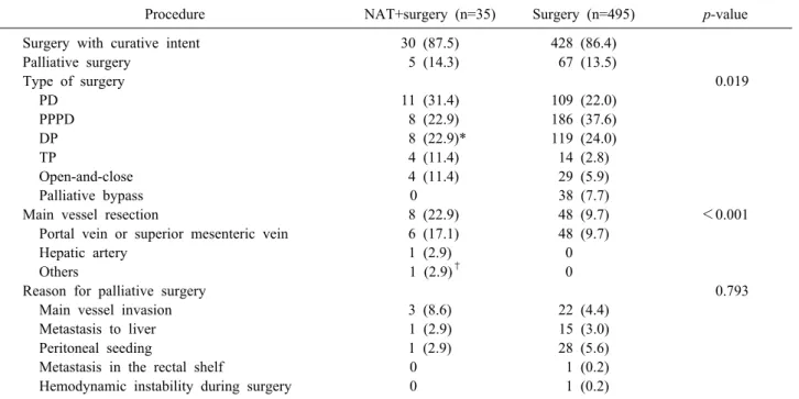
Surgery without NAT was the dominant treatment modal- ity in the RPC group (434/495, 87.6%), NAT+surgery in the BRPC group (28/35, 80.0%), and CTx or palliative care in the LAPC (285/794, 35.9%) and MPC groups (449/794, 56.5%). The surgical procedures performed in the NAT+surgery and surgery groups are summarized in Table 3. Overall, 30 patients (85.7%) in the NAT+surgery group and 428 patients (86.4%) in the surgery group un- derwent surgical resection with curative intent. There were significant differences between these two groups in terms of the type of surgery (p=0.019) and main vessel resection (p<0.001), but not the reason for palliative sur- gery (p=0.793). Of 495 patients in surgery group, 353 pa- tients underwent adjuvant treatment after surgical re- section and 142 patients did not undergo adjuvant treat- ment after surgery.
Table 3. Surgical procedures in the NAT plus surgery and Surgery (no NAT) groups
Procedure NAT+surgery (n=35) Surgery (n=495) p-value
Surgery with curative intent 30 (87.5) 428 (86.4)
Palliative surgery 5 (14.3) 67 (13.5)
Type of surgery 0.019
PD 11 (31.4) 109 (22.0)
PPPD 8 (22.9) 186 (37.6)
DP 8 (22.9)* 119 (24.0)
TP 4 (11.4) 14 (2.8)
Open-and-close 4 (11.4) 29 (5.9)
Palliative bypass 0 38 (7.7)
Main vessel resection 8 (22.9) 48 (9.7) <0.001
Portal vein or superior mesenteric vein 6 (17.1) 48 (9.7)
Hepatic artery 1 (2.9) 0
Others 1 (2.9)† 0
Reason for palliative surgery 0.793
Main vessel invasion 3 (8.6) 22 (4.4)
Metastasis to liver 1 (2.9) 15 (3.0)
Peritoneal seeding 1 (2.9) 28 (5.6)
Metastasis in the rectal shelf 0 1 (0.2)
Hemodynamic instability during surgery 0 1 (0.2)
Values are expressed as n (%)
NAT, neoadjuvant therapy; PD, pancreaticoduodenectomy; PPPD, pylorus-preserving pancreaticoduodenectomy; DP, distal pan- createctomy; TP, total pancreatectomy
*One patient with pancreatic tail cancer and liver metastasis underwent palliative distal pancreatectomy after NAT due to pan- creatico-colonic fistula; †One patient with pancreatic head cancer underwent PPPD and partial resection of the inferior vena cava and left renal vein
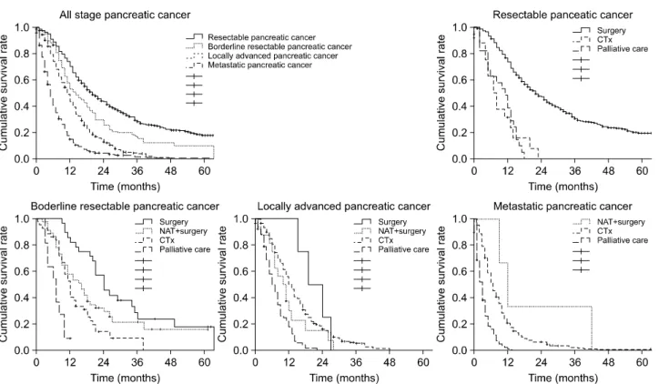
Median survival time according to clinical stage
We analyzed the median survival time of the patients according to clinical stage and treatment modality (Table 4). Kaplan-Meier plots for survival according to clinical stage are presented in Fig. 1. In the RPC patients, the me- dian survival time in the surgery group (22 months) was significantly longer than those in the CTx (8 months, p
<0.001) and palliative care (11 months, p<0.001) groups. In the BRPC patients, the median survival time in the surgery group (16 months) was significantly differ- ent from those in the NAT+surgery (24 months, p=0.049) and palliative care group (7 months, p<0.001) but was not significantly longer than that in the CTx group (12 months, p=0.091). Similarly, in LAPC patients, the me- dian survival time was not significantly different between the surgery and CTx groups (10 and 13 months, re- spectively, p=0.142). In the MPC patients, the median sur- vival time in the CTx group (7 months) was not sig- nificantly different from that in the NAT+surgery group (12 months, p=0.138) but was longer than that in the pal-
liative care group (3 months, p<0.001). In RPC and BRPC groups, the median survival time in the surgery with adjuvant therapy group (25 and 17 months, re- spectively) was significantly different from those in the surgery without adjuvant therapy (15 and 8 months, re- spectively, both p<0.001).
Pathologic profiles and types of NAT
The stage of pancreatic cancer in the 35 patients who received NAT+surgery was borderline resectable in 24, locally advanced in 4 (11.4%), and metastatic in 3 (8.6%).
Two patients had single liver metastasis, and one had para-aortic lymph node metastasis; these three patients showed improvements following NAT and then under- went surgical resection. Twenty patients (57.2%) under- went concurrent chemoradiotherapy and 15 patients (42.8%) underwent CTx only. The CTx regimens were gemcitabine in 24 patients (68.6%), FOLFIRINOX (a combination of 5-fluorouracil, oxaliplatin, irinotecan, and leucovorin) in 5 patients (14.3%), gemcitabine plus erloti- nib in 4 patients (11.4%), and 5-fluorouracil plus cisplatin
Table 4. Median survival of patients with pancreatic cancer
Stage and treatment n (%) 2-year/5-year (%) MST
(months) 95% CI p-value
All stages
Surgery 495 (30.1) 44.6/18.5 20.0 17.3-22.7 Reference
Surgery with adjuvant therapy 353 (21.4) 48.1/20.2 23.0 Surgery without adjuvant therapy 142 (8.7) 35.9/11.2 13.0
NAT+surgery 35 (2.1) 42.2/13.8 24.0 20.8-27.2 0.985
CTx 794 (48.2) 9.8/0.7 9.0 8.3-9.6 <0.001
Palliative care 322 (19.6) 0.0/0.0 4.0 3.5-4.5 <0.001
All patients 1646 (100.0) 19.4/6.6 10.0 9.4-10.6
RPC
Surgery 434 (91.4) 47.2/19.6 22.0 19.1-24.9 Reference
Surgery with adjuvant therapy 315 (66.3) 50.2/19.9 25.0 Surgery without adjuvant therapy 119 (25.1) 39.5/12.9 15.0
NAT+surgery 0 (0.0) - - - -
CTx 17 (3.6) 0.0/0.0 8.0 5.1-10.8 <0.001
Palliative care 24 (5.1) 0.0/0.0 11.0 6.5-15.4 <0.001
All patients 475 (100.0) 43.9/18.2 20.0 17.4-22.5
BRPC
Surgery 45 (34.9) 29.3/16.0 16.0 10.5-21.5 Reference
Surgery with adjuvant therapy 29 (22.4) 33.7/23.1 17.0 Surgery without adjuvant therapy 16 (12.5) 22.5/0.0 8.0
NAT+surgery 28 (21.7) 45.7/17.9 24.0 20.9-27.0 0.049
CTx 43 (33.3) 14.0/0.0 12.0 10.2-13.7 0.091
Palliative care 13 (10.1) 0.0/0.0 7.0 3.8-10.1 <0.001
All patients 129 (100.0) 24.1/9.3 13.0 9.4-16.5
LAPC
Surgery 16 (4.2) 15.0/0.0 10.0 6.0-13.9 Reference
Surgery with adjuvant therapy 9 (2.3) 11.1/0.0 10.0 Surgery without adjuvant therapy 7 (1.9) 0.0/0.0 11.0
NAT+surgery 4 (1.0) 25.0/0.0 19.0 10.2-27.8 0.186
CTx 285 (74.2) 14.8/0.0 13.0 11.8-14.1 0.142
Palliative care 79 (20.6) 1.4/0.0 6.0 4.3-7.6 0.025
All patients 384 (100.0) 12.2/0.0 11.0 9.9-12.0
MPC
Surgery 0 (0.0) - - -
NAT+surgery 3 (0.5) 0.0/0.0 12.0 7.2-16.8 0.138
CTx 449 (68.2) 4.6/0.8 7.0 6.2-7.7 Reference
Palliative care 206 (31.3) 0.0/0.0 3.0 2.5-3.4 <0.001
All patients 658 (100.0) 3.3/0.5 5.0 4.5-5.5
MST, median survival time; CI, confidence interval; NAT, neoadjuvant therapy; CTx, chemotherapy; RPC, resectable pancreatic cancer; BRPC, borderline resectable pancreatic cancer; LAPC, locally advanced pancreatic cancer; MPC, metastatic pancreatic cancer
in 2 patients (5.7%). The tumor response after NAT was classified as partial response in 11 patients (31.4%), stable disease in 20 patients (57.2%), and progressive disease in 4 patients (11.4%). The CA 19-9 level after NAT was normalized in 15 patients (42.9%), stable in 16 patients (45.7%), and elevated in 4 patients (11.4%).
In the NAT+surgery and surgery groups in particular, there was no difference in terms of TNM residual tumor
classification, which was classified as R0 in 26 patients (86.7%) in the NAT+surgery group and 362 patients (84.6%) in the surgery group. However, there were sig- nificant differences in terms of the pathologic T stage (p
<0.001), pathologic N stage (p=0.016), and AJCC patho- logic stage (p=0.004) between these two groups. The mean number of lymph nodes removed was 15.42 in the NAT+surgery group and 17.32 in the surgery group
Fig. 1. Overall survival according to clinical stage and treatment.
(p=0.387). The mean number of metastatic lymph nodes was 0.71 in the NAT+surgery group and 1.87 in the sur- gery group (p<0.001).
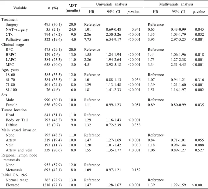
Prognostic factors for survival according to clinical stage
Table 5 lists the results of the Cox proportional hazards regression analyses. The NAT+surgery group conferred a 35% lower hazard of mortality compared with the surgery group (hazard ratio [HR]=0.65, p=0.045). In contrast, the CTx (HR=1.35, p=0.032) and palliative care (HR=3.95, p<
0.001) groups were associated with greater risk compared with the surgery group. The risk of mortality was greater in patients aged 71-80 years (HR=1.39, p<0.001) or 81-100 years (HR=1.51, p=0.002) at diagnosis than in pa- tients aged 18-60 years at diagnosis. The risk of mortality was lower in females than in males (HR=0.89, p=0.035) and was higher in patients with elevated CA 19-9 levels than in patients with normal CA 19-9 levels (HR=1.39, p<0.001). Main vessel invasion, regional lymph node metastasis at the time of diagnosis, and tumor location showed no significant effects on survival rate.
Among the RPC patients, compared with the surgery group, regional lymph node metastasis (HR=1.42, p=
0.004) and an elevated initial CA 19-9 level (HR=2.09, p<0.001) were associated with higher risk in terms of survival. In BRPC patients, compared with the surgery group, there were no significant differences in risk in the NAT+surgery or CTx group but a higher risk in the pal- liative care group (HR=3.08, p<0.001). In LAPC pa- tients, the NAT+surgery, CTx, and palliative care groups did not show significant differences in risk compared with the surgery group. In MPC patients, the NAT+surgery group did not show a significant difference in risk com- pared with CTx group, but the palliative care (HR=3.23, p<0.001) group was associated with a higher risk com- pared with the CTx group.
DISCUSSION
This retrospective cohort study aimed to categorize pa- tients according to clinical stage using uniform imaging diagnostic criteria based on high-resolution imaging and to evaluate the impact of treatment modalities on the sur- vival of pancreatic cancer patients. Of the 1,646 total pa- tients, 475 (28.9%) were diagnosed with RPC, 129 (7.8%) with BRPC, 384 (23.3%) with LAPC, and 658 (40.0%) with MPC. There were 530 (32.2%) surgically treatable
Table 5. Multivariate analysis of prognostic factors for survival of patients with pancreatic cancer
Variable n (%) MST
(months)
Univariate analysis Multivariate analysis
HR 95% CI p-value HR 95% CI p-value
Treatment
Surgery 495 (30.1) 20.0 Reference Reference
NAT+surgery 35 (2.1) 24.0 1.01 0.69-0.48 0.941 0.65 0.43-0.99 0.045
CTx 794 (48.2) 9.0 2.86 2.50-3.26 <0.001 1.35 1.03-1.79 0.032
Palliative care 322 (19.6) 4.0 7.75 6.54-9.17 <0.001 3.95 2.97-5.25 <0.001 Clinical stage
RPC 475 (29.1) 20.0 Reference Reference
BRPC 129 (7.6) 13.0 1.55 1.24-1.94 <0.001 1.44 1.06-1.96 0.018
LAPC 384 (23.3) 11.0 2.26 1.94-2.64 <0.001 1.73 1.27-2.38 0.001
MPC 658 (40.0) 5.0 4.51 3.92-5.18 <0.001 3.34 2.51-4.45 <0.001
Age, years
18-60 585 (35.5) 12.0 Reference Reference
61-70 584 (35.5) 11.0 1.01 0.88-1.13 0.936 1.07 0.94-1.21 0.316
71-80 401 (24.4) 8.0 1.29 1.13-1.48 <0.001 1.39 1.21-1.60 <0.001
81-100 76 (4.6) 6.0 1.81 1.41-2.33 <0.001 1.51 1.16-1.97 0.002
Sex
Male 990 (60.1) 10.0 Reference Reference
Female 656 (39.9) 10.0 1.11 0.99-1.23 0.051 0.89 0.80-0.99 0.035
Tumor location
Head 841 (51.1) 11.0 Reference
Body or Tail 793 (48.2) 9.0 1.29 1.16-1.43 <0.001
Diffuse 12 (0.7) 9.0 1.32 0.72-2.39 0.358
Main vessel invasion
None 795 (48.3) 11.0 Reference Reference
Artery 319 (19.4) 10.0 1.47 1.27-1.69 <0.001 0.84 0.71-1.01 0.055
Vein 193 (11.7) 10.0 1.20 1.01-1.42 0.030 1.18 0.98-1.44 0.088
Artery and vein 339 (20.6) 8.0 1.55 1.35-1.77 <0.001 1.06 0.89-1.27 0.527 Regional lymph node
metastasis
None 953 (57.9) 12.0 Reference
Metastasis 693 (42.1) 8.0 1.09 0.97-1.21 0.152
Initial CA 19-9
Normal range 362 (22.9) 13.0 Reference Reference
Elevated 1218 (77.1) 10.0 1.47 1.28-1.67 <0.001 1.39 1.22-1.59 <0.001
MST, median survival time; HR, relative hazard ratio; CI, confidence interval; NAT, neoadjuvant therapy; CTx, chemotherapy;
RPC, resectable pancreatic cancer; BRPC, borderline resectable pancreatic cancer; LAPC, locally advanced pancreatic cancer;
MPC, metastatic pancreatic cancer; CA 19-9, carbohydrate antigen 19-9
pancreatic cancer patients, which is more than reported previously, and 35 (2.1%) patients underwent surgery af- ter NAT.3 Of the 475 RPC patients, 438 (91.4%) under- went surgery, and R0 resection was achieved in 388 (81.0%).
According to the annually reported summary staging of pancreatic cancer by the Korea National Cancer Registry, there were 2,576 (10.9%) localized, 7,492 (31.8%) region- al, and 10,735 (45.6%) distant cases of metastasis, and 2,715 (11.7%) unknown cases in 2014, 5-year prevalence (5,948 were newly diagnosed with pancreatic cancer in
2014; however, this report did not include the incidence of pancreatic cancer according to clinical stage).3 Sum- mary staging is the most basic method of categorizing how far a cancer has spread from its point of origin, but it does not reflect the clinical stage, which has been essen- tial for determining resectability the suitability for NAT.15 A large retrospective cohort study using the 2003-2011 dataset from the National Cancer Database based in the U.S. reported that of 18,332 pancreatic cancer patients, 7,095 (38.7%) were clinical stage I, 9,760 (53.2%) clinical stage II, and 1,477 (53.2%) clinical stage III.10 However,
that study has several limitations in that patients with clin- ical stage IV or unknown stage pancreatic cancer or those who did not undergo surgical resection were excluded. In addition, BRPC and LAPC were not distinguished in clin- ical stage III patients, and the multicenter data were het- erogenous because of patient collection from over 1500 facilities. Most of the classification systems focus on ana- tomical findings and there is still debate whether these are a solid classification, due to a lack of prospective studies in this regard. Guidelines that also incorporate biological features are needed to help predict early recurrence, even in resectable pancreatic cancer, and to support indications for NAT in select patients with resectable pancreatic cancer.7,9,16,17
Another recent meta-analysis showed that of 4,394 (from 111 studies) pancreatic cancer patients, 46.9% who were initially staged as unresectable underwent surgical exploration.18 Of these patients, 69.9% were resected suc- cessfully, leading to a resectability rate after NAT of 33.2% (comparable R0 resection rate to that in the ini- tially resectable group), suggesting that patients with pan- creatic cancer who can undergo surgical resection are in- creasing more than one out of five in the past by applying NAT at an advanced stage.18 Furthermore, an increase in the early diagnosis rate, detection of various low-malig- nancy-risk pancreatic cancers, and development of surgi- cal techniques have accelerated the surgical resection rate of pancreatic cancer.3,10
Katz et al. conducted a prospective, multicenter, sin- gle-arm pilot study in 2013 of 22 patients with borderline resectable pancreatic cancer who were treated with a modified FOLFIRINOX regimen and chemoradiotherapy as NAT. The median survival was 21.7 months, and R0 resection was achieved in 20 (93%) patients.19 In 2014, Tzeng et al. published the results of a retrospective study, in which 84 patients received gemcitabine or 5-fluorour- acil-based NAT followed by surgery, and 57 patients un- derwent NAT without surgery. The R0 resection rate was 92% in the NAT+surgery group. The median survival was 30.9 months in the NAT+surgery group versus 12 months in the NAT only group.20 In our study, among the patients with BRPC, the median survival was 24 months in the NAT+surgery group, 16 months in the surgery group, 12 months in the CTx group, and 7 months in the palliative care group. Only 32 (6.2%) patients with BRPC or LAPC
underwent surgery after NAT, and 61 (12.0%) underwent surgery without NAT. Few studies have assessed the over- all median survival in patients with pancreatic cancer stratified by treatment modality and disease stage in the same period of time.
Among patients who underwent curative resection, the rate of R0 resection was comparable between the NAT+
surgery and surgery groups, even though the proportion of patients with advanced cancer was greater in the former group. We recognize that the NAT+surgery group tended to undergo more aggressive surgical procedures than did the surgery group; for example, the main vessel resection rate was greater in the former group (22.9% vs. 9.7%; p<
0.001). Since the dense stroma associated with pancreatic cancer may result in little change on computed tomo- graphic imaging, despite an excellent cellular response, there may be some discrepancy between the Response Evaluation Criteria in Solid Tumors criteria and tumor re- gression grade; hence, adjuvant surgery may be necessary to identify a pathological response.21
Tumor response after neoadjuvant treatment were as- sessed radiologically with pancreatobiliary protocol CT and were categorized according to the new response eval- uation criteria in RECIST guidelines in this study. Also, serum concentrations of CA 19-9 were measured at the first visit to the hospital, after neoadjuvant treatment and after surgery. Kim et al. reported a retrospective study in 2017 of 40 BRPC patients who were treated with NAT followed by resection that the RECIST criteria and re- duced serum CA 19-9 concentration were associated with biological response.22 Several recent studies were con- ducted that Positron Emission Tomography/Computer to- mography (PET/CT) could increase the chance of detect- ing patients with progressive pancreatic cancer after neo- adjuvant therapy compared to the conventional anatomi- cal-based assessment of RECEIST criteria.23,24
We also conducted multivariate analysis using the Cox proportional hazards model to determine prognostic fac- tors for survival in patients with pancreatic cancer. The results showed surgery after NAT compared with surgery or CTx alone, female sex, resectable stage, a CA 19-9 lev- el within the normal range at diagnosis, and tumor loca- tion in the pancreatic body or tail to be prognostic factors for improved survival in patients with pancreatic cancer.
Tzeng et al. also reported that serum CA 19-9 is a dynam-
ic preoperative marker of tumor biology and the response to NAT, and provides prognostic information in patients with unresected or resected BRPC.20 In addition, another study revealed that patients with advanced pancreatic can- cer whose serum tumor marker levels had normalized af- ter NAT may be appropriate candidates for tumor resection.25
Some strengths and limitations of this study also war- rant discussion. First, because this was a retrospective analysis of patients treated at a single institution, the re- sults are subject to the biases and limitations inherent to retrospective studies. Nevertheless, this is one of the larg- est reported cohorts of patients with resectable, borderline resectable, locally advanced, or metastatic pancreatic can- cer in which the cancer was staged using objective radio- graphic criteria and classified using consensus guidelines.
Second, we divided the patients into four groups based on their treatment modalities. Unfortunately, the numbers of patients varied among the groups, with some groups being too small for detailed analyses. As a result of eth- ical issues and difficulty in recruiting patients, no random- ized clinical trials of prior surgery compared with NAT have been performed. Nevertheless, several phase I/II tri- als and on-going phase III clinical trials, such as NEOPAC (NCT01521702) and Prep-02/JSAP-05 (UMIN- No. 000009634) are underway and will hopefully yield further insight into the benefits of NAT.19,26-28 Third, this study included heterogenous regimens of NAT, which showed different oncologic outcomes.
In conclusion, based on the results of this retrospective, single-center, large cohort study, categorizing all pancre- atic cancer patients according to clinical stage using uni- form imaging diagnostic criteria with high-resolution im- ages, the rates of resectable and surgically treatable pan- creatic cancer were 29.1% and 32.2%, which are higher than those reported previously. In the RPC group, 434 (91.4%) patients underwent surgery, and R0 resection was achieved in 388 (81.0%). Although our findings support the use of NAT in eligible patients with borderline resect- able pancreatic cancer, the survival rate of advanced-stage patients is still low; thus, more effective systemic thera- pies are required. In addition, further studies are needed to evaluate the effects of specific NAT regimens and to update consensus guidelines with biological features.
ACKNOWLEDGEMENTS
This research was supported by the Collaborative Genome Program for Fostering New Post-Genome Indus- try of the National Research Foundation funded by the Ministry of Science and ICT (NRF-2017M3C9A5031597).
REFERENCES
1. Siegel RL, Miller KD, Jemal A. Cancer statistics, 2015. CA Cancer J Clin 2015;65:5-29.
2. Tran KT, Smeenk HG, van Eijck CH, Kazemier G, Hop WC, Greve JW, et al. Pylorus preserving pancreaticoduodenectomy versus standard Whipple procedure: a prospective, randomized, multicenter analysis of 170 patients with pancreatic and peri- ampullary tumors. Ann Surg 2004;240:738-745.
3. Jung KW, Won YJ, Oh CM, Kong HJ, Lee DH, Lee KH. Cancer statistics in Korea: incidence, mortality, survival, and prevalence in 2014. Cancer Res Treat 2017;49:292-305.
4. Tempero MA, Malafa MP, Behrman SW, Benson AB 3rd, Casper ES, Chiorean EG, et al. Pancreatic adenocarcinoma, ver- sion 2.2014: featured updates to the NCCN guidelines. J Natl Compr Canc Netw 2014;12:1083-1093.
5. Conroy T, Desseigne F, Ychou M, Bouché O, Guimbaud R, Bécouarn Y, et al. FOLFIRINOX versus gemcitabine for meta- static pancreatic cancer. N Engl J Med 2011;364:1817-1825.
6. Liu GF, Li GJ, Zhao H. Efficacy and toxicity of different chemo- therapy regimens in the treatment of advanced or metastatic pan- creatic cancer: a network meta-analysis. J Cell Biochem 2018;
119:511-523.
7. Varadhachary GR, Tamm EP, Abbruzzese JL, Xiong HQ, Crane CH, Wang H, et al. Borderline resectable pancreatic cancer: defi- nitions, management, and role of preoperative therapy. Ann Surg Oncol 2006;13:1035-1046.
8. Callery MP, Chang KJ, Fishman EK, Talamonti MS, William Traverso L, Linehan DC. Pretreatment assessment of resectable and borderline resectable pancreatic cancer: expert consensus statement. Ann Surg Oncol 2009;16:1727-1733.
9. Katz MH, Marsh R, Herman JM, Shi Q, Collison E, Venook AP, et al. Borderline resectable pancreatic cancer: need for standardization and methods for optimal clinical trial design.
Ann Surg Oncol 2013;20:2787-2795.
10. Mirkin KA, Hollenbeak CS, Wong J. Survival impact of neo- adjuvant therapy in resected pancreatic cancer: a Prospective Cohort Study involving 18,332 patients from the National Cancer Data Base. Int J Surg 2016;34:96-102.
11. Goonetilleke KS, Siriwardena AK. Systematic review of carbo- hydrate antigen (CA 19-9) as a biochemical marker in the diag- nosis of pancreatic cancer. Eur J Surg Oncol 2007;33:266-270.
12. Eisenhauer EA, Therasse P, Bogaerts J, Schwartz LH, Sargent D, Ford R, et al. New response evaluation criteria in solid tu- mours: revised RECIST guideline (version 1.1). Eur J Cancer 2009;45:228-247.
13. Wittekind C, Compton CC, Greene FL, Sobin LH. TNM residual tumor classification revisited. Cancer 2002;94:2511-2516.
14. Washington MK, Berlin J, Branton PA, Burgart LJ, Carter DK, Compton CC, et al. Protocol for the examination of specimens from patients with carcinoma of the perihilar bile ducts. Arch Pathol Lab Med 2010;134:e19-24.
15. National Cancer Institute (NIH). Surveillance, Epidemiology, and
End Results Program (SEER) Summary staing manual 2018.
Bethesda: NIH; 2018 [cited 2018 May 2]. Available from:
https://seer.cancer.gov/tools/ssm/SSM2018-DIGESTIVE-AND-H EPATOBILIARY-SYSTEMS.pdf.
16. Katz MH, Pisters PW, Evans DB, Sun CC, Lee JE, Fleming JB, et al. Borderline resectable pancreatic cancer: the importance of this emerging stage of disease. J Am Coll Surg 2008;206:833- 846.
17. Vauthey JN, Dixon E. AHPBA/SSO/SSAT consensus conference on resectable and borderline resectable pancreatic cancer: ration- ale and overview of the conference. Ann Surg Oncol 2009;16:
1725-1726.
18. Gillen S, Schuster T, Meyer Zum Büschenfelde C, Friess H, Kleeff J. Preoperative/neoadjuvant therapy in pancreatic cancer:
a systematic review and meta-analysis of response and resection percentages. PLoS Med 2010;7:e1000267.
19. Katz MH, Shi Q, Ahmad SA, Herman JM, Marsh Rde W, Collisson E, et al. Preoperative modified FOLFIRINOX treat- ment followed by capecitabine-based chemoradiation for border- line resectable pancreatic cancer: alliance for clinical trials in on- cology trial A021101. JAMA Surg 2016;151:e161137.
20. Tzeng CW, Balachandran A, Ahmad M, Lee JE, Krishnan S, Wang H, et al. Serum carbohydrate antigen 19-9 represents a marker of response to neoadjuvant therapy in patients with bor- derline resectable pancreatic cancer. HPB (Oxford) 2014;16:430- 438.
21. Evans DB, George B, Tsai S. Non-metastatic pancreatic cancer:
resectable, borderline resectable, and locally advanced-defi- nitions of increasing importance for the optimal delivery of mul- timodality therapy. Ann Surg Oncol 2015;22:3409-3413.
22. Kim HS, Jang JY, Han Y, Lee KB, Joo I, Lee DH, et al.
Survival outcome and prognostic factors of neoadjuvant treat-
ment followed by resection for borderline resectable pancreatic cancer. Ann Surg Treat Res 2017;93:186-194.
23. Dalah E, Tai A, Oshima K, Hall WA, Erickson B, Li XA.
PET-based treatment response assessment for neoadjuvant che- moradiation in pancreatic adenocarcinoma: an exploratory study.
Transl Oncol 2018;11:1104-1109.
24. Sakane M, Tatsumi M, Hori M, Onishi H, Tsuboyama T, Nakamoto A, et al. Volumetric parameters of 2-deoxy-2-[18F]
fluoro-d-glucose positron emission tomography/computed tomog- raphy can predict histopathologic treatment response after neo- adjuvant chemoradiotherapy in pancreatic adenocarcinoma. Eur J Radiol 2017;94:64-69.
25. Murakami Y, Uemura K, Sudo T, Hashimoto Y, Kondo N, Nakagawa N, et al. Prognostic impact of normalization of serum tumor markers following neoadjuvant chemotherapy in patients with borderline resectable pancreatic carcinoma with arterial contact. Cancer Chemother Pharmacol 2017;79:801-811.
26. Versteijne E, van Eijck CH, Punt CJ, Suker M, Zwinderman AH, Dohmen MA, et al. Preoperative radiochemotherapy versus im- mediate surgery for resectable and borderline resectable pancre- atic cancer (PREOPANC trial): study protocol for a multicentre randomized controlled trial. Trials 2016;17:127.
27. Heinrich S, Pestalozzi B, Lesurtel M, Berrevoet F, Laurent S, Delpero JR, et al. Adjuvant gemcitabine versus NEOadjuvant gemcitabine/oxaliplatin plus adjuvant gemcitabine in resectable pancreatic cancer: a randomized multicenter phase III study (NEOPAC study). BMC Cancer 2011;11:346.
28. Motoi F, Kawaguchi K, Aoki T, Kudo K, Yabuuchi S, Fukase K, et al. [Efficacy of neoadjuvant chemotherapy for resectable pancreatic carcinoma]. Gan To Kagaku Ryoho 2013;40:1632- 1636. Japanese.