Journal of the Korean Surgical Society 43
pISSN 2233-7903 •eISSN 2093-0488
Primary leiomyosarcoma of the thyroid
Bahadır Ege, Sezai Leventoğlu
1Clinic of General Surgery, Private Koru Hospital, Ankara,
1Department of General Surgery, Gazi University School of Medicine, Ankara, Turkey
CASE REPORT
A 56-year-old male with primary leiomyosarcoma of the thyroid is presented. The paucity of diagnostic maneuvers, including tumor markers, fine needle aspiration, and frozen section biopsy, are stressed, in addition to the fulminate course of the disease.
Journal of the Korean Surgical Society
JKSS
INTRODUCTION
Primary leiomyosarcoma of the thyroid (PLT) is rare [1]. The differential diagnosis includes medullary thyroid cancer, anaplastic thyroid cancer, and primary versus metastatic sarcoma [2,3]. Histologically, PLT is composed of characteristic spindle cells [4]. It is an aggressive malignancy with poor long-term prognosis [1-4]. We report a 56-year-old male with PLT, who presented with enlargement of the neck, hoarseness and dysphagia.
CASE REPORT
A 56-year-old male presented with 4 months history of a rapidly enlarging left anterior neck mass, hoarseness, and dysphagia. He was referred from another city with an initial diagnosis of thyroid cancer. No comorbidities existed, and the medical history was unremarkable. A history of radiation exposure did not exist, either. Physical examination revealed a hard mass of about 3 cm in the left lobe of the thyroid gland. There were no palpable cervical or supraclavicular lymph nodes. He was clinically euthyroid. Serum thyroid stimulating hormon, serum T3 (triiodothyronine) and T4 (thyroxinne) were within normal levels. Ultrasound of the neck showed a 2.71 cm × 2.59 cm solid nodule arising from the left thyroid lobe, with small contralateral nodules measuring 0.5 and 1.1 cm (Fig. 1). There was no invasion to adjacent tissues.
Fine-needle aspiration (FNA) biopsy of the thyroid mass was then performed.
FNA biopsy reported malignant spindle-shaped cells. Computed tomography of the chest and abdomen showed no distant metastases. Serum calcitonin, carcinoembryonic antigen, and thyroglobulin were within normal levels. Initially, the left thyroid lobe was excised and sent to pathology for frozen section examination,
Corresponding Author Bahadır Ege
Clinic of General Surgery, Private Koru Hospital, Oguzlar Mah. 1377 sok. No 21, Balgat, Ankara, Turkey
TEL : +903122879797 FAX : +903122879898 E-mail : mdbahadirege@gmail.com Keywords
Thyroid, Leiomyosarcoma
Received May 15, 2012 Revised January 21, 2013 Accepted February 7, 2013 J Korean Surg Soc 2013;85:43-46 http://dx.doi.org/10.4174/jkss.2013.85.1.43
Copyright © 2013, the Korean Surgical Society
cc Journal of the Korean Surgical Society is an Open Access Journal. All articles are distributed under the terms of the Creative Commons Attribution Non-Commercial License (http://
creativecommons.org/licenses/by-nc/3.0/) which permits unrestricted non-commercial use, distribution, and reproduction in any medium, provided the original work is properly cited.
thesurgery.or.kr
44
JKSS
Bahadır Ege and Sezai Leventoğlu: Thyroidal leiomyosarcoma
which reported poorly-differentiated tumor suggesting a
‘malignant spindle cell tumor’. Total thyroidectomy and central neck dissection were performed. No complications occurred, and the patient was discharged on the first postoperative day.
On gross examination, the tumor measured 3 cm × 3 cm × 2.5 cm. Histologically, the tumor cells infiltrated the thyroid parenchyma, with no invasion to the thyroid
capsule. Vascular invasion was present. Hematoxylin-eosin staining demonstrated spindle-shaped cell bundles. The tumor cells showed a disordered fascicular growth pattern,
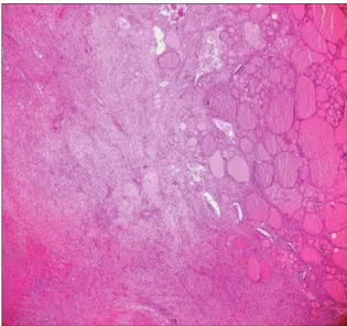
Fig. 1. Ultrasound of neck showed 2.71 cm × 2.59 cm solid nodule arising from left thyroid lobe.
Fig. 2. Hematoxylin-eosin staining showed spindle-shaped cells with hyperchromatic, blunt-ended nuclei and abundant eosinophilic cytoplasm (H&E,
×10).
Fig. 3. The tumor was strongly positive for smooth muscle actin, positive for desmin, and negative for pancytokeratin, suggesting primary leiomyosarcoma of thyroid. (A) Positive for desmin (x10). (B) Negative for pancytokeratin (x10). (C) Strongly positive for smooth muscle actin (x10).
Bahadır Ege and Sezai Leventoğlu: Thyroidal leiomyosarcoma
Journal of the Korean Surgical Society 45 round or spindle configuration with hyperchromatic, blunt-
ended nuclei and abundant eosinophilic cytoplasm (Fig. 2).
Immunohistochemical staining with pancytokeratin, desmin, and smooth muscle actin was performed. The tumor was strongly positive for smooth muscle actin, positive for desmin, and negative for pancytokeratin (Fig. 3). No tumor-bearing lymph nodes were detected.
The oncology department did not suggest any adjuvant modalities. The patient was free of disease at three- and six- month’s follow-ups. About eight months after the operation, his family doctor informed us that he had developed sudden respiratory symptoms and died in the intensive care unit with numerous pulmonary metastases.
DISCUSSION
Although the the incidence of thyroid cancer rate is different in the Korea population (the age-standardized incidence rate of thyroid cancer in 2007 was 32.8 per 100,000 [9.9 per 100,000 men and 55.6 per 100,000 women]) [5,6], thyroid cancer represents less than 1% of all malignancies in the world. The 90% to 95% of thyroid cancer cases are categorized as well-differentiated tumors arising from the follicular cells [7,8]. However, PLT gland is extremely rare, and it is the most aggressive form of thyroid cancer [2]. Only a few cases have been reported [1,3].
In our patient, FNA biopsy of the thyroid mass was the initial diagnostic step. The unusual FNA biopsy report of malignant spindle-shaped cells lead us to consider unusual tumors of the thyroid. Computed tomography of the chest and abdomen did not add to the diagnosis, similar to several tumor markers within normal ranges. The frozen section examination also failed to define the exact pathology, although the definition of a poorly differentiated tumor with malignant spindle cells weighed in favor of our suspicion of PLT. Still, possibilities such as anaplastic thyroid cancer, and primary versus metastatic sarcoma existed [1,3]. The final diagnosis could only be done by immunohistochemical staining.
Spindle cell anaplastic carcinomas resemble sarcomas.
Focal keratin positivity is comon in anaplastic carcinomas.
Immunohistochemically, leiomyosarcomas are reactive for desmin or vimentin and actin [9]. In our patient, the diag- nosis of leiomyosarcoma was favored because smooth muscle markers and desmin were positive and immunostains for keratins were negative. PLT showed smooth muscle cells with atypia and mitotic figures. Smooth muscle mar kers were uniformly positive, confirming the diagnosis. Leio- myosarcomas are malignant mesenchymal tumors with
smooth muscle differentiation that can arise in the smooth muscle of any organ; in the thyroid, this includes the muscular vessel walls located in the thyroid capsule [6]. Primary thyroid leiomyosarcomas tend to occur in older populations, equally in both genders. They tend to be larger than their benign counterparts with malignant histologic features characterized by pleomorphism, prominent mitotic activity, necrosis, hemor- rhage, and invasive and/or extrathyroidal growth. Thyroid leiomyosarcomas are generally fatal, although follow-up was limited in the published reports [10]. Leiomyosarcomas of the thyroid can also be metastatic in origin, with primary sites in soft tissue, stomach, and pelvis, all being excluded in our patient by preoperative imaging.
The mainstay of treatment for PLT is oncologic resection, as exemplified again in this case. Chemotherapy is of limited benefit. The disease is invariably fatal, and survival rates are reported to be 5% to 10% at 1 year [3]. In spite of the silent and optimistic short-term course, our patient also experienced a fulminating and fatal tumor attack sooner than a year.
CONFLICTS OF INTEREST
No potential conflict of interest relevant to this article was reported.
REFERENCES
1. Adachi M, Wellmann KF, Garcia R. Metastatic leiomyosarcoma in brain and heart. J Pathol 1969;98:294-6.
2. Wang TS, Ocal IT, Oxley K, Sosa JA. Primary leiomyosarcoma of the thyroid gland. Thyroid 2008;18:425-8.
3. Mansouri H, Gaye M, Errihani H, Kettani F, Gueddari BE.
Leiomyosarcoma of the thyroid gland. Acta Otolaryngol 2008;
128:335-6.
4. Chetty R, Clark SP, Dowling JP. Leiomyosarcoma of the thyroid:
immunohistochemical and ultrastructural study. Pathology 1993;25:203-5.
5. Han MA, Choi KS, Lee HY, Kim Y, Jun JK, Park EC. Current status of thyroid cancer screening in Korea: results from a nation wide interview survey. Asian Pac J Cancer Prev 2011;
12:1657-63.
6. Chung KW, Kim SW, Kim SW. Gene expression profiling of papillary thyroid carcinomas in Korean patients by oligonu- cleotide microarrays. J Korean Surg Soc 2012;82:271-80.
7. Geeto L, Orlo HC. Thyroid, parathyroid, and Adrenal. In:
Brunicardi FC. Schwartz’s principles of surgery. New York:
McGraw-Hill Co.; 2005. p.1395-471.
8. John BH. Thyroid. In: Courtney M. Sabiston textbook of surgery.
thesurgery.or.kr
46
JKSS
Bahadır Ege and Sezai Leventoğlu: Thyroidal leiomyosarcoma
Philadelphia: Elseiver; 2004. p.947-85.
9. Li YF, Yu CP, Wu ST, Dai MS, Lee HS. Malignant mesen chymal tumor with leiomyosarcoma, rhabdomyosarcoma, chondro- sarcoma, and osteosarcoma differentiation: case report and
literature review. Diagn Pathol 2011;6:35-41.
10. Thompson LD, Wenig BM, Adair CF, Shmookler BM, Heffess CS. Primary smooth muscle tumors of the thyroid gland. Cancer 1997;79:579-87.