Malignant lymphoma is the generic term given to tumors of the lymphoid system. This lesion can involve the lymph nodes, spleen, and sometimes the non-hematopoietic tis- sues. This tumor can be divided into two major catego- ries: Hodgkin’s lymphoma and non-Hodgkin’s lymphoma (NHL). NHL is a heterogeneous group of malignancies characterized by an abnormal clonal proliferation of T cells, B cells, or both. The majority of the adult NHLs are of B cell origin.1Palatal and nasal lymphomas are rare, and the majority of the lymphomas in this region origi- nate from B cells. Since early detection of hard palate tu- mors is difficult by clinical examination, the vast majority of such tumors are detected after maxillary or sphenoid bone invasion.2The present report showed a case of B cell lymphoma in a 60-year-old male patient manifested as an ulcerative growth on the right maxillary alveolar ridge extending on to the hard palate, but on imaging it was an extensive lesion involving maxillary sinus, nasal cavity, and orbit.
Case Report
A 60-year-old man visited the Oral Medicine and Ra- diology Department, complaining of a mild intermittent pain associated with sudden ulcerated growth on the right maxillary edentulous alveolar ridge since 1 month earlier.
The patient gave a history of extraction of teeth in the same region one year before. On examination, a diffuse mild extra oral swelling was present on the right cheek, with proptosis and watery discharge from the right eye (Fig. 1). On palpation, extraorally the swelling was mil- dly tender and discontinuity was felt on the right infra or- bital margin. Intraoral examination revealed a soft well defined ulcerated growth on the right maxillary edentul- ous alveolar ridge, extending from the right maxillary first molar region to the maxillary tuberosity region posterior- ly. The surface of the growth was covered with a yello- wish necrotic material. It was slightly tender on palpation (Fig. 2).
The clinical differential diagnosis included the most com- mon malignancies in the oral cavity such as squamous cell carcinoma (SCC), minor salivary gland tumor, and carcinoma of the maxillary sinus. Panoramic radiograph
─ 111 ─
Imaging characteristics of diffuse large cell extra nodal non-Hodgkin’s lymphoma involving the palate and maxillary sinus: a case report
Lakshmi Kavitha Nadendla, Venkateswarlu Meduri, Geetha Paramkusam
Department of Oral and Medicine and Radiology, Kamineni Institute of Dental Sciences, Nalgonda, India ABSTRACT
Non-Hodgkin’s lymphomas are a group of highly diverse malignancies and have a strong tendency to affect organs and tissues that do not ordinarily contain lymphoid cells. Primary extra nodal lymphoma of the hard palate is rare.
Here, we present a case of diffuse large B cell lymphoma in a 60-year-old male patient that manifested as slightly painful ulcerated growth on the edentulous right maxillary alveolar ridge extending onto the palate, closely resem- bling carcinoma of the alveolar ridge. Computed tomography images showed the involvement of the maxillary sinus and right nasal cavity, along with destruction of hard palate, superiorly extending into the orbit. This case report highlights the importance of imaging to evaluate the exact extent of such large malignant lesions, which is essential for treatment planning. (Imaging Sci Dent 2012; 42 : 111-4)
KEY WORDS : Lymphoma, Non-Hodgkin; Palate; Maxillary Sinus; Lymphoma, Large B-Cell, Diffuse
Received January 9, 2012; Revised February 6, 2012; Accepted March 9, 2012 Correspondence to : Dr. Lakshmi Kavitha Nadendla
Department of Oral and Medicine and Radiology, Kamineni Institute of Dental Sciences, Sreepuram, Narketpally, Nalgonda, Andhara Pradesh, India
Tel) 91-9949-678533, Fax) 91-8682-272296, E-mail) [email protected]
Imaging Science in Dentistry 2012; 42 : 111-4 http://dx.doi.org/10.5624/isd.2012.42.2.111
Copyright ⓒ 2012 by Korean Academy of Oral and Maxillofacial Radiology
This is an Open Access article distributed under the terms of the Creative Commons Attribution Non-Commercial License (http://creativecommons.org/licenses/by-nc/3.0) which permits unrestricted non-commercial use, distribution, and reproduction in any medium, provided the original work is properly cited.
Imaging Science in Dentistry∙pISSN 2233-7822 eISSN 2233-7830
revealed severe bone destruction of the right maxilla distal to the canine. The floor of the right maxillary sinus was also destroyed. Computed tomography (CT) scan was per-
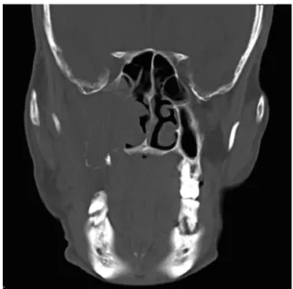
formed and sections were taken in the axial, coronal, and sagittal planes. Coronal section revealed a large soft tissue density lesion with its epicentre in the maxillary sinus, causing destruction of all walls of the maxillary sinus, extending medially into the lateral wall of the nasal sep- tum, left nasal cavity involving the inferior and middle tur- binates, laterally into the pterygopalatine fossa, superiorly into the orbit, inferiorly causing destruction of the hard palate and alveolar processes of the right maxilla in the molar region. There was no evidence of calcification (Fig.
3). Three dimensional CT reconstructions using the bone and soft tissue settings demonstrated a large lytic lesion in the right maxillary sinus destroying all walls of the maxillary sinus, hard palate, and alveolar process in the
─ 112 ─
Imaging characteristics of diffuse large cell extra nodal non-Hodgkin’s lymphoma involving the palate and maxillary sinus: a case report
Fig. 2. Intraoral photograph shows an ulcerative growth in the right maxillary alveolar ridge extending on to the palate.
Fig. 3.Coronal CT image shows a large soft tissue density lesion with destruction of all walls of the right maxillary sinus, hard pal- ate, and extension into the nasal cavity.
Fig. 4. Three-dimensional CT recon- struction image (bone setting) shows destruction of the maxilla, hard pal- ate, and maxillary alveolar ridge on the right side.
Fig. 5.Three-dimensional CT recon- struction image (soft tissue setting) shows facial asymmetry due to the swelling and proptosis of the eye on the right side.
Fig. 1.Mild diffuse swelling on the right cheek and proptosis of the right eye is seen.
4 5
molar region (Figs. 4 and 5).
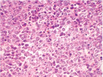
The patient was advised to undergo routine haema- tological examination, and an incisional biopsy was per- formed which revealed a cellular connective tissue stroma predominantly made up of lymphocytes, the majority being large cells and a few small cells showing vesicular nuclei with prominent nucleoli. Scattering foamy macrophages were seen amongst these cells. It was finally diagnosed as diffuse large cell lymphoma (Fig. 6). The patient was referred to the oncology department, who started chemo- therapy; unfortunately, the patient passed away after two cycles of chemotherapy.
Discussion
Malignant lymphoma can appear in all parts of the body, and may have varied radiographic manifestations.3 24- 84% of NHLs arise from extra nodal sites. The head and neck is the second most common region for extra nodal lymphoma after the gastro intestinal tract.4 NHL is the second most common neoplasm of the head and neck region after SCC and the third most common group of malignant lesions of the oral region after SCC and sali- vary gland neoplasm.5,6
NHL commonly involves oropharyngeal lymphoid tis- sue comprising Waldeyer’s ring, but occasionally involves other oral tissues.6 NHL occurs more frequently in the head and neck region in the pediatric age group,7however our patient was a 60-year-old male. Sinuses are the pri- mary site of NHL in 90% of the cases, most commonly invaded by diffuse large B cell lymphoma.7 In our case,
the epicentre of the lesion seemed to be in the right max- illary sinus on the CT images.
The etiological factors for primary lymphoma of the oral region are unclear. A few cases of oral lymphomas were reported in association with acquired immune defi- ciency syndrome (AIDS), and it might even be the first presentation of AIDS in certain individuals.8In our case, the patient was human immunodeficiency virus (HIV) negative. Primary oral and para-oral lymphoma most com- monly presents as a painless local mass that gradually increases in size with superficial ulceration.9Diagnosis of NHL in oral cavity may result from mucosal tissue/gin- gival swelling/masses. When oral soft tissue lesions first appear, they are relatively soft and often have an overly- ing ulceration, and are often characterized by absence of other symptoms. If bone is the primary site, tooth mobili- ty and alveolar bone loss are often noted. Pain, swelling, numbness of the lip, and pathologically related fractures may be associated with the bone lesion.5In our case, the patient presented with a slightly painful ulcerated growth on the maxillary edentulous alveolar ridge resembling SCC.
Clinical signs and symptoms of lymphomas of the para- nasal sinuses include a mass in the nasal cavity, facial pain, paresthesia, recurrent sinusitis, nasal discharge, eyelid swelling, and proptosis if orbital invasion has occurred.10 Our patient showed an extraoral swelling in the right maxillary sinus region and also proptosis of the eye sug- gestive of extension of the lesion into the sinus and orbit.
Radiological imaging is vital for many reasons, includ- ing assessment of tumor extension, assessment of bony destruction, evidence of mucosal thickening, and choice of biopsy site and route. CT is the best imaging modality for demonstrating fine bony detail.10 Lymphoma can cause nonspecific bony destruction at and around the paranasal sinuses. The maxillary sinuses are most frequently affect- ed, followed by the ethmoidal and frontal sinuses. The sphenoid sinuses are rarely affected. As the process of de- struction continues, the bony margins of the maxillary sinu- ses can become eroded, especially the medial and poster- ior walls. Erosion of the anterior wall of the maxillary sinus was also reported.11The epicentre was found to be in the right maxillary sinus on CT images in our case also.
All walls of the maxillary sinus were eroded by the soft tissue lesion.
Lymphomas are usually submucosal, and on gross ap- pearance, differ from SCC which is usually ulcerative.12 Our case clinically manifested as an ulcerated growth close- ly resembling SCC, and it was very difficult to differen- tiate both of the lesions clinically. Biopsy should be per-
─ 113 ─
Lakshmi Kavitha Nadendla et al
Fig. 6. Histopathological examination shows connective tissue stroma made up of predominantly large lymphocytes, few small cells, and scattered foamy macrophages (H&E stain, ×400).
formed to ensure the accurate diagnosis and histological grading of lymphoma. Management also varies depending on the stage of lymphoma. Paranasal lymphomas have a poor prognosis, which is usually worse than that associat- ed with lymphomas in other sites in the body.10
In conclusion, this report focused on the importance of CT in assessing the exact tumor extent and staging for large, clinically doubtful lesions, which was essential for diagnosis and treatment planning.
References
1. Jayakrishnan R, Thomas G, Kumar A, Nair R. Non-Hodg- kin’s lymphoma of the hard palate. J Oral Maxillofac Pathol 2008; 12 : 85-7.
2. Dalirsani Z, Mohtasham N. T-cell lymphoma of palate with nose and maxillary sinus involvement: a case report. Iran J Med Sci 2010; 35 : 254-8.
3. Ueda F, Suzuki M, Matsui O, Minato H, Furukawa M. MR findings of nine cases of palatal tumor. Magn Reson Med Sci 2005; 4 : 61-7.
4. Teh CS, Chong SY. An unusual presentation of lymphoma of the head and neck region. Med J Malaysia 2011; 66 : 264-5.
5. Villa A, Mariani U, Villa F. T-cell lymphoma of the oral cavity:
a case report. Aust Dent J 2010; 55 : 203-6.
6. Vaswani B, Shah M, Shah PM, Parikh BJ, Anand AS, Sharma G. Non hodgkin’s lymphoma of tongue -a case report. Indian J Med Paediatr Oncol 2008; 29 : 59-61.
7. Zagolski O, Dwivedi RC, Subramanian S, Kazi R. Non-Hodg- kin’s lymphoma of the sino-nasal tract in children. J Cancer Res Ther 2010; 6 : 5-10.
8. Maheshwari GK, Baboo HA, Shah NM, Patel MH, Shah R.
Primary non-Hodgkin’s lymphoma of the oral tongue. Turk J Cancer 2001; 31 : 121-4.
9. Essadi I, Ismaili N, Tazi E, Elmajjaoui S, Saidi A, Ichou M, et al. Primary lymphoma of the head and neck: two case reports and review of the literature. Cases J 2008; 1 : 426.
10. Park YM, Cho JH, Cho JY, Huh JS, Ahn JY. Non-Hodgkin’s lymphoma of the sphenoid sinus presenting as isolated oculo- motor nerve palsy. World J Surg Oncol 2007; 5 : 86.
11. Al-Hakeem DA, Fedele S, Carlos R, Porter S. Extra nodal NK/
T-cell lymphoma, nasal type. Oral Oncol 2007; 43 : 4-14.
12. Chalastras T, Elefteriadou A, Giotakis J, Soulandikas K, Kor- res S, Ferekidis E, et al. Non-Hodgkin’s lymphoma of nasal ca- vity and paranasal sinuses. A clinicopathological and immun- histochemical study. Acta Otorhinolaryngol Ital 2007; 27 : 6- 9.
─ 114 ─
Imaging characteristics of diffuse large cell extra nodal non-Hodgkin’s lymphoma involving the palate and maxillary sinus: a case report