174
혈관면역 T세포 림프종 치료 후 발생한 엡스타인바 바이러스 양성 미만성 큰 B세포 림프종 1예
원자력병원 내과
최윤희ㆍ김효석ㆍ남승범ㆍ강혜진ㆍ나임일ㆍ양성현ㆍ류백렬
A Case of Epstein-Barr Virus-positive Diffuse, Large B-cell Lymphoma after Angioimmunoblastic T-cell Lymphoma
Yoon Hee Choi, M.D., Hyo Seog Kim, M.D., Seung Bum Nam, M.D., Hye Jin Kang, M.D., Im Il Na, M.D., Sung Hyun Yang, M.D. and Baek-Yeol Ryoo, M.D.
Department of Internal Medicine, Korea Cancer Center Hospital, Seoul, Korea
Angioimmunoblastic T-cell lymphoma (AITL) is a systemic lymphoproliferative disorder that presents with profound immune dysfunction and immunodeficiency. As in many other immunodeficiencies, Epstein-Barr virus (EBV) associated B-cell lymphoid proliferation can occur in AITL but few cases of EBV-positive B-cell lymphoma have been reported in patients with preexisting AITL. We report a case of AITL in which EBV-positive diffuse large B-cell lymphoma (DLBCL) developed 13 months after the initial diagnosis of AITL. Although the exact mechanisms remain unclear, Epstein-Barr virus may have played a role in the pathogenesis of the secondary DLBCL. (Korean J Hematol 2008;43:174-178.)
Key Words: Angioimmunoblastic T-cell lymphoma, Diffuse large B-cell lymphoma, Epstein-Barr virus
접수:2007년 10월 7일, 수정:2008년 7월 18일 승인:2008년 7월 22일
교신저자:류백렬, 서울시 노원구 공릉동 215-4
139-706, 원자력병원 내과 Tel: 02-970-1208, Fax: 02-970-2410 E-mail: [email protected]
Correspondence to:Baek-Yeol Ryoo, M.D.
Department of Internal Medicine, Korea Cancer Center Hospital 215-4, Gongneung-dong, Nowon-gu, Seoul 139-706, Korea Tel: +82-2-970-1208, Fax: +82-2-970-2410
E-mail: [email protected] 서 론
혈관면역T세포림프종(angioimmunoblastic T-cell lym- phoma, AITL)은 조직학적으로는 다형성 세포 침윤과 여포상가지돌기세포(follicular dendritic cells)의 증식, 내피세정맥(endothelial venules)들이 얽어져 림프절 구 조의 파괴가 나타나고 임상적으로는 전반적인 림프절 병, 간비장종대를 보이며 흔히 면역기능장애가 나타나 는 질환이다.1,2) AITL의 경우 50∼97%에서 엡스타인 바 바이러스(Epstein-Barr virus, EBV)가 감염된 림프 구들이 관찰되었다는 보고가 있고,3-5) 대부분 비종양 성 B세포에서 관찰되었는데,2,4) 드물게 AITL에 속발 또는 병발한 미만성큰B세포림프종(diffuse large B-cell
lymphoma, DLBCL)이 국외에서 종종 보고되었다. 저 자들은 AITL에 대한 복합항암화학요법 후 완전관해에 이르러 추적 관찰 중에 DLBCL이 속발한 예를 경험하 였기에 보고하는 바이다.
증 례
환 자: 노○○, 남자, 78세
주 소: 우측 편도주위 통증과 종창
현병력: 13개월 전 좌측 경부 림프절종대를 주소로 내원하여 종격동 림프절과 복강 내 림프절 침범을 보 이는 혈관면역T세포림프종, 병기 IIIA로 진단 받고 CHOP (cyclophophamide, doxorubicin, vincristine, pre- dnisone) 복합항암화학요법을 6회 시행 받았다. 직후
촬영한 전산화단층촬영(CT)과 18F-fluorodeoxyglucose 양전자방출단층촬영(18F-FDG PET)에서 완전관해에 도달하였고 경과관찰 8개월째 우측 편도주위 통증과 종창 소견으로 내원하였다. 발열, 야간 발한 및 체중 감소는 없었다.
과거력 및 가족력: 당뇨와 고혈압으로 투약 중이었 으며 그 외 특이 소견 없었다.
신체검사소견: 혈압 120/80mmHg, 맥박수 80회/분, 호흡수 20회, 체온 37oC, 의식은 명료하였고 ECOG활 동도는 2였다. 우측 편도에서 궤양을 동반한 융기병변 이 관찰되었으며, 촉지되는 경부, 액와부, 서혜부 림프 절은 없었고 간 비장비대는 관찰되지 않았다.
검사실검사소견: 내원 13개월 전 혈관면역T세포림프 종 진단 당시 일반혈액검사에서는 백혈구 6,240/μL, 혈색소 12.3g/dL, 혈소판 278,000/μL을 보여주었고 젖 산탈수소효소(lactate dehydrogenage)는 4,15IU/L, 간기 능과 콩팥기능은 정상이었다. 당시 골수 침범은 보이 지 않았다. 최근 우측 편도부 종창과 통증으로 내원하
여 시행한 일반혈액검사에서 백혈구 5,440/μL, 혈색 소 11.3g/dL, 혈소판 247,000/μL였고 혈청생화학검사 에서 젖산탈수소효소(lactate dehydrogenage) 5,48IU/L, AST 17IU/L, ALT 16IU/L, 총 빌리루빈 0.6mg/dL였다.
B형, C형 간염에 대한 항체와 사람면역결핍바이러스 항체(anti-HIV)는 음성이었고 골수도말 및 생검에서 악성림프종의 침범 소견은 발견되지 않았다.
방사선검사소견: 경부 CT에서 우측 구개편도가 커 져있고 18F-FDG PET소견으로 양측 경부 림프절들과 종격동 림프절, 대동맥 주위, 대정맥 주위, 좌측 서혜부 림프절에서 18F-FDG대사능 항진 병소들이 관찰되었 다.
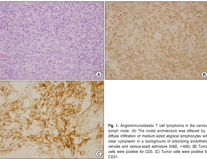
조직검사소견: 13개월 전 시행한 좌측 경부 림프절 생검에서는 림프절의 구조가 소실되었고 다수의 내피 세정맥들과 중간 크기의 종양세포들 사이에 정상 림프 구들이 섞여 있는 것이 관찰되었다(Fig. 1A). 면역조직 화학염색 결과는 CD3 (Fig. 1B), CD10, CD21 (Fig. 1C) 에서는 양성이었고 CD20은 음성을 보여 AITL을 진단
Fig. 1. Angioimmunoblastic T cell lymphoma in the cervical lymph node. (A) The nodal architecture was effaced by a diffuse infiltration of medium-sized atypical lymphocytes with clear cytoplasm in a background of arborizing endothelial venules and various-sized admixture (H&E, ×400). (B) Tumor cells were positive for CD3. (C) Tumor cells were positive for CD21.
하였다. 당시 EBV 제자리부합법(EBV In situ hybrid- ization)은 음성이었다. 최근 발생한 우측 편도 주위 종 창부에서 시행한 생검에서 중간 혹은 큰 크기의 림프 구들이 미만성 침윤을 보이고(Fig. 2A) 면역조직화학 염색에서는 CD20양성(Fig. 2B), CD3, CD4, CD8은 음 성을 나타내어 DLBCL을 진단하였다. 당시 EBV 제자 리부합법(EBV In situ hybridization)은 양성을 보였다 (Fig. 2C).
경과: AITL에 속발한 DLBCL 진단 후 R-DHAP (ri- tuximab, cisplatin, cytarabine, dexamethasone) 복합항 암화학요법을 시행하였고 2주기 후 추적 검사에서 완 전관해에 이르렀다. 이후 불량한 전신상태로 2주기만 더 시행하고 경과를 관찰하였다. 완전관해 도달 4개월 째 구강 내 통증을 동반한 연구개 종괴가 발현하였고 조직검사에서 CD3, CD10, CD21은 양성, CD20은 음 성, EBV는 음성으로 AITL의 재발이 확인되었다. 18F- FDG PET/CT에서 우측 연구개, 구인두와 좌측경부 림 프절, 액와림프절의 침범소견을 보였고 재발한 림프종
의 골수 침범은 없었다. 이후 4주기의 IMEP (ifosfa- mide, methotrexate, leucovorin, etoposide) 복합항암화 학요법을 시행하여 부분반응을 보였으나 전신상태 불 량으로 항암치료를 중단하였고 현재 2개월째 대증치 료 중이다.
고 찰
AITL는 비호지킨림프종 중 10% 미만에서 발생하는 말초T세포림프종(peripheral T-cell lymphoma)의 아형 으로 주로 나이든 성인에서 발병하며, 림프절병증, 전 신염증증상, 간비장비대, 피부발진, 면역학적 장애를 특징으로 한다. 예후는 일반적으로 나쁘며 평균 생존 기간은 3년 이내로 심각한 면역학적 결핍과 기회감염 의 증가를 보인다.1,2) 조직학적으로 종양침범부는 악성 전환을 보이는 T림프구, 반응 T림프구, 반응 B림프구, 형질세포, 조직구, 호염기구 등의 침윤을 보이고, 불규 칙한 여포상가지돌기세포 그물(follicular dendritic cell Fig. 2. The tonsilar biopsy showed a diffuse large B cell lymphoma that developed 13 months after the diagnosis of angioimmunoblastic T cell lymphoma. (A) Diffuse infiltration of large-sized lymphoid cells were shown (H&E, ×400). (B) Tumor cells were diffusely positive for CD20. (C) In situ hybridization of EBV showed the positivity of a few large lymphocytes.
meshworks)이 증가되고, 내피세정맥들이 나뭇가지 형 태로 나타나는 등의 다양성을 지닌다.1,2)
AITL와 병발하거나 혹은 속발하는 B세포림프종들 이 국외에서 종종 보고되었다. 1993년에 Abruzzo 등6) 은 AITL 진단 2년 후 부검에서 EBV연관 B세포림프종 을 확인하였고 Knecht 등7)은 AITL에서 속발된 EBV연 관 DLBCL을 2예 보고하고 이후 Matsue 등8)이 2예를 더 보고하였다.
Zettl 등9)은 AITL을 포함한 말초T세포림프종 17증 례를 조사하여 EBV관련 B세포 증식과 B세포림프종 발생을 보고하였는데 이 중 10예에서 AITL 침윤 내에 EBV에 감염된 비종양성 B림프구들을 확인하였고 이 후 속발된 DLBCL이 발생하고 EBV감염이 확인된 경 우는 2예로 EBV에 감염된 큰B세포가 DLCBL의 전구 병변임을 제시하였다. Xu 등10)도 AITL병변 속에 EBV 양성인 DLBCL 침윤이 있음을 보고하였다. 보고된 증 례들은 보통 AITL 진단 후 2개월에서 10년 이내 속발 한 B세포림프종이 발생하였고 대부분 EBV에 양성을
보였다.6-11)
최근 Attygalle 등12)은 31예의 AITL 환자에서 연속 적으로 조직검사를 시행하여 AITL의 경과를 살펴보았 는데 23%에서 EBV관련 B세포 림프종(5예는 DLBCL, 2예는 호지킨림프종)이 발생함을 확인하였다. 이들 중 1예에서는 AITL 치료 후 완전 관해를 보였으나 8개월 째 AITL이 재발과 동시에 EBV양성 DLBCL이 발생하 였고 다른 1예에서는 본 증례와 유사하게 AITL 진단 8개월째 EBV양성 DLBCL이 나타났고 항암요법 후 완 전관해에 이르렀다가 7개월 후 AITL이 재발하였다. 따 라서 AITL의 경과 중에 B세포림프종들이 발생할 수 있으며 EBV가 B세포의 비정상적인 증식에 관여할 것 으로 여겨진다.6-12)
본 증례의 환자는 초기 AITL 진단 시 EBV감염 증거 가 없었으나 항암화학치료 후 진단 13개월째 EBV 양 성인 DLBCL이 발생하였다. 이전 연구들이 설명한 바
같이,9-12) EBV관련 B세포림프종의 발생이 AITL에서
나타나는 면역결핍과 항암제치료로 인한 면역억제상 태로 인해 EBV가 감염되거나 혹은 재활성화되고 이러 한 EBV가 감염된 B세포들이 증식되면서 클론진화되 어 공격적인 B세포림프종이 발생했을 것으로 생각된 다.
근래에는 혈액학적 종양연구와 진단에 DNA에 기초 한 방법들이 이용되고 있다. T세포 수용체(T-cell re- ceptor, TCR)와 면역글로불린중쇄(immunoglobulin heavy chain, IHC)에 대한 중합효소연쇄반응(polymerase chain
reaction, PCR)을 통해 AITL의 같은 조직 표본에서 B 세포와 T세포 클론들이 둘 다 존재함을 확인하였다.
이는 한 클론에서 TCR유전자와 면역글로불린 유전자 재조합의 결과로 설명할 수 있고 미성숙 종양에서 더 흔히 관찰되었다.13) Tan 등5)의 연구에서 AITL에서 TCRγ와 IHC에 대한 PCR을 통해 B세포 클론이 34%
에서 나타났고 EBV와 관련이 높음을 보여 주었다. 또 한 최근 AITL의 종양 T세포에서 나타나는 CXCL13, CXCR5이 B세포 귀소(homing)와 여포상가지돌기세포 들의 증식과 유도에 관여한다는 게 밝혀졌다.14,15) 이러 한 환경 또한 B세포 증식과 종양성 변화에 관여하리라 여겨진다.
아직 명확한 기전들이 밝혀지지 않았지만 결국 AITL에서 B세포림프종의 발생은 별개의 질환이 아닌 AITL의 진행과정 중의 하나로 이해할 수 있고, 이러한 관점에서 AITL의 치료와 AITL에서 병발 혹은 속발된 B세포림프종 치료에 대한 연구가 진행된다면 더 좋은 결과를 기대할 수 있을 것으로 여겨진다.
요 약
AITL은 중증의 면역기능장애와 면역결핍을 지닌 림 프세포증식질환이다. 다른 면역결핍질환들과 유사하 게, AITL의 경우에도 EBV와 관련된 B세포 림프증식 이 발생할 수 있다. 그러나, AITL에 속발하거나 병발 한 B세포 림프종에 대한 일부 증례와 연구들만 보고되 었다. 저자들은 AITL 진단 13개월 후 DLBCL이 속발 한 증례를 경험하였다. 명확한 기전은 알려지진 않았 지만 EBV가 속발한 DLBCL의 발생에 관여했을 것으 로 여겨진다.
참 고 문 헌
1) Dogan A, Attygalle AD, Kyriakou C. Angioimmuno- blastic T-cell lymphoma. Br J Haematol 2003;121:
681-91.
2) Khan G, Norton AJ, Slavin G. Epstein-Barr virus in angioimmunoblastic T-cell lymphomas. Histopatho- logy 1993;22:145-9.
3) Weiss LM, Jaffe ES, Liu XF, Chen YY, Shibata D, Medeiros LJ. Detection and localization of Epstein- Barr viral genomes in angioimmunoblastic lympha- denopathy and angioimmunoblastic lymphadenop- athy-like lymphoma. Blood 1992;79:1789-95.
4) Ohshima K, Takeo H, Kikuchi M, et al. Heteroge- neity of Epstein-Barr virus infection in angioimmu-
noblastic lymphadenopathy type T-cell lymphoma.
Histopathology 1994;25:569-79.
5) Tan BT, Warnke RA, Arber DA. The frequency of B- and T-cell gene rearrangements and Epstein-Barr virus in T-cell lymphomas: a comparison between angioimmunoblastic T-cell lymphoma and periph- eral T-cell lymphoma, unspecified with and without associated B-cell proliferations. J Mol Diagn 2006;8:
466-75.
6) Abruzzo LV, Schmidt K, Weiss LM, et al. B-cell lym- phoma after angioimmunoblastic lymphadenopathy:
a case with oligoclonal gene rearrangements asso- ciated with Epstein-Barr virus. Blood 1993;82:241-6.
7) Knecht H, Martius F, Bachmann E, et al. A deletion mutant of the LMP1 oncogene of Epstein-Barr virus is associated with evolution of angioimmunoblastic lymphadenopathy into B immunoblastic lymphoma.
Leukemia 1995;9:458-65.
8) Matsue K, Itoh M, Tsukuda K, Kokubo T, Hirose Y. Development of Epstein-Barr virus-associated B cell lymphoma after intensive treatment of patients with angioimmunoblastic lymphadenopathy with dy- sproteinemia. Int J Hematol 1998;67:319-29.
9) Zettl A, Lee SS, Rüdiger T, et al. Epstein-Barr vi- rus-associated B-cell lymphoproliferative disorders in angioimmunoblastic T-cell lymphoma and periph- eral T-cell lymphoma, unspecified. Am J Clin Pathol 2002;117:368-79.
10) Xu Y, McKenna RW, Hoang MP, Collins RH, Kroft SH. Composite angioimmunoblastic T-cell lympho- ma and diffuse large B-cell lymphoma: a case report and review of the literature. Am J Clin Pathol 2002;
118:848-54.
11) Hawley RC, Cankovic M, Zarbo RJ. Angioimmuno- blastic T-cell lymphoma with supervening Epstein- Barr virus-associated large B-cell lymphoma. Arch Pathol Lab Med 2006;130:1707-11.
12) Attygalle AD, Kyriakou C, Dupuis J, et al. Histologic evolution of angioimmunoblastic T-cell lymphoma in consecutive biopsies: clinical correlation and insights into natural history and disease progression. Am J Surg Pathol 2007;31:1077-88.
13) van Dongen JJ, Wolvers-Tettero IL. Analysis of im- munoglobulin and T cell receptor genes. part II:
Possibilities and limitations in the diagnosis and management of lymphoproliferative diseases and re- lated disorders. Clin Chim Acta 1991;198:93-174.
14) Krenacs L, Schaerli P, Kis G, Bagdi E. Phenotype of neoplastic cells in angioimmunoblastic T-cell lym- phoma is consistent with activated follicular B helper T cells. Blood 2006;108:1110-1.
15) Grogg KL, Attygalle AD, Macon WR, Remstein ED, Kurtin PJ, Dogan A. Angioimmunoblastic T-cell lym- phoma: a neoplasm of germinal-center T-helper cells? Blood 2005;106:1501-2.