This is an Open Access article distributed under the terms of the Creative Commons Attribution Non-Commercial License (http://creativecommons.org/licenses/by-nc/3.0) which permits unrestricted non-commercial use, distribution, and reproduction in any medium, provided the original work is properly cited.
BLOOD RESEARCH
VOLUME 49ㆍNUMBER 2 June 2014CASE REPORT
A case of chronic myeloid leukemia with features of essential thrombocythemia in peripheral blood and bone marrow
Young Jae Byun, Byeong-Bae Park, Eun Sung Lee, Kyung Soo Choi, Dae Sung Lee
Department of Internal Medicine, Hanyang University College of Medicine, Seoul, Korea
p-ISSN 2287-979X / e-ISSN 2288-0011 http://dx.doi.org/10.5045/br.2014.49.2.127 Blood Res 2014;49:127-9.
Received on July 27, 2012 Revised on September 19, 2012 Accepted on May 8, 2014
Abstract
Chronic myeloid leukemia (CML) is a myeloproliferative neoplasm characterized by over- production of myeloid white blood cells. Philadelphia chromosome is an essential finding for CML diagnosis. Generally, a clinical diagnosis of essential thrombocythemia (ET) can be established from isolated marked thrombocytosis in peripheral blood. However, Philadelphia chromosome-positivity or bcr/abl rearrangement with isolated thrombocy- tosis should be diagnosed as CML, not ET, according to World Health Organization diag- nostic criteria. Therefore, CML should not be excluded before confirming the presence of the Philadelphia chromosome or bcr/abl rearrangement in cases of isolated thrombocy- tosis in peripheral blood. We report a case of CML with clinical features of ET in a patient successfully treated with imatinib.
Key Words Chronic myeloid leukemia, Philadelphia chromosome, Essential thrombocythemia
Correspondence to Byeong-Bae Park, M.D.
Department of Internal Medicine, Hanyang University College of Medicine, 222, Wangsimni-ro, Seongdong-gu, Seoul 133-791, Korea
Tel: +82-2-2290-8335 Fax: +82-2-2298-9183 E-mail: [email protected]
Ⓒ 2014 Korean Society of Hematology
INTRODUCTION
Chronic myeloid leukemia (CML) is a myeloproliferative neoplasm with clonal hyperproliferation of myeloid cells in the bone marrow. Clinical findings in chronic-phase CML include fatigue, decreased appetite, and splenomegaly, and hematological findings include leukocytosis, thrombocytosis, neutrophilia, and decreased leukocyte alkaline phosphatase (LAP) scores [1].
Isolated thrombocytosis is assumed to be essential throm- bocythemia (ET) rather than chronic-phase CML in most cases. According to the 2008 World Health Organization diagnostic criteria, a diagnosis of ET is routinely made, if platelet counts exceed 450×109/L with proliferation of mega- karyocytes in the bone marrow and JAK2 V617F mutation, excluding any evidence of other myeloproliferative neo- plasms [2]. Therefore, cases of isolated thrombocytosis with Philadelphia chromosome or bcr/abl rearrangement, which are diagnostic markers for CML, should not be diagnosed as ET. In previous studies, however, patients have been diag- nosed with Philadelphia chromosome-positive ET [3, 4].
Other contradictory results described them as variants of
CML [5, 6].
We experienced a case of isolated thrombocytosis, initially suggestive of ET, positive for the Philadelphia chromosome and bcr/abl rearrangement. The patient was eventually diag- nosed with chronic-phase CML, and was successfully treated with imatinib. Here, we report our case and include a liter- ature review.
CASE REPORT
A 21-year-old woman presented to the outpatient clinic with lower abdominal pain. Seven days before presentation, she had a ruptured corpus luteal cyst, which was detected on abdominal computed tomography (CT) at another clinic.
Her initial platelet count was estimated to be 3,777×109/L at our clinic. Because thrombocytosis appeared to be secon- dary to bleeding, the patient’s blood cell counts were only monitored during her clinical course. However, we decided to perform further evaluation because of thrombocytosis per- sisted for 2 weeks with no decrease in the platelet count.
Her medical history was unremarkable, and she had no family history of hematologic disease or genetic disorders. Her vital
Blood Res 2014;49:127-9. bloodresearch.or.kr
128 Young Jae Byun, et al.
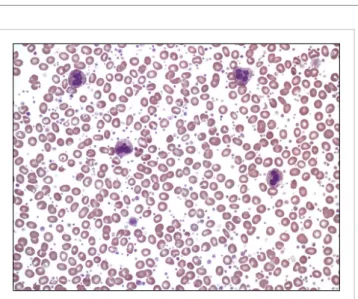
Fig. 1. Thrombocytosis without obvious morphologic abnormalities of the white blood cells and erythrocytes in peripheral blood smear (Wright Giemsa stain, ×400).
Fig. 2. Bone marrow core biopsy sample showing hypercellular bone marrow for age with expanded myelopoiesis and small megakaryo- cytes with decreased nuclear lobation (H&E stain, ×200).
signs were normal at admission. Except for mild lower ab- dominal tenderness, the patient had no other positive find- ings on physical examination. Complete blood count (CBC) revealed a hemoglobin level of 10.1 g/dL, hematocrit level of 30.7%, white blood cell (WBC) count of 10×109/μL (differential count: neutrophils 63%, lymphocytes 33%, eosi- nophils 1%, basophils 3%, and monocytes 0%), and platelet count of 3,294×109/L. A serum biochemistry panel showed the following: total protein, 7.2 g/dL; albumin, 3.9 g/dL;
total bilirubin, 1.2 mg/dL; aspartate aminotransferase, 11 IU/L; alanine aminotransferase, 13 IU/L; blood urea nitrogen, 6 mg/dL; creatinine, 0.6 mg/dL; lactic dehydrogenase, 410 IU/L; and C-reactive protein, 2.0 mg/dL. A peripheral blood smear showed thrombocytosis. In addition, serum iron level was 73 μg/dL, total iron binding capacity was 267 μg/dL, and ferritin level was 206.5 ng/mL. The patient had an LAP score of 127 points, which was within the normal range.
Abdominal and pelvic CT showed a small amount of hemo- peritoneum resulting from the previous ruptured ovarian cyst. Bone marrow aspiration and biopsy revealed a high number of megakaryocytes, but no cells undergoing malig- nant transformation (Fig. 1, Fig. 2). A cytogenetic abnormal- ity was detected with the karyotype 46,XX,t(9;22)(q34;q11.2) on bone marrow. We also observed a bcr/abl rearrangement in the bone marrow using reverse transcriptase PCR, which also showed amplified products from the b3a2 mRNA dele- tion in the major bcr gene. Results were negative for the JAK2 V617F mutation. Because the patient had isolated thrombocytosis (3,294×109/L), she was tentatively diagnosed with ET before the results of the cytogenetic and molecular studies were available, even if results for the JAK2 V617F mutation were unknown. Hydroxyurea was administered to the patient at a dose of 2,000 mg/day for 14 days to lower her platelet count. A follow-up CBC showed persistent thrombocytosis, platelet counts of 2,206×109/L, and leukocy- topenia (1.1×10⁹/L). We stopped hydroxyurea and identified the Philadelphia chromosome and bcr/abl rearrangement,
but no JAK2 V617F mutation. This led to the final diagnosis of chronic-phase CML, for which the patient received imatinib. In the 6 days following the treatment with imatinib, the patient’s platelet count normalized to 438×109/L. She is currently followed up to confirm complete molecular re- sponse against bcr/abl rearrangement. In the 3 months after treatment with imatinib, a major molecular response (3-log reduction of transcript levels) was observed.
DISCUSSION
Recently, a therapeutic advance in the treatment of CML was achieved with the advent of tyrosine kinase inhibitors for the bcr/abl protein. The long-term survival rate of patients with CML has improved to 89% [7]. To determine a distinc- tive diagnosis, we should distinguish CML from ET, which might have similar laboratory findings at initial diagnosis because of the different therapeutic approaches and clinical outcomes between the two diseases.
If severe thrombocytosis and hyperproliferation of mega- karyocytes in the bone marrow occurs in the absence of splenomegaly, it is difficult to make a differential diagnosis between CML and ET without cytogenetic and molecular studies. Although the JAK2 V617F mutation is observed in approximately 50% of ET patients, JAK2 V617F mutation does not indicate a definitive diagnostic of ET, because ET should be confirmed with an exclusive diagnostic process.
Therefore, the Philadelphia chromosome or bcr/abl re- arrangement is a significant diagnostic tool for distinguishing CML from ET under these hematologically abnormal conditions.
A previous study showed mild basophilia and a Philadel- phia chromosome in 1 of 121 patients with ET [8]. This patient had a Philadelphia chromosome and isolated throm- bocytosis that progressed to the accelerated phase, which is similar to the clinical course observed in a patient with
bloodresearch.or.kr Blood Res 2014;49:127-9.
Chronic myeloid leukemia with features of essential thrombocythemia 129
CML. Patients with a Philadelphia chromosome and isolated thrombocytosis considered a variant of CML or an initial presentation of CML in the chronic phase were refractory to hydroxyurea, but showed improvement in symptoms and laboratory values after administration of imatinib [6].
Imatinib mesylate (Gleevec) is a bcr-abl tyrosine kinase inhibitor and the primary treatment agent for patients with CML. By binding to bcr-abl protein tyrosine kinase and inhibiting the bcr/abl pathway, imatinib reduces the pro- liferation of bcr/abl-positive CML cells [9]. In patients ini- tially diagnosed with chronic-phase CML, the rate of com- plete cytogenetic remission has increased to 76.2% in the imatinib era [10]. In addition, the overall survival rate sig- nificantly improved after treatment with imatinib in ear- ly-phase CML [7]. Therefore, imatinib is currently approved as a standard initial treatment for patients with chronic-phase CML.
In conclusion, our case indicates that CML could not be completely excluded until the presence of the Philadelphia chromosome or bcr/abl rearrangement was confirmed, which would be clinically sufficient to diagnose CML.
AuthorsÊ Disclosures of Potential Conflicts of Interest
No potential conflicts of interest relevant to this article were reported.
REFERENCES
1. Sawyers CL. Chronic myeloid leukemia. N Engl J Med 1999;340:
1330-40.
2. Tefferi A, Thiele J, Orazi A, et al. Proposals and rationale for revision of the World Health Organization diagnostic criteria for polycythemia vera, essential thrombocythemia, and primary myelofibrosis: recommendations from an ad hoc international expert panel. Blood 2007;110:1092-7.
3. Kwong YL, Chiu EK, Liang RH, Chan V, Chan TK. Essential thrombocythemia with BCR/ABL rearrangement. Cancer Genet Cytogenet 1996;89:74-6.
4. Stoll DB, Peterson P, Exten R, et al. Clinical presentation and natural history of patients with essential thrombocythemia and the Philadelphia chromosome. Am J Hematol 1988;27:77-83.
5. Emilia G, Marasca R, Zucchini P, et al. BCR-ABL rearrangement is not detectable in essential thrombocythemia. Blood 2001;97:
2187-9.
6. Michiels JJ, Berneman Z, Schroyens W, et al. Philadelphia (Ph) chromosome-positive thrombocythemia without features of chronic myeloid leukemia in peripheral blood: natural history and diagnostic differentiation from Ph-negative essential thrombo- cythemia. Ann Hematol 2004;83:504-12.
7. Druker BJ, Guilhot F, O'Brien SG, et al. Five-year follow-up of patients receiving imatinib for chronic myeloid leukemia. N Engl J Med 2006;355:2408-17.
8. Damaj G, Delabesse E, Le Bihan C, et al. Typical essential thrombocythaemia does not express bcr-abelson fusion trans- cript. Br J Haematol 2002;116:812-6.
9. Savage DG, Antman KH. Imatinib mesylate-a new oral targeted therapy. N Engl J Med 2002;346:683-93.
10. O'Brien SG, Guilhot F, Larson RA, et al. Imatinib compared with interferon and low-dose cytarabine for newly diagnosed chronic- phase chronic myeloid leukemia. N Engl J Med 2003;348:994- 1004.