Current Practices in Breast Magnetic Resonance Imaging: a Survey Involving the Korean Society of Breast Imaging
This is an Open Access article distributed under the terms of the Creative Commons Attribution Non-Commercial License (http://creativecommons.org/licenses/
by-nc/3.0/) which permits unrestricted non-commercial use, distribution, and reproduction in any medium, provided the original work is properly cited.
Received: August 7, 2017 Accepted: September 11, 2017 This study was supported by a fund for breast magnetic resonance imaging (MRI) study group from the Korean Society of Magnetic Resonance in Medicine.
This research was supported by Basic Science Research Program through the National Research Foundation of Korea(NRF) funded by the Ministry of Education(No. 2017R1D1A1B03033975) Correspondence to:
Sun Mi Kim, M.D.
Department of Radiology, Seoul National University Bundang Hospital, 82, Gumi-ro 173 Beon- gil, Bundang-gu, Seongnam-si, Gyeonggi-do 13620, Korea.
Tel. +82-31-787-7617 Fax. +82-31-787-4070 E-mail: [email protected]
Copyright © 2017 Korean Society of Magnetic Resonance in Medicine (KSMRM)
Original Article
Purpose: To report on the current practices in breast magnetic resonance imaging (MRI) in Korea.
Materials and Methods: We invited the 68 members of the Korean Society of Breast Imaging who were working in hospitals with available breast MRI to participate in a survey on how they performed and interpreted breast MRI. We asked one member from each hospital to respond to the survey. A total of 22 surveys from 22 hospitals were analyzed.
Results: Out of 22 hospitals, 13 (59.1%) performed at least 300 breast MRI examinations per year, and 5 out of 22 (22.7%) performed > 1200 per year. Out of 31 machines, 14 (45.2%) machines were 1.5-T scanners and 17 (54.8%) were 3.0-T scanners. All hospitals did contrast-enhanced breast MRI. Full-time breast radiologists supervised the performance and interpreted breast MRI in 19 of 22 (86.4%) of hospitals. All hospitals used BI-RADS for MRI interpretation. For computer-aided detection (CAD), 13 (59.1%) hospitals sometimes or always use it and 9 (40.9%) hospitals did not use CAD. Two (9.1%) and twelve (54.5%) hospitals never and rarely interpreted breast MRI without correlating the mammography or ultrasound, respectively. The majority of respondents rarely (13/21, 61.9%) or never (5/21, 23.8%) interpreted breast MRI performed at an outside facility. Of the hospitals performing contrast-enhanced examinations, 15 of 22 (68.2%) did not perform MRI- guided interventional procedures.
Conclusion: Breast MRI is extensively performed in Korea. The indication and practical patterns are diverse. The information from this survey would provide the basis for the development of Korean breast MRI practice guidelines.
Keywords: Breast neoplasms; Diagnosis; Magnetic resonance imaging; Surveillance;
South Korea
Bo La Yun1, Sun Mi Kim1, Mijung Jang1, Bong Joo Kang2, Nariya Cho3, Sung Hun Kim2, Hye Ryoung Koo4, Eun Young Chae5, Eun Sook Ko6, Boo-Kyung Han6
1Department of Radiology, Seoul National University Bundang Hospital, Seongnam-si, Korea
2Department of Radiology, Seoul St. Mary's Hospital, College of Medicine, The Catholic University of Korea, Seoul, Korea
3Department of Radiology, Seoul National University College of Medicine, Seoul, Korea
4Department of Radiology, Hanyang University Hospital, Seoul, Korea
5Department of Radiology and Research Institute of Radiology, Asan Medical Center, University of Ulsan, College of Medicine, Seoul, Korea
6Department of Radiology, Samsung Medical Center, Sungkyunkwan University School of Medicine, Seoul, Korea
INTRODUCTION
According to statistics from the Ministry of Health and Welfare, breast cancer is the second most common cancer among Korean women following thyroid cancer.
The incidence of breast cancer increased steadily, reaching 5,744 per 100,000 in 1999, but tripled to 18,381 in 2014. In addition, the 5-year relative survival rate of breast cancer patients in the last 5 years (2010-2014) is 92%, showing a high survival rate along with thyroid cancer and prostate cancer (1). Breast magnetic resonance imaging (MRI) combined with mammography has been reported to be 90%
to 100% sensitive in detecting breast cancer. Its usefulness has been reported not only in high-risk screening but also in pre-operative examination, evaluation of neoadjuvant chemotherapy response, and post-operative screening (2- 5). The use of breast MRI has been rapidly growing in Korea for the last 10 years. However, there are several problems associated with breast MRI. There are recommendations for breast MRI from the American Cancer Society (ACS) (6), the medical environment is different in Korea. Thus, it is necessary to know the types of indications that are actually used. Since there is no standard imaging protocol, hospitals use different planes and sequences, making it difficult to read images taken by other institutions. Furthermore, unlike the United States, which has a breast MRI accreditation program, there is no regulation in Korea, and image quality is not well managed. Regarding the standardization of imaging interpretation, it seems that the Breast Imaging Reporting and Data System (BI-RADS) (7) from the American College of Radiology (ACR) is often used for interpretation; it is not clear how many hospitals actually use it. Some hospitals that perform breast MRI do not perform the MRI-guided biopsy for lesions that are visible only on MRI. The purpose of our study was to report on the current practices in breast MRI in Korea. The questionnaire included the characteristics of the hospitals in which the breast MRI was performed, type of practices, indications, techniques and protocols used, type of interpretation, clinician’s understanding of breast MRI, and availability of breast MRI intervention. The information from this survey would provide the basis for the development of Korean breast MRI practice guidelines.
MATERIALS AND METHODS
From October 2015 to January 2016, we invited the 68
members of the Korean Society of Breast Imaging (KSBI) working in breast MR-available hospitals to participate in a survey regarding how they performed and interpreted breast MR. We asked one member from each hospital to respond to the survey. The survey was conducted by email.
Two follow-up emails were sent to non-respondents. A total of 22 surveys from 22 hospitals were analyzed. The respondents returned the survey within 3 months from the first contact. The questionnaire used in this study was developed with reference to the questionnaire developed in the United States (8) by collaboration of breast radiologists who were familiar with the recent trends in breast MRI.
This survey consisted of 28 questions divided into 5 categories (Appendix). It covered baseline characteristics of respondents, breast MRI indication, protocol and technique, interpretation, and breast MRI-guided intervention. The results of the survey were evaluated by all of the authors.
RESULTS
Baseline Characteristics of Respondents
Most respondents were below 40 years of age (95.5%), and all were female. The type of hospital in this survey included 21 university hospitals and 1 secondary hospital.
Thirteen hospitals (59%) performed more than 300 breast MRI exams per year, of which 5 hospitals (23%) performed more than 1200 exams a year. Table 1 shows the detailed baseline characteristics of the respondents.
Breast MR Indications
For the indications for diagnostic breast MRI, the results are shown in Figure 1. All respondents perform diagnostic MRI in their hospitals. The most common indications were to follow up for response to chemotherapy (21, 95.5%) and pre-operative evaluation (21, 95.5%). In 14 hospitals, pre-operative MRI is performed on breast cancer patients regardless of operation method. Four hospitals perform MRI only for breast cancer patients who are scheduled for breast conservation surgery. In contrast, 2 hospitals perform MRI only for patients who undergo mastectomy. One hospital performs pre-operative MRI for patients who have dense breasts.
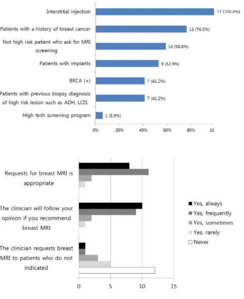
Out of 22 hospitals, 17 perform screening breast MRI.
Figure 2 shows the responses from these 17 hospitals. The most common indication for screening was patients with interstitial injection, followed by patients with a history of breast cancer, patients who ask for MRI screening, and
patients with implants. The proportion of MRI screening for patients with BRCA mutation and those with a history of high-risk lesions are lower compared to that in the United States. Figure 3 shows the responses about clinicians’
MRI prescriptions. Respondents said that most clinicians prescribed breast MRI appropriately.
Breast MRI Protocols and Technique
In 22 hospitals, 31 MRIs were used for breast examinations;
17 machines were 3-T scanners, and the remaining 14 machines were 1.5-T scanners. Contrast-enhanced breast MRI is available in all of the hospitals. Non-contrast breast MRI is not performed in 3 hospitals. Gadovist (Bayer Schering Pharma AG, Berlin, Germany), Dotarem (Guerbet, Paris, France), Prohance (Bracco, Milan, Italy), Uniray (Dongkook Pharm, Seoul, Korea), and Multihance (Bracco, Milan, Italy) were used for contrast-enhanced breast MRI. Machine injection is preferred over manual injection. MRI examinations in all except one hospital do not take menstrual cycles into account, even if they are for screening. Five hospitals record menstrual cycles for MRI interpretation. For breast MRI dynamic protocols, all respondents performed bilateral breast MRI. Fifteen (68.2%) hospitals performed dynamic scan in the axial plane, five (22.7%) in the sagittal plane, and two (9.1%) in the mixed plane. For the dynamic series, 18 hospitals preferred using fat saturation and 1 preferred non-fat saturation, while the other 3 used both of them. All hospitals performed T2 weighted image (T2WI), 14 (64.6%) with fat saturation and 8 (36.4%) without fat saturation. Half of the hospitals used pre-contrast non-fat saturated images to evaluate fat-containing lesions and subcutaneous fat. T1 weighted (T1WI) (n = 7, 40.9%) and T2WI (n = 6, 36.4%) images were used for this non-fat saturated sequence. Two hospitals did non-fat saturated for both T1WI and T2WI images. Out of 22 hospitals, 19 performed reformatted orthogonal plane images with dynamic series. Maximum intensity projection was the most commonly used reconstruction. In addition, diffusion weighted images were performed in 18 hospitals and MR spectroscopy was performed in 1 hospital.
Table I. Characteristics of Korean Breast MR Research Society survey responders and hospitals
Characteristics Number (%)
Gender
Male 0 (0)
Female 22 (100)
Age (years)
30-39 1 (4.5)
40-49 12 (54.5)
>50 9 (40.9)
Practice type
University hospital 21 (95.5)
General hospital (secondary hospital) 1 (4.5) Private clinic
Breast MR exams per year
>1200 5 (22.7)
900-1200 1 (4.5)
600-900 4 (18.2)
300-600 4 (18.2)
100-300 8 (36.4)
<100 2 (9.1)
Fig. 1. Graph shows responses to the question “What is the general indication for a diagnostic breast MRI in your institution?” (Check all that apply) (n = 83).
Breast MRI Interpretation
Full-time breast radiologists supervised the performance and interpretation of breast MRI in 19 of 22 (86.4%) hospitals. All hospitals used BI-RADS for MRI interpretation.
Regarding the use of computer-aided diagnosis (CAD),
“sometimes or always” was the answer in 13 (59.1%) hospitals and “no” was the answer in 9 (40.9%) hospitals.
Out of 22, 14 practices (63.6%) “never or rarely” interpreted breast MRI without correlating mammography or ultrasound and the other 8 practices “sometimes or frequently” did. The majority of respondents (16/21, 76.2%) interpreted breast MRI performed at an outside facility, but there were several hospitals that never interpreted MRI (5/21, 23.8%) that was performed in another hospital. Out of 22, 17 (77.3%) hospitals did not audit a breast MR practice.
MRI Guided Interventions
Out of 21 hospitals, 19 performed “second-look”
ultrasound for suspicious breast MRI findings and 2 “rarely”
did. Of the hospitals that perform breast MRI examinations, 15 of 22 (68.2%) did not perform MRI-guided interventional procedures. Six hospitals performed MRI-guided biopsy based only on their images and one hospital did biopsy not only based on their images but also based on images taken in other institutions. From 12G to 8G vacuum- assisted devices used for MRI guided biopsy and none of the respondents used core gun for MRI-guided biopsy.
DISCUSSION
According to the statistical data provided by the Health Insurance Review and Assessment Service in Korea, breast MRI increased more than threefold from 13,692 cases in 2010 to 40,286 cases in 2016 (9). Although breast MRI examinations performed at primary hospitals or private Fig. 3. Graph shows responses to the question “Your clinician's response to the MRI prescription”.
Fig. 2. Graph shows responses to the question “What is the general indication for a screening breast MRI in your institution?” (Check all that apply) (n = 64).
17 (100.0%)
clinics continue to increase, the majority (97.9%) of the examinations were performed in tertiary hospitals or secondary hospitals until 2016. There is a possibility that the use of breast MRI will continue to increase due to an increase in breast MRI-available centers, an increase in the prevalence of breast cancer, and the attention of people and media.
From this survey, 3 common indications for diagnostic MRI are evaluation of chemotherapy response, pre- operative evaluation, and workup for cancer of unknown primary origin. These tests are for patients with confirmed cancer. Inconclusive findings in conventional imaging, which is one of the indications for diagnostic MRI in the United States and Europe, were only performed in some hospitals in Korea. This is probably because the threshold for the expense of an MRI examination is relatively low in breast cancer patients because Korean health insurance supports the cost of breast MRI by the special law, Cancer Patient Registry. Three major conditions for the use of breast MRI screening are interstitial injection, personal history of breast cancer, and patients who ask for breast MR screening. According to the ACS, breast MRI screening is recommended for women who have an approximately 20- 25% or greater lifetime risk, BRCA mutation, and untested first-degree relative of BRCA carrier based on evidence.
Furthermore, the ACS panel recommended against MRI screening for patients with a personal history of breast cancer who have an estimated absolute lifetime risk of 10%
(6). In Korea, however, screening breast MR is more active in cancer patients because of insurance coverage for the 5 years after diagnosis. High-risk women, such as women with BRCA mutations, who need MRI screening, are not supported by insurance. It is necessary to expand insurance coverage for high-risk groups.
In this survey, 3-T scanners are more frequently used for breast MRI than 1.5-T scanner because almost all respondents worked in a tertiary hospital. Bilateral axial plane where it is easy to compare background parenchymal enhancement, is preferred for dynamic image. For contrast- enhanced breast MRI, dynamic image and T2WI are included in all hospitals. However, there are various preferences for acquisition direction or fat saturation images in each hospital. In addition, there are various additional images for each hospital.
Breast MRI results are likely to be interpreted by specialized personnel, and about half of which use CAD.
More than one-third of hospitals sometimes or frequently interpret breast MRI without correlating conventional
images. Because there is no standardized protocol, breast MRI taken at another institution is difficult to read, but most of the hospitals read outside exams for the clinicians’ or patients’
needs. To reduce unnecessary repetitive examinations, outside hospital MRI interpretation is essential but a standardized protocol would reduce the error of reading from the unfamiliar MR protocols.
In this questionnaire, 68.2% of the respondents said that they did not perform MRI-guided biopsy even though they were referral hospitals. MRI is a very sensitive test and because of the high rate of cancers that are not detected in other modalities such as ultrasound or mammography (10- 12), it is not enough to perform a second look ultrasound or mammography correlation. MRI-guided biopsy is necessary. If MRI-guided biopsy cannot be performed in the hospital, there should be a system that can create a referral arrangement with a cooperating facility. In Korea, this system is insufficient. The ACR guideline also states that “facilities performing breast MRI should have the capacity to perform mammographic correlation, directed breast ultrasound, and MRI-guided intervention, or create a referral arrangement with a cooperating facility that could provide these services” (13). Additionally, only 1 of the 7 hospitals providing MRI-guided biopsy performs a biopsy on the basis of an outside image, and the remaining 6 hospitals perform a biopsy after re-imaging. Repeated exams waste medical resources and increase costs, so image standardization is required.
This study has several limitations. First, only 36.8% of those surveyed responded. There is a possibility of selection bias because this study included only those who voluntarily answered. In addition, all respondents worked in secondary or tertiary hospitals and did not reflect the situation in private clinics. Second, the survey was also made only for KSBI members. The practices of other doctors including breast surgeons who have big private breast clinics were not reflected, and hence it is difficult to represent current breast imaging practices.
Despite these limitations, this study could be important in providing information on the current practices of experts regarding their screening or diagnostic MRI indications and protocols. In addition, this study could provide the basic data for the development of Korean breast MRI guidelines and quality control goals. This study will help create reasonable guidelines for Korean medical environments by providing information on current practices of hospitals that perform breast MRI. The results of this study may also be helpful to hospitals that are currently performing or
planning on performing breast MRI.
Acknowledgments
This study was supported by a fund for breast MRI study group from the Korean Society of Magnetic Resonance in Medicine. We are most grateful to the members of the KSBI who provided their information on the questionnaire.
The list of respondents is as follows: Yoon Jung Choi, Sungkyunkwan University Kangbuk Samsung Hospital; Won Hwa Kim, Kyungpook National University Hospital; Bora Kwon, Keimyung University Dongsan Medical Center; Youme Kim, Dankook University Hospital; Jin Hwa Lee, Dong-A University Medical Center; Sun Hye Jeong, Soonchunhyang University Bucheon Hospital, Nariya Cho, Seoul National University; Ann Yi, SNUH Gangnam Health Care Center;
Young Mi Park, Busan Paik Hospital, Inje University College of Medicine; Ajung Chu, SMG-SNUH Boramae Medical Center; Hye-Won Kim, Wonkwang University Hospital; Min Jung Kim, Severance Hospital, Yonsei University; Ki Seok Choo, Kyung Jin Nam, Pusan National University Yangsan Hospital; Chang Suk Park, Incheon St. Mary’s Hospital, The Catholic University of Korea; Hye Young Choi, Gyeongsang National University Hospital; Jeong Seon Park, Hye Ryoung Koo, Hanyang University College of Medicine; Boo-Kyung Han, Samsung Medical Center, Sungkyunkwan University School of Medicine, Sung Hun Kim, Seoul St. Mary's Hospital, The Catholic University of Korea; Hye Shin Ahn, Chung-Ang University Hospital; Eun Ju Son, Gangnam Severance Hospital; Jin You Kim, Pusan National University Hospital
REFERENCES
1. National Cancer Statistics in Korea, 2014. Korean statistical information service Web site. http://kosis.kr. Published December 30, 2016. Accessed September 24, 2017
2. Kuhl CK, Schrading S, Leutner CC, et al. Mammography, breast ultrasound, and magnetic resonance imaging for surveillance of women at high familial risk for breast cancer. J Clin Oncol 2005;23:8469-8476
3. Plana MN, Carreira C, Muriel A, et al. Magnetic resonance imaging in the preoperative assessment of patients with primary breast cancer: systematic review of diagnostic accuracy and meta-analysis. Eur Radiol 2012;22:26-38 4. Hylton NM, Gatsonis CA, Rosen MA, et al. Neoadjuvant
chemotherapy for breast cancer: functional tumor volume by mr imaging predicts recurrence-free survival-results from the ACRIN 6657/CALGB 150007 I-SPY 1 TRIAL.
Radiology 2016;279:44-55
5. Cho N, Han W, Han BK, et al. Breast cancer screening with mammography plus ultrasonography or magnetic resonance imaging in women 50 years or younger at diagnosis and treated with breast conservation therapy.
JAMA Oncol 2017 [Epub ahead of print]
6. Saslow D, Boetes C, Burke W, et al. American Cancer Society guidelines for breast screening with MRI as an adjunct to mammography. CA Cancer J Clin 2007;57:75-89 7. Morris EA, Comstock CE, Lee CH, et al. ACR BI-RADS®
Magnetic Resonance Imaging. In: ACR BI-RADS® Atlas, Breast Imaging Reporting and Data System. Reston, VA, American College of Radiology; 2013
8. Bassett LW, Dhaliwal SG, Eradat J, et al. National trends and practices in breast MRI. AJR Am J Roentgenol 2008;191:332-339
9. Healthcare Bigdata Hub. Website. http://opendata.hira.or.kr.
Accessed September 24, 2017
10. Warner E, Plewes DB, Shumak RS, et al. Comparison of breast magnetic resonance imaging, mammography, and ultrasound for surveillance of women at high risk for hereditary breast cancer. J Clin Oncol 2001;19:3524-3531 11. Kuhl CK, Schmutzler RK, Leutner CC, et al. Breast MR
imaging screening in 192 women proved or suspected to be carriers of a breast cancer susceptibility gene: preliminary results. Radiology 2000;215:267-279
12. Stoutjesdijk MJ, Boetes C, Jager GJ, et al. Magnetic resonance imaging and mammography in women with a hereditary risk of breast cancer. J Natl Cancer Inst 2001;93:1095-1102
13. ACR practice parameter for the performance of contrast- enhanced magnetic resonance imaging (MRI) of the breast.
Website. https://www.acr.org/~/media/2a0eb28eb590 41e2825179afb72ef624.pdf. Published 2013, Accessed September 24, 2017
Appendix
Korean Breast MR Research Society: MR Status National Survey
All responses in this survey are confidential. If you have any questions about the contents, please contact us at the following address.
1. Sex: Male / Female
2. Age group: 30s / 40s / 50s / 60s
3. Does your institution perform breast magnetic resonance imaging (MRI)?
① No, we do not. (go to question 4)
② Yes, we only perform tests without contrast for evaluation of implants. (go to question 4)
③ Yes, we only perform contrast-enhanced tests to evaluate breast cancer. (go to question 5)
④ Yes, we have both breast cancer evaluation and examination for implant evaluation. (go to question 5) 4. If your institution does not perform contrast-enhanced
breast magnetic resonance imaging, please indicate the reason(s) from the items below.
① MRI equipment shortage
② There is a lack of a breast MRI specialists in radiology
③ It has not yet been implemented.
④ I do not feel the need for breast MRI.
⑤ Other (explanation):
5. Which type of radiological facility do you work for?
① Private clinic
② Secondary hospital
③ Tertiary hospital
④ University hospital
⑤ Other (explanation):
6. Who monitors and reads breast MRI results at your institution? (Check all that apply)
① Radiologist(s) who does breast-related work for 100%
of working time
② Radiologist(s) who does breast-related work for more than 50% of working time
③ Radiologist(s) who does breast-related work less than 50% of working time
④ Other (explanation):
* Please provide details of breast MRI implementation status. If you have difficulties in one-year analysis due to the large number of cases, please let us know by adjusting the number of months.
7. How many breast MRIs do you perform in a year?
① < 100
② 100-300
③ 300-600
④ 600-900
⑤ 900-1200
⑥ 1200 or more
8. How many breast MRI screening exams are performed per year (including post-operative patients in BI-RADS category 2)?
How many diagnostic MRIs are performed per year?
9. How often do you perform MRI alone without mammography and/or ultrasound? (Check one)
① Never
② Rarely
③ Sometimes
④ Frequently
⑤ Always
10. Please check all your breast MRI machine(s):
① ≤ 1 Tesla
② 1.5 Tesla
③ ≥ 3 Tesla
④ Other
11. Do you consider the menstruation cycle of the patient for breast MRI?
① No
② Yes, I will consider the case of screening MR.
③ Yes, all MRs are considered.
12. Do you record the menstruation cycle of the patient for breast MRI?
① Yes
② No
13. What contrast agents do you use?
14. Which method do you use to inject contrast agent?
① Mainly hand injection
② Mainly used injector machine
③ Both methods are mixed.
15. Please mark all the sequences used for breast MRI.
Multiple responses are possible.
15-1. Sequence for evaluating axilla
① Pre-contrast non-fat saturation T1
② Contrast-enhanced fat saturation T1
③ Fat saturation T2
④ Not performed
⑤ Others
15-2. Pre-contrast sequence for breast evaluation
① T2 fat saturation
② T2 non-fat saturation
③ T1 fat saturation
④ T1 non-fat saturation
⑤ Not performed
⑥ Others
15-3. Dynamic contrast enhance sequence for breast evaluation
① T1 fat saturation
② T1 non-fat saturation
③ Not performed
④ Other
15-4. Dynamic contrast-enhanced sequence plane. Check all that apply to the test.
① Axial plane
② Sagittal plane
③ Coronal plane
④ Unilateral breast
⑤ Bilateral breast
⑥ Others
15-5. Reconstruction of enhancement image
① Axial plane
② Sagittal plane
③ Maximum intensity projection (MIP)
④ Not performed
⑤ Other
15-6. Additional sequences for breast assessment
① Diffusion weighted image
② Spectroscopy
③ Not performed
④ Other
16. Do you use computer assisted diagnosis program (CAD)
for MRI interpretation?
① No, CAD is not used.
② Yes, I sometimes use CAD.
③ Yes, I use CAD almost every time.
④ Other
17. Do you use BI-RADS for interpreting MRI results?
① No
② Yes, I use BI-RADS from time to time.
③ Yes, I use BI-RADS almost every time.
④ Other
18. What is the general indication for a diagnostic breast MRI in your institution? (Check all that apply)
① Preoperative evaluation
② Chemotherapy baseline
③ Follow-up examination for evaluation of chemotherapy response
④ To find the primary lesion (unknown primary origin)
⑤ Mammogram result with abnormal findings (without any other additional imaging)
⑥ After addition examination of abnormal findings, for problem solving
⑦ Other
19. What is the pre-operative breast MRI accreditation of your institution? (Check all)
① We do not perform pre-operative breast MRI
② Breast conservation surgery is scheduled
③ For dense breasts
④ Mastectomy is scheduled
⑤ Others
20. Do you perform screening breast MRI (including post op screening) at your institution?
① Yes (go to question 21)
② No (go to question 22)
21. What is the general indication for screening breast MRI in your institution? (Check all that apply)
① BRCA mutation (+) patient
② Women with a mother or sister who had breast cancer before menopause
③ Women with a mother or sister who had breast cancer after menopause
④ Screening of patients diagnosed with atypical ductal hyperplasia, lobular carcinoma in situ, and atypical lobular hyperplasia by previous biopsy.
⑤ Women who are not in a high-risk group but want to take an MRI screening
⑥ As part of the high tech Screening Program
⑦ Screening for patients with a history of breast cancer
⑧ Screening for breast cancer in patients with implants
⑨ Screening for patients with interstitial injection
⑩ Other
22. Please check your clinician's response to the MRI prescription.
22-1. Requests for breast MRI is appropriate.
① Never
② Rarely
③ Sometimes
④ Frequently
⑤ Always
22-2. The clinician will follow your opinion if you recommend breast MRI.
① Never
② Rarely
③ Sometimes
④ Frequently
⑤ Always
22-3. The clinician requests breast MRI to patients without indication:
① Never
② Rarely
③ Sometimes
④ Frequently
⑤ Always
23. Do you perform a medical audit of your breast MRI?
① Yes
② No
24. Do you perform MRI-guided interventional breast procedures in your institution?
① No (go to question 27)
② Yes, only MRI-guided localization is performed. (go to question 25)
③ Yes, only MRI-guided biopsy is performed. (go to question 25)
④ Yes, both are not performed. (go to question 25) 25. If you perform MRI-guided biopsy, which gauge devices
do you use?
26. How often do you perform MRI-guided biopsies with outside MRI?
① Never
② Rarely
③ Sometimes
④ Frequently
⑤ Always
⑥ Other
27. How often do you perform a second look ultrasound on suspicious lesions seen on MRI?
① Never
② Rarely
③ Sometimes
④ Frequently
⑤ Always
⑥ Other
28. Do you read breast examination results performed at an outside hospital?
① Never
② Rarely
③ Sometimes
④ Frequently
⑤ Always
⑥ Other