*Received: March 4, 2013 / Revised: June 27, 2013 Accepted: July 3, 2013
†Corresponding author: Su-Jin Kim. Department of Cosmeceutical Science, Daegu Hanny University, Yugok‐dong, Kyungsan 712‐715, Korea.
Tel: +82‐53‐819‐1389, e-mail: [email protected]
ⒸThe Korean Society for Biomedical Laboratory Sciences. All rights reserved.
J. Exp. Biomed. Sci. 2013, 19(3): 188~194 pISSN : 1738-3226
The Beneficial Effect of Adenophorae Radix on DSS‐induced Colitis in Mice
Ji‐Wook Jung1, Sa‐Rang Oh2, Eun‐Mi Ahn3, Eun‐Ju Yang4 and Su‐Jin Kim5,†
1Department of Herbal Medicinal Pharmacology, College of Herbal Bio‐industry, Daegu Haany University, Gyeongsan 712‐715, Korea
2College of Pharmacy, Keimyung University, Sindang‐dong, Dalseo‐gu, Dae‐gu 704‐701, Korea
3Department of Herbal Foodceutical Science, College of Health and Welfare, Daegu Haany University, Gyeongsan 712‐715, Korea
4Department of Clinical Laboratory Science, Daegu Haany University, Gyeongsan‐si 712‐715, Korea
5Department of Cosmeceutical Science, Daegu Hanny University, Yugok‐dong, Kyungsan 712‐715, Korea
Ulcerative colitis (UC) is an inflammatory bowel disease, which is a chronic gastrointestinal disorder. Adenophorae Radix (AR) has been used as a traditional medicine for various diseases including strengthening cardiac function, allaying a fever, and easing pain and cough. However, the regulatory effects of AR in intestinal inflammation are not yet understood.
This study attempted to determine the effect of AR in dextran sulfate sodium (DSS) ‐ induced colitis in mice. The colitis mice were induced by drinking water containing 5% DSS for 7 days. The results showed that mice treated with DSS showed remarkable clinical signs, including weight loss, and reduced colon length. Administration of AR attenuated weight loss, colon shortening and inhibited the levels of interleukin (IL) ‐ 6 in DSS ‐ treated colon tissues. These results provide experimental evidence that AR might be a useful therapeutic medicine for patients with UC.
Key Words: Adenophorae radix, Ulcerative colitis, Dextran sulfate sodium, Interleukin‐6
INTRODUCTION
Ulcerative colitis (UC) is a typical inflammatory intestinal disease belonging to the inflammatory bowel diseases (IBD). It is characterized by bloody diarrhea, colonic mucosal ulceration and, in severe cases, systemic symptoms. During the last decades, the incidence of IBD in the Western countries has been increasing. The incidence and prevalence rates of IBD in Korea are still low compared with those of the Western countries, but
have been increasing rapidly during the past decades (Yang, 2002). Unfortunately, despite many years of extensive research implicating immune dysfunction, genetic susceptibility, and bacterial flora within the intestinal environment as possible factors associated with development of the disease, its pathogenesis is still poorly understood.
Recent studies have reported that pro‐inflammatory cytokines are involved in the initiation of the inflammatory response in colitis (Li et al., 2010). Other studies have reported that interleukin (IL)‐6 level is elevated in inflammatory bowel conditions, and it suggests that IL‐6 plays an integral role in the pathogenesis of these conditions (MacDonald and Murch, 1994; Zheng et al., 2005). Hence, there is a strong interest in the development of agents that can block the generation or actions of inflammatory cytokines.
Original Article
Traditional herbal medicine has been the subject of increased interest for its potential in the treatment of inflammation (Talhouk et al., 2007; Lin et al., 2009).
Adenophorae Radix (AR) is a dry root of Umbelliferae adenophora triphilla var. japonica Hara. It is grown and harvested commercially in Japan, China, and many regions of tropical south Asia. It was reported that AR has the actions of strengthening cardiac function, allaying a fever and easing pain and cough. However, there has been no information on whether AR regulates intestinal inflammation. In this study, we were interested in determining whether AR has the effect in inflammatory diseases and chose the dextran sulfate sodium (DSS)‐
induced mouse colitis model as the subject for this study.
This model resembles human IBD, and is used for pharmacological analysis of potentially effective anti‐
inflammatory agents (Copper et al., 1993; Camuesco et al., 2005; Ramakers et al., 2007). To provide experimental evidence that AR might be a useful therapeutic drug for patients with UC, we examined the effects of AR on colitis induced by DSS. The specific aims were as follows: (I) to assay the effect of AR on clinical signs of colitis including weight loss, colon shortening, diarrhea, and occult/gross bleeding; (II) to investigate the effect of AR on IL‐6 levels in DSS‐treated colon tissues.
MATERIALS AND METHODS Animals and Reagents
Female BALB/c mice (six weeks old) were obtained from SamTaco animal facility (Gyeonggi, Korea). The mice were housed in a pathogen‐free environment for at least one week to allow them to adapt the environmental changes and were euthanized using CO2 inhalation at the end of the study. DSS (mol wt; 36,000–50,000) was purchased from MP Biomedicals (Solon, OH, USA).
Purified anti‐mouse IL‐6, recombinant mouse (rm) IL‐6 and biotinylated anti‐mouse IL‐6 was obtained from BD‐
Pharmingen (San Diego, CA). Sulfasalazine and other chemical reagents were obtained from Sigma‐Aldrich Co.
(St. Louis, MO, USA).
Preparation of AR
The dried roots of AR were purchased from the Human herb (Gyeongbuk, Korea). The roots (400 g) were chopped using a blender with 4 L of 70% aqueous ethanol solution under room temperature for 24 h and then concentrated under a vacuum. Then the extract solution obtained was filtered, concentrated on a water bath under vacuo, frozen and lyophilized to yield ethanol extracts (yield: 16.1%).
Induction of Colitis by DSS and Experimental Procedures
Acute colitis in mice was induced by providing 5% (w/v) DSS drinking water ad libitum for seven days. The mice (n=6) were examined daily for weight loss, stool consistency, and the presence of gross bleeding. The mice were randomized into groups (n=6/ group) receiving AR (200 mg/kg), sulfasalazine (150 mg/kg) as a positive control, or water as a negative control. AR and sulfasalazine diluted with water (200 μl) were administered orally once a day from day 0 of DSS treatment. The mice were euthanized and assessed after seven days of DSS treatment.
Disease activity index (DAI)
Intestinal disease activity was assessed based on weight loss, the presence of diarrhea accompanied by blood and mucus, and colonic shortening (Hendrickson et al., 2002). DAIs were calculated by scoring weight loss, diarrhea, and rectal bleeding, based on a previous scoring system (Table 1) as described by Murthy et al. (1993) Weight loss was defined as the difference between initial and final weights, and diarrhea as the absence of fecal pellet formation and the presence of continuous fluid fecal material in the colon. Rectal bleeding was assessed based on the presence of diarrhea containing visible blood and on the presence of gross rectal bleeding. DAI values were calculated using the following formula: DAI
= (weight loss score) + (diarrhea score) + (rectal bleeding score). The clinical parameters used in the present study were chosen to represent the subjective clinical symptoms
Score Weight loss (%) Stool consistency Bloodstain or gross bleeding
0 Non Normal Negative
1 1~5 Loose stool Negative
2 5~10 Loose stool Positive
3 10~15 Diarrhea Positive
4 15< Diarrhea Gross bleeding
observed in human ulcerative colitis.
Cytokine Assay
Distal colon specimens were homogenized in 20mM sodium‐phosphate buffer (pH 7.4) and centrifuged for 30 min.
The supernatants were used as the cytokine containing extract. The levels of IL‐6 were measured using a modification of the enzyme‐linked immunosorbent assay (ELISA). Ninety‐six well plates were coated with 100 μl aliquots of anti‐mouse IL‐6 monoclonal Abs at 1.0 μg/ml in phosphate‐buffered saline (PBS) at pH 7.4 and incubated overnight at 4℃. Additional washes were performed and 100 μl of the sample or the IL‐6 standards were added and incubated at 37℃ for 2 h. After incubation, the wells were washed and 0.2 μg/ml of biotinylated anti‐mouse IL‐6 were added and incubated at 37℃ for 2 h. After the wells were washed, avidin‐peroxidase was added. After incubating for 30 min at 37℃, the wells were washed and 2,2`‐azino‐bis‐(3‐ethylbenzthiazoline‐6‐sulfonic acid) was added. Color development was measured at 405 nm using an automated microplate ELISA reader. A standard curve was run for each assay plate using the recombinant IL‐6 in serial dilutions. Protein concentration was measured using a bicinchoninic acid protein assay reagent (Sigma, USA).
Histological processing
All trimmed rectums were fixed in 10% neutral buffered formalin. After paraffin embedding, 4‐μm sections were prepared. Representative sections were stained with hematoxylin and eosin (H&E) for examination under light microscopy.
Statistical analysis
The results are presented as the mean ± S.E.M. of at least three experiments. The results were examined using an independent t‐ tests and ANOVA with a Tukey post hoc test. A P value < 0.05 of was considered significant.
RESULTS
Effects of AR on clinical signs of DSS‐induced colitis in mice
DSS‐induced colitis in mice has a phenotype similar to that of human acute and chronic UC. The inhibitory effects of AR on the intestines of mice in DSS‐induced experimental colitis were evaluated. The physiological signs (weight loss, colon shortening, diarrhea, and occult/gross bleeding) induced by 5% DSS treatment for 7 days were observed, and their DAIs were calculated.
Mice treated with DSS showed a significant weight loss compared to the control. However, the groups administrated with AR showed a significant attenuation of body weight loss caused by DSS (Fig. 1). As sulfasalazine has been used as a treatment for colitis, it was used as a positive control in this study. Any other side effects except colitic symptoms did not detected in AR treated group.
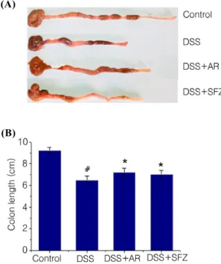
The DSS‐induced model of colitis is associated with a significant decrease in colon length (Fiocchi 1998;
Hendrickson et al, 2002) and colon length measurement has often been used as a morphological parameter for the degree of inflammation in DSS colitis. To assess colon length in the present study, mice from each group were killed at days 7. At day 7 following DSS treatment, we
Fig. 1. Effect of AR on DSS‐induced the body weight loss in mice. Experimental colitis in mice (n = 6/ group) was induced by a 5% DSS dissolved in the drinking water for 7days. AR was administered orally at doses of 200 mg/kg once a day for 7 days prior 5% DSS supplement. Body weight of mice was measured. Data were represented in the mean ± S.E.M. from triplicate experiments (#P < 0.05 vs. control,
*P < 0.05 vs. DSS alone).
Fig. 3. Effect of AR on DSS‐induced the DAI increase in mice. DAI was calculated as described in Materials and Methods. SFZ (150 mg/kg) was used as a positive control.
Data were represented in the mean ± S.E.M. (n = 6) from triplicate experiments (#P < 0.05 vs. control, *P < 0.05 vs.
DSS alone).
Fig. 2. Effect of AR on DSS‐induced the colon length shortening in mice. (A) Experimental colitis in mice was induced by 5% DSS dissolved in the drinking water for 7 days. AR was administered orally at doses of 200 mg/kg once a day for 7 days prior to 5% DSS supplement. The colons were removed at day 7 after DSS treatment, and the colon lengths were measured. (B) Relative colon lengths were represented. SFZ (150 mg/kg) was used as a positive control.
Data were represented in the mean ± S.E.M. (n = 6) from triplicate experiments (#P < 0.05 vs. control, *P < 0.05 vs.
DSS alone).
found that colon length in the DSS‐administered mice was significantly shorter than that of control. However, AR treatment alleviated DSS effects on colon shortening (Fig. 2A). Relative colon lengths are shown in Fig. 2B.
Another common feature of the DSS‐induced model of colitis is an increase in DAI. Increased DAI score was remarkably inhibited in the group administered with AR compared to the group with DSS (Fig. 3).
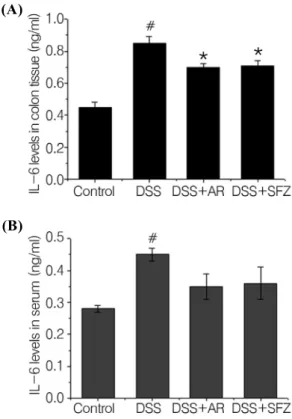
Effect of AR on IL‐6 levels in DSS‐induced colitis IL‐6 is considered important inflammatory mediators that play a key role in the pathogenesis of IBD. To determine the effect of AR on major inflammatory cytokine in mouse serum and tissues of colitis, ELISA was performed. At the end of the experiment, colon tissues were excised and homogenized. As shown in Fig.
4A, the levels of IL‐6 were significantly increased in the colon tissues of DSS‐treated mice compared to those of the control group. However, administration of AR reduced these levels induced by DSS. The rate of inhibition of IL‐6 production by AR was 25.11 ± 4.8%.
We also evaluated the effects of AR on serum levels of IL‐6. Blood samples were collected and serum IL‐6 levels were measured using ELISA. IL‐6 levels were significantly increased as a result of DSS exposure.
Elevated IL‐6 levels were reduced in the AR group although the decrease was not significant (Fig. 4B).
(A)
(B)
Fig. 4. Effect of AR on the IL‐6 levels in DSS‐induced colitis. (A) Experimental colitis was induced by 5% DSS drinking water for seven days in mice. AR (200 mg/kg) was administered orally once a day for seven days prior to 5%
DSS supplementation. SFZ (150 mg/kg) was used as a positive control. At the end of the experiment, the colon tissues were excised and homogenized. The levels of IL‐6 were quantified by ELISA. (B) Blood samples were collected and the levels of serum IL‐6 in the indicated groups were measured via ELISA. All data are expressed as the means
± S.E.M of three independent experiments (#P < 0.05 vs.
control, *P < 0.05 vs. DSS alone).
Fig. 5. Effects of AR on epithelial injury in DSS‐induced colitis. Paraffin sections of colonic tissue were stained with hematoxylin and eosin and analyzed by light microscopy (100×) (SL, submucosa layer; ML, mucosa layer).
Effects of AR on epithelial injury in DSS‐ induced colitis
Epithelial injury was detected in DSS control as compared with intact control. However, these histopathological changes induced by DSS treatments were inhibited by treatment of AR (Fig. 5).
DISCUSSION
Although traditional herbal medicines have long been used effectively based on traditional knowledge, the pharmacologic mechanisms of most herbal prescription have not been discovered. In this study, we attempted to provide experimental evidence that AR might be a useful
therapeutic drug for patients with UC. The finding of this study first demonstrated that AR inhibits inflammatory response and colon injury provoked by DSS treatment, and suggested an important effect by which AR attenuated intestinal inflammation.
UC is an idiopathic disease characterized by the development of intestinal inflammation (Bouma and Strober, 2003). Approximately ten individuals per 100,000 per year are diagnosed with UC. The pathogenesis of UC remains uncertain; however, it likely depends on the interaction between local immune reactions and environmental factors in genetically susceptible individuals. The signs and symptoms of UC include abdominal pain, weight loss, and bloody diarrhea (Ardizzone and Bianchi, 2005; Rufo and Bousvaros, 2006; Sato et al., 2007). Most therapies for UC include glucocorticosteroids, sulfasalazine, and other such drugs (Sandborn and Targan, 2002; Ishiguro et al., 2006).
However, these treatments cause serious side effects.
Consequently, there is a need for anti‐inflammatory agents that cause fewer side effects. Recently, traditional herbal medicine has been increased interest for the treatment of these disorders. This study investigated the effect of AR on DSS‐induced colitis. Treatment with AR reduced the weight loss and colon shortening caused by DSS. In addition, the DAI, scored using three major clinical signs (weight loss, diarrhea, and rectal bleeding), was remarkably inhibited in the group given AR. The inhibitory effect of AR on colon shortening and DAI was similar to the sulfasalazine group. These results suggest that AR effectively inhibits the symptoms of colitis caused by DSS.
(B) (A)
Pro‐inflammatory cytokines are involved in the initiation of the inflammatory response in colitis.
Accumulated experimental evidence shows that IL‐6 is strongly associated with the pathogenesis and progression of intestinal inflammatory disorders. It was also reported that the level of IL‐6 is remarkably elevated in UC patients (Li et al., 2010) and that it plays an integral role in their pathogenesis (Zheng et al., 2005). Therefore, research on new biological therapies for UC has focused on blocking components of the inflammatory cascade such as cytokines. In this study, we observed that the levels of IL‐6 increased in DSS treated‐colon tissues and serum compared with those of the control, and that treatment with AR reduced these levels. These results indicated that the anti‐inflammatory effect of AR is attributable to the regulation of inflammatory mediator in DSS‐induced colitis.
Although AR attenuated the IL‐6 level, the effects of AR on the other mediators or pathways involving colitis were not determined. Other study reported that AR attenuated the airway inflammation through suppression of IL‐13, IL‐5, eosinophils and CCR3 expression (Roh et al., 2008). Therefore, further studies will be necessary in order to clarify more precisely the role of AR on the intestinal inflammation.
In conclusion, we demonstrated at first that a treatment of AR could reduce significantly the clinical signs and the levels of inflammatory mediators in a colitis model caused by DSS treatment. Therefore, these results suggested that AR may be a useful therapeutic candidate for colitis. However, the further studies must be performed to elucidate the precise mechanism of AR for the treatment of intestinal inflammatory disorders.
Acknowledgements
This research was supported by Basic Science Research Program through the National Research Foundation of Korea (NRF) funded by the Ministry of Education, Science and Technology (2011‐0022703).
REFERENCE
Ardizzone S, Bianchi PG. Biologic therapy for inflammatory bowel disease. Drugs 2005. 65: 2253‐2286.
Bouma G, Strober W. The immunological and genetic basis of inflammatory bowel disease. Nat Rev Immunol. 2003.
3: 521‐533.
Camuesco D, Gálvez J, Nieto A, Comalada M, Rodríguez‐ Cabezas ME, Concha A, Xaus J, Zarzuelo A. Dietary olive oil supplemented with fish oil, rich in EPA and DHA (n‐3) polyunsaturated fatty acids, attenuates colonic inflammation in rats with DSS‐induced colitis. J Nutr.
2005. 135: 687‐694.
Copper HS, Murthy SNS, Shah RS, Sedergran DJ.
Clinicopathologic study of dextran sulfate sodium experimental murine colitis. Lab Invest. 1993. 69: 238‐249.
Fiocchi C. Inflammatory bowel disease: etiology and pathogenesis. Gastroenterology. 1998. 115: 182‐205.
Hendrickson BA, Gokhale R, Cho JH. Clinical aspects and Pathophysiology of inflammatory bowel disease. Clin Microbiol Rev. 2002. 15: 79‐94.
Ishiguro K, Ando T, Maeda O, Hasegawa M, Kadomatsu K, Ohmiya N, Niwa Y, Xavier R, Goto H. Paeonolattenuates TNBS‐induced colitis by inhibiting NF‐κB and STAT1 transactivation. Toxicol Appl Pharmacol. 2006. 217: 35‐42.
Kim SJ, Kim YG, Kim DS, Jeon YD, Kim MC, Kim HL, Kim SY, Jang HJ, Lee BC, Hong SH, Um JY. Oldenlandia diffusa Ameliorates Dextran Sulfate Sodium‐Induced Colitis Through Inhibition of NF‐kappa B Activation. Am J Chin Med. 2011. 39: 957‐969.
Kim SJ, Kim KW, Kim DS, Kim MC, Jeon YD, Kim SG, Jung HJ, Jang HJ, Lee BC, Chung WS, Hong SH, Chung SH, Um JY. The Protective Effect of Cassia obtusifolia on DSS‐Induced Colitis. Am J Chin Med. 2011. 39: 565‐ 577.
Li Y, de Haar C, Chen M, Deuring J, Gerrits MM, Smits R, Xia B, Kuipers EJ, van der Woude J. Disease‐related expression of the IL‐6/STAT3/SOCS3 signaling pathway in ulcerative colitis and ulcerative colitis‐related carcinogenesis. Gut. 2010. 59: 227‐235.
Lin TY, Liu YC, Jheng JR, Tsai HP, Jan JT, Wong WR, Horng JT. Anti‐enterovirus 71 activity screening of Chinese herbs with anti‐infection and inflammation activities. Am J Chin Med. 2009. 37: 143‐158.
MacDonald TT, Murch SH. Aetiology and pathogenesis of
chronic inflammatory bowel disease. Baillieres Clin.
Gastroenterol. 1994. 8: 1‐34.
Murthy SN, Cooper HS, Shim H, Shah RS, Ibarahim SA, Sedergran DJ. Treatment of dextran sulfate sodium‐induced murine colitis by intracolonic cyclosporin. Dig Dis Sci.
1993. 38: 1722‐1734.
Ramakers JD, Verstege MI, Thuijls G, Te Velde AA, Mensink RP, Plat J. The PPARgamma agonist rosiglitazone impairs colonic inflammation in mice with experimental colitis. J Clin Immunol. 2007. 27: 275‐283.
Roh SS, Kim SH, Lee YC, Seo YB. Effects of radix adenophorae and cyclosporine A on an OVA‐induced murine model of asthma by suppressing to T cells activity, eosinophilia, and bronchial hyperresponsiveness. Mediators Inflamm. 2008. 2008: 781425.
Rufo PA, Bousvaros A. Current therapy of inflammatory bowel disease in children. Paediatr Drugs. 2006. 8: 279‐ 302.
Sato K, Ohkura S, Kitahara Y, Ohama T, Hori M, Sato M, Kobayashi S, Sasaki Y, Hayashi T, Nasu T, Ozaki H.
Involvement of CPI‐17 downregula in the dysmotility of the colon from dextran sodium sulfate‐induced experimental colitis in a mouse model. Neurogastroenterol Motil. 2007. 19: 504‐514.
Sandborn, WJ, Targan SR. Biologic therapy of inflammatory bowel disease. Gastroenterology. 2002. 122: 1592‐1608.
Talhouk RS, Karam C, Fostok S, El‐Jouni W, Barbour EK.
Anti‐inflammatory bioactivities in plant extracts. J Med Food. 2007. 10: 1‐10.
Yang SK. Current status and clinical characteristics of inflammatory bowel disease in Korea. Korean J Gastroenterol. 2002. 40: 1‐14.
Zheng P, Niu FL, Liu WZ, Shi Y, Lu LG. Anti‐inflammatory mechanism of oxymatrine in dextran sulfate sodium‐ induced colitisofrats. World J Gastroenterol. 2005. 11: 4912
‐4915.