전기침이 급만성 결박스트레스로 유도된 스트레스호르몬 반응에 미치는 영향
정은우*ㆍ김현식*ㆍ이상관*ㆍ김민수**ㆍ조장희***ㆍ성강경*
*원광대학교 한의과대학 내과학교실
**전남대학교 통계학과
***가천의과학대학교 뇌과학연구소
목적 : 침은 다양한 질환의 치료에 보편적으로 쓰이고 있으며 고혈압, 당뇨, 정신질환 등을 포함한 스트레 스성 질환에도 활용되고 있다. 결박 스트레스는 스트레스 호르몬(코르티코스테론, 멜라토닌)의 혈장 농도를 크게 증가시키는 간단하고 효과적인 스트레스 요인이다. 본 연구는 결박 스트레스를 시행한 백서의 스트레 스 호르몬의 혈중 농도에 대한 전기침의 효과를 조사하였다.
방법 : 결박 그룹은 2시간의 결박 스트레스를 받았으며 결박 스트레스 및 고주파수 전침그룹과 결박 스 트레스 및 저주파수 전침그룹은 결박 스트레스와 고주파수 전침, 또는 결박 스트레스와 저주파수 전침을 동 시에 각각 시행하였다. 급성 스트레스 유발 시에는 결박 스트레스를 1차례, 만성 스트레스 유발 시에는 7차 례 시행하였다. 전기침 자극에는 우측 족삼리(ST36)를 사용하였다. 결박 스트레스 및 전기침으로 유도된 코 르티코스테론과 멜라토닌의 농도를 측정하기 위해서 결박 스트레스 및 전기침 자극이 시작된 30분, 60분, 90 분, 120분 후에 백서를 단두하여 혈액 샘플을 채취하였다.
1)
Effects of Electroacupuncture on Plasma Stress Hormone Responses to Acute and Chronic
Immobilization Stress
Jeong Eun-woo
*, Kim Hyun-sik
*, Lee Sang-kwan
*, Kim Min-soo
**, Cho Zang-hee
***and Sung Kang-keyng
**
Department of Oriental Medicine & Neuroscience, College of Oriental Medicine, Wonkwang Uinversity
**
Department of Statistics, Chonnam National University
***
Neuroscience Research Institute, Gachon University of Medicine and Science
* This paper was supported by Wonkwang University in 2008
․Acceptance : 2010. 9. 10. ․Adjustment : 2010. 9. 28. ․Adoption : 2010. 9. 28.
․Corresponding author : Sung Kang-keyng, 543-8, Juwol-Dong, Nam-Gu, Gwangju-si 503-310. Republic of Korea Tel. 82-062-670-6412 E-mail : [email protected]
국문초록
Original Article
결과 : 급성 스트레스 유발 시에는 고주파수 전침그룹의 코르티코스테론 혈장 농도가 증가하였고 멜라토 닌 농도의 시간적 패턴을 변화시켰으나 저주파수 전침그룹에서는 유의한 변화가 없었다. 만성 스트레스 유 발 시에는 고주파수 전침그룹의 혈장 코르티코스테론과 멜라토닌 농도가 유의하게 감소되었으나 저주파수 전침그룹에서는 변화가 없었다.
결론 : 이러한 결과는 전침이 결박 스트레스로 유도된 스트레스 호르몬의 혈장 농도 및 시간적 분비패턴 을 변화시키는 효과가 있으나 스트레스 호르몬 반응을 변화시키는 데 있어서 주파수에 따른 유의한 차이가 있다는 것을 의미한다.
핵심 단어 : 결박 스트레스, 전기침, 고주파수, 스트레스 호르몬, 코르티코스테론, 멜라토닌
Ⅰ. Introduction
Acupuncture has been used for the treatment of various diseases including stress-induced diseases such as hypertension, diabetes mellitus(DM), psy- chiatric disease. It is well known that acupuncture plays a role in maintenance of homeostatic balanc- ing when homeostatic potentialities are crushed by acute or chronic stress condition
1). According to results of animals and clinical studies, the action of acupuncture for compensating physiological malf- unction in stress and painful condition is evoked by the autonomic nervous system and endocrine system
2-4). In some studies that investigated the effect of acupuncture on stress response, acupuncture reduced norepinephrine (NE) levels in perfusate of brain regions as well as in the circulating blood
2). Also, the secretion of adrenal hormones in animals exposed to immobilization stress
5), induced by long- lasting cardiovascular and behavioral depression in spontaneous hypertensive rats (SHR)
6), and produced an anxiolytic effect in animals exposed to restraint- induced stress
7).
Although it has been reported that acupuncture has a controlling effect on stress hormones, how- ever the evidence responsible for these effects in acute and chronic stress status was not enough concluant.
To investigate the controlling effect of EA on corticosterone and melatonin responses changed by
acute and chronic immobilization stress, the present study carried out measurement of the plasma corticosterone and melatonin concentration at 30, 60, 90, or 120min after the beginning of the immo- bilization stress and high(100Hz) and low frequency (2Hz) electro acupuncture stimulation at Zusanli acupuncture point.
Ⅱ. Materials and methods
1. Animal handling
Healthy adult males Sprague-Dawley rats weighing 250~300g were used. All procedures were performed in accordance with the National Institutes of Health Guidelines for Animal Research(Guide for the Care and Use of Laboratory Animals) and approved by the Institutional Animal Care and Use Committee at Wonkwang University. Animals were housed in groups of three in a vivarium with 12-h light/dark cycle(lights on at 8 : 00), 50~60% humidity, and free access to food and water. Animals acclimatized to the laboratory seven days before the beginning of experiments.
2. Immobilization stress and electro acupuncture treatment
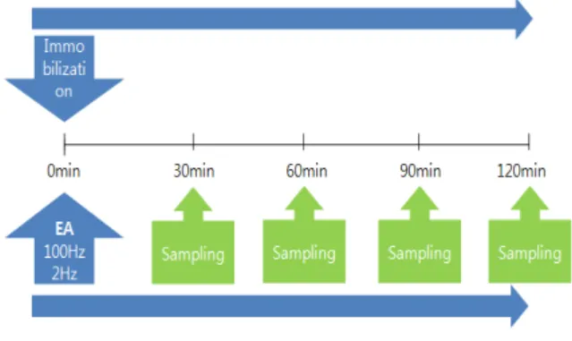
The experimental design of the present study is
shown in Fig. 1. Rats were randomly assigned to 3 groups(Immobilization group ; acute : 27 and chronic : 20, Immobilization + High frequency EA group ; acute : 24 and chronic : 23, Immobilization + Low frequency EA group ; acute : 22 and chronic : 26).
The immobilization groups were given 2h of immobilization stress. The Immobilization + High frequency EA group and Immobilization + Low EA were given simultaneously 2h immobilization stress with high frequency(100Hz) or low frequency(2Hz) electro acupuncture stimulation. The immobilization stress was given by attaching the four limbs of each animal in a prone position to a wooden board with adhesive tape.
Immobilization stress was carried out once in acute stress condition and 7 times in chronic stress condition, once a day between 09 : 00 AM and 13 : 00 PM. All the experiments were completed between 09 : 00 and 13 : 00 to minimize variability due to circadian rhythm.
Right ST
36(Zusanli) was chosen for acupun- cture stimulation. This point is between the head of fibula and the tibial tuberosity of the rat which corresponds to human ST
36. A pair of stainless steel pins of 0.3mm diameter were inserted with a depth of 5mm into the right ST
36and a point about 5mm far from the right ST
36. The two needles were connected with the output terminals of
Fig. 1. Experimental protocol
Acute Stress Group : Experiments were carried out once on 1st day. Immobilization, (EA), decapitation and sam- pling were carried out.
Chronic Stress Group : Experiments were carried out for 7 days. Immobilization, (EA) were carried out from 1st day to 6th day. Immobilization, (EA), decapitation and sampling were carried out on 7th day.
an EA device(Dual impedance research stimulator, Harvard Apparatus, USA). The frequency was 2Hz in low frequency EA group and 100Hz in high frequency EA group, while other conditions were same. Pulse duration and width was 1ms, current was 3mA, time duration was 30, 60, 90 and 120min.
In acute or chronic stress condition, EA was car- ried out once.
3. Blood sample collection and storage
We measured corticosterone and melatonin levels in separate groups of rats given immobilization stress and the electro acupuncture procedure as described above. To determine plasma concentra- tions of corticosterone and melatonin induced by immobilization stress and EA, blood samples were taken by decapitation at 30, 60, 90, or 120min after the beginning of the immobilization stress and EA stimulation.
Sampled blood was allowed to clot for 30min before centrifugation for 10min at 1000xg. Plasma was separated and stored at ≤-20°C.
4. Corticosterone and melatonin immuno assay
A Luminex 100, an endocrine Multiplex Immu- noassay(LINCO Research, St Charles, MO), was utilized to measure plasma corticosterone and mel- atonin. Corticosterone and melatonin levels were obtained by commercially available radioimmunoassay kits(LINCO Research, St Charles, MO). All samples were analyzed in duplicate, with the coefficient of variation ranging from 3 to 4.5%.
5. Statistical analysis
All values are means ± stand error of the mean
(S.E.M). The effects of immobilization, high frequency
EA and low frequency EA on corticosterone and
melatonin were analyzed using a two-way analysis
of varience(2-way ANOVA) followed by Duncan’s
post hoc test. All statistical analyses were perfor-
med using SAS system.
Ⅲ. Results
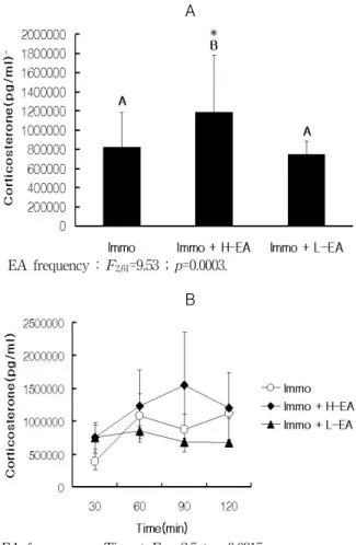
1. Effects of EA on plasma corticosterone concentration in acute stress
There was a significant effect of High frequency EA on elevating plasma corticosterone level relative to immobilization stress in acute stress states.
Analysis of corticosterone responses to immobi- lization stress, high frequency EA and low frequency EA showed main effect of treatment (F=9.53, p<
0.0003) and interaction of treatment with time(F=
2.5 ; df 6,61 ; p<0.0315).
Fig. 2A shows the total corticosterone level for 2 hr in the 3 groups with the respective treatment.
Plasma corticosterone concentration of high fre- quency EA was higher than that of immobilization stress and low frequency EA, while low frequency EA was similar to immobilization stress. Plasma corticosterone level to immobilization stress was 800000 ± 000pg/ml. In high frequency EA, cortico- sterone level was 1200000 ± 0000pg/ml. In low freq- uency EA, corticosterone level was 800000 ± 000pg/ml.
Fig. 2B shows the corticosterone levels over time in the groups in response to immobilization, high frequency EA and low frequency EA. Both tre- atment group and time influenced the response as indicated by the treatment group × response time interaction. Corticosterone response to treatment differed by each group and response time. At all time point, the level of plasma corticosterone of high frequency EA group was the highest among the groups. At 90min, while plasma corticosterone of immobilization and low frequency EA group had decreased relatively to 60min, that of high freq- uency EA has more sharply increased than cortico- sterone level at 60min. There was a biggest gap between high frequency EA and immobilization or low frequency EA at this time point. At 120min.
The level of plasma corticosterone of high freque-
✳
A
EA frequency : F2,61=9.53 ; p=0.0003.
B
EA frequency × Time : F6,61=2.5 ; p=0.0315.
Fig. 2. Effects of electro acupuncture on plasma corticosterone concentration in acute immobilization stress
A : Comparison of total plasma corticosterone concent- ration in rat treated with immobilization stress, immo- bilization stress + high frequency acupuncture and immobilization stress + low frequency acupuncture for 2 hours. Values represent means±SE(n=). Results(F- and
p-values) from ANOVA with treatment(Immo, Immo+
H-EA and Immo + L-EA) are given in the figure.
Different letters and asterisk indicate significant differ- ences at ✳ ; p<0.05 level(Duncan’s post-hoc test) bet- ween the groups.
B : Temporal profile of plasma corticosterone concentration to immobilization stress, immobilization stress + high frequency acupuncture and immobilization stress + low frequency acupuncture for 2 hours. Values represent means±SE (n=). There are significant Interaction(F- and P-values) from 2-way ANOVA with treatment(Immo, Immo+H-EA and Immo+L-EA) and immo-time(30, 60, 90 and 120min) as given in the figure.
ncy EA had finally decreased relatively to 90min and was similar with immobilization, but low fre- quency EA had not changed from 90min and was the lowest level. By Duncan’s post-hoc test(p<
0.05), the corticosterone concentration of high
frequency EA showed significant differences with that of immobilization stress and low frequency EA.
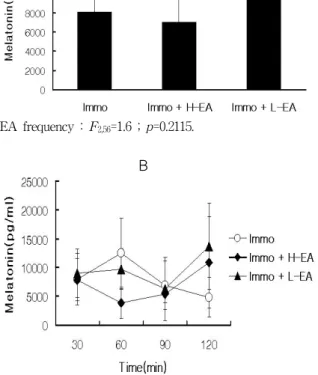
2. Effects of EA on plasma melatonin concentration in acute stress
Although there was no significant effect of EA treatment on total plasma melatonin level compared to immobilization stress, EA changed temporal pat-
A
EA frequency : F2,56=1.6 ; p=0.2115.
B
EA frequency × Time : F6,56=2.5 ; p=0.0323.
Fig. 3. Effects of electro acupuncture on plasma melatonin concentration in acute immobilization stress
A : Comparison of total plasma melatonin concentration inrat treated with immobilization stress, immobilization stress + high frequency acupuncture and immobilization stress + low frequency acupuncture for 2 hours. Values represent means±SE(n=). Results(F- and p-values) from ANOVA with treatment(Immo, Immo+H-EA and Immo+
L-EA) are given in the figure. Same letters indicate that there are no significant differences at p<0.05 level(Duncan’s post-hoc test) between the groups.
B : Temporal profile of plasma corticosterone concentration to immobilization stress, immobilization stress + high frequency acupuncture and immobilization stress + low frequency acupuncture for 2 hours. Values represent means±SE(n=). Interaction(F- and p-values) from 2- way ANOVA with treatment(Immo, Immo+H-EA and Immo+L-EA) and immo-time(30, 60, 90 and 120min) are given in the figure.
tern of melatonin concentration in acute stress states.
Analysis of melatonin responses to immobilization stress, high frequency EA and low frequency EA showed no main effect of treatment (F=1.6, p<
0.2115). However, there was interaction of treatment with time(F=2.5 ; df 6,56 ; p<0.0323).
Fig. 3A shows total melatonin level for 2 hr in the 3 groups with the respective treatment. Low frequency EA tends to increase plasma melatonin concentration relative to immobilization stress and high frequency EA. Plasma melatonin level to acute immobilization stress was 8000 ± 000pg/ml. In high frequency EA, melatonin level was 7000 ± 0000pg/ml. In low frequency EA, melatonin level was 10000 ± 000pg/ml.
Fig. 3B shows the melatonin levels over time in the groups in response to immobilization, high frequency EA and low frequency EA. Both treatment group and time influenced the response as indicated by the treatment group × response time interaction. Melatonin response to treatment differed in each group and response time. The level of plasma melatonin of each group was similar at 30min, but the big gap between plasma melatonin of high frequency EA and immobilization or low frequency EA was made at 60min. At 90min. The level of plasma melatonin of each group was was similar again, and then the level of plasma melatonin of high and low frequency EA was rapidly increased at 120min, while that of immobilization was slightly decreased.
By Duncan’s post-hoc test (p<0.05), the melatonin concentration between all groups did not show significant differences.
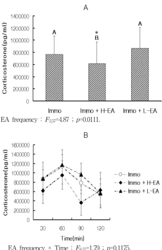
3. Effects of EA on plasma corticoster- one concentration in chronic stress
There was a significant effect of High frequency
EA on decreasing plasma corticosterone level re-
lative to immobilization stress in chronic stress
states, whereas low frequency EA did not change
plasma corticosterone concentration induced by immo-
bilization stress. Analysis of corticosterone responses
✳
A
EA frequency : F2,57=4.87 ; p=0.0111.
B
EA frequency × Time : F6,57=1.79 ; p=0.1175.
Fig. 4. Effects of electro acupuncture on plasma corticosterone concentration in chronic immobilization stress
A : Comparison of total plasma corticosterone concentration in rat treated with immobilization stress, immobilization stress + high frequency acupuncture and immobilization stress + low frequency acupuncture for 2 hours after 7 days immobilization stress for 2 hours. Values represent means±SE(n=). Results(F- and P-values) from ANOVA with treatment(Immo, Immo+H-EA and Immo+ L-EA) are given in the figure. Different letters and asterisk indicate significant differences at ✳ ; p<0.05 level(Duncan’s post- hoc test) between the groups.
B : Temporal profile of plasma corticosterone concentration to immobilization stress, immobilization stress + high frequency acupuncture and immobilization stress + low frequency acupuncture for 2 hours. Values represent means ± SE(n=). Interaction(F- and p-values) from 2-way ANOVA with treatment(Immo, Immo+H-EA and Immo+L- EA) and immo-time(30, 60, 90 and 120min) are given in the figure. Fig. 4. Effects of Electro Acupuncture on Plasma Corticosterone Concentration in Chronic Immobi- lization Stress.
to immobilization stress, high frequency EA and low frequency EA showed main effect of treatment (=4.87, p<0.0111). However there was no interaction of treatment with time (F=1.79 ; df ; p<0.1175).
Fig. 4A shows total corticosterone level for 2 hr in the 3 groups with the respective treatment. Pla- sma corticosterone concentration of high frequency EA was lower than that of immobilization stress and low frequency EA, while low frequency EA was similar to immobilization stress. Plasma corticoster- one level to immobilization stress was 800000 ± 000pg/ml. In high frequency EA, corticosterone level was 1200000 ± 0000pg/ml. In low frequency EA, corticosterone level was 800000 ± 000pg/ml.
Fig. 4B shows the corticosterone levels over time in the groups in response to immobilization, high frequency EA and low frequency EA. Both treatment and time did not influence the response as indicated by the treatment group × response time interaction, although the level of plasma corticosterone of high frequency EA group remained the lowest level at 30, 60 and 90min. And the big gap between high frequency EA and immobilization or low frequency EA was made at 90min.
By Duncan’s post-hoc test(p<0.05), the cortico- sterone concentration of high frequency EA showed significant differences with that of immobilization stress and low frequency EA.
4. Effects of EA on plasma melatonin concentration in chronic stress
There was a significant effect of high frequency EA on decreasing plasma melatonin level induced by immobilization stress in chronic stress states, whereas low frequency EA did not change plasma melatonin concentration induced by immobilization stress. Analysis of melatonin responses to immo- bilization stress, high frequency EA and low freq- uency EA showed main effect of treatment(F=10.39, p<0.0002) However, there was no significant inter- action(F=2.26 ; df ; p<0.052) of treatment with time.
Fig. 5A shows the total melatonin level for 2hr in the 3 groups with the respective treatment.
High frequency EA decreased plasma melatonin
concentration relative to immobilization stress and
low frequency EA. Plasma melatonin level to
✳
A
EA frequency : F2,51=10.39 ; p=0.0002.
B
EA frequency × Time : F6,51=2.26 ; p=0.052.
Fig. 5. Effects of electro acupuncture on plasma melatonin concentration in chronic immobilization stress
A : Comparison of total plasma corticosterone concentrationin rat treated with immobilization stress, immobilization stress + high frequency acupuncture and immobilization stress + low frequency acupuncture for 2 hours after 7 days immobilization stress for 2 hours. Values represent means±SE(n=). Results(F- and P-values) from ANOVA with treatment(Immo, Immo+H-EA and Immo+ L-EA) are given in the figure. Different letters and asterisk indicate significant differences at ✳ ; p<0.05 level(Duncan’s post- hoc test) between the groups.
B : Temporal profile of plasma corticosterone concentration to immobilization stress, immobilization stress + high frequency acupuncture and immobilization stress + low frequency acupuncture for 2 hours. Values represent means ± SE(n=). Interaction(F- and p-values) from 2-way ANOVA with treatment(Immo, Immo+H-EA and Immo+
L-EA) and immo-time(30, 60, 90 and 120min) are given in the figure. Fig. 4. Effects of Electro Acupuncture on Plasma Corticosterone Concentration in Chronic Immo- bilization Stress.
chronic immobilization stress was 3000 ± 000pg/ml.
In high frequency EA, melatonin level was 1000 ± 0000pg/ml. In low frequency EA, melatonin level was 3000 ± 000pg/ml.
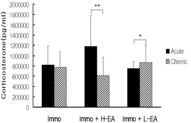
Immo ; Acute-Chronic : F1,39=0.21 ; p=0.6491.
Immo + H-EA ; Acute-Chronic : F1,39=15.86 ; p=0.0003.
Immo + L-EA Acute-Chronic : F1,40=5.35 ; p=0.026.
Fig. 6. Comparison of plasma corticosterone con- centration to immobilization stress, immobilization stress + high frequency acupuncture and immobilization stress + low frequency acupuncture for 2hours in acute and chronic immobilization stress
Values represent means±SE (n=). Results(F- and P-values) from ANOVA with treatment(Immo, Immo+H-EA and Immo+L-EA) are given in the figure. Asterisk indicate significant differences at ✳ ; p<0.05, ✳✳ ; p<0.0001 level.
Immo ; Acute-Chronic : F1,35=39.24 ; p=0.0001.
Immo + H-EA ; Acute-Chronic : F1,32=2.25 ; p=0.143.
Immo + L-EA Acute-Chronic : F1,40=25.32 ; p=0.0001.
Fig. 7. Comparison of plasma melatonin concen- tration to immobilization stress, immobilization stress + high frequency acupuncture and immobilization stress + low frequency acupuncture for 2 hours in acute and chronic immobilization stress
Values represent means±SE (n=). Results(F- and P-values) from ANOVA with treatment(Immo, Immo+H-EA and Immo+
L-EA) are given in the figure. Asterisk indicate significant differences at ✳✳ ; p<0.0001 level.