VOLUME 6, NUMBER 1, JUNE 2001
Mercury Injury of Hand from Broken Thermometer
-Three cases report-
Eugene Kim, M.D., Juno Yoon, M.D.*, Jae Dong Um, M.D.
Department of Orthopaedic Surgery, Asan Foundation Kang Nung Hospital, Kannung, Korea
Department of Orthopaedic Surgery, Ulsan University, Asan Medical Center, Seoul,Korea*
We report on three patients with soft tissue hand injuries by mercury from broken thermometers; acute and chronic inflammation and foreign body granuloma.
After excision of the granuloma, foreign body, and abscess, wound was healed and revealed no systemic intoxication on minimal 18month-follow-up.
Key Words : Hand, Mercury injury
INTRODUCTION
Digital thermometer is familiar, but the conventional thermometer with mercury is used broadly until now. Hand injury by
metallic mercury following an accident with a thermometer is probably more common than the paucity of reports in the literature would lead one to believe. To our knowledge, a few cases described by dermatologist and pathologist but never done by orthopedic hand surgeon. The purpose of this report is to describe an instance of soft-tissue hand injury resulting from and accident with a mercury thermometer, and to provide more details about the pathologic findings.
CASE REPORT
Case 1
A 38-year-old woman was admitted to the hospital on April 5, 1996, with pain, tender- ness, erythema, and edema on her dorsum of hand. Two weeks ago, she cared her baby at a hospital, and a nurse injured her hand with broken thermometer shaking before checking the baby’s fever by accident. The wound was 0.3 cm size and treated by simple dressing. Any other pain or sign were complained. Several days after, her hand was getting swollen and erythematous. She tried acupuncture two times but symptom was aggravated.
On examination tenderness, erythema and local heat were on the dorsum of fourth metacarpal area. Preoperative chest x-ray,
깨진 체온계의 수은으로 인한 수부 연부조직 손상 - 증례 보고 -
아산재단 강릉병원 정형외과학교실 울산대학교 의과대학 서울중앙병원 정형외과학교실*
김유진・윤준오*・엄재동
통신저자 : 김 유 진
강원도 강릉시 사촌면 방동리 415 아산재단 강릉병원 정형외과학교실 TEL : 033-610-3242, FAX : 033-641-8050 E-mail : [email protected]
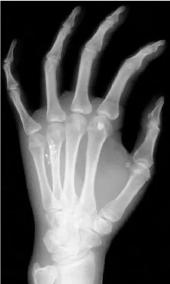
urinalysis, and complete blood count, ESR (Erythrocyte sedimentation rate), CRP(C- reactive protein) were all within normal limits. The serum mercury level was within normal limits at 0.2 μg/dl. X-rays of the hand(Fig. 1) showed metallic densities in soft tissues.
Debridement was performed around pre- vious wound. An abscess on the hand was incised and drained of pus and globular metalic material.
On the pathologic findings, mixed inflam- matory infiltrate composed of polymorpho- nuclear leukocytes, lymphocytes, and his- tiocytes was present in the vicinity of the metallic mercury globules. In addition, there were focal areas containing anaerobic
debris and polymorphonuclear leukocytes suggestive of micro-abscess formation.
Central spaces with intraluminal opaque black globules and surrounding collagen necrosis were findings(Fig. 2).
After surgery, the patient recovered com- pletely and wound was healed. She had no evidence of renal, nervous system, or gas- trointestinal disease on 2-year-follow-up.
Case 2
A 29-year-old woman was admitted to the hospital on December 27, 1996, with a growing mass on the radial side of her left index finger. It was firm, mild tender without any infectious sign and measured 1 c m×0.7 cm. X-ray of patient’s hand showed scattered areas of metallic density in soft tissues(Fig. 3).
She injured her finger by broken thermo- meter six weeks ago remaining minimal wound. Preoperative chest x-ray, urina- lysis, and complete blood count, ESR, CRP were all within normal limits. The serum mercury level was within normal limits at 0.3 μg / d l .
The mass was surgically removed and the biopsy showed a diffuse fibrotic reaction in the to deep dermis.
It also revealed foreign body with giant
Fig. 1. Multiple sand like foreign bodies in the soft tissue around the 4th metacarpal bone.
Fig. 2. Black spherical materials and suppurative inflammation with foreign body reaction in the deep dermis(Hematoxylin- eosin stain, original magnification ×100)
cell and scattered lymphocytes, polymor- phonuclear leukocytes and numerous his- tiocytes. It is adequate to be diagnosed foreign body granuloma(Fig 4).
After surgery, the patient recovered completely and wound was healed. She had no evidence of renal, nervous system, or gastrointestinal disease on 18 month- follow-up.
Case 3
A 22-year-old nurse was admitted on March 27, 1996, with pain, tenderness,
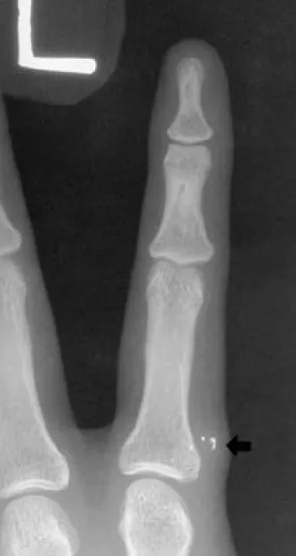
erythema, and edema on radial aspect of proximal interphalalgial joint of her left middle finger. Ten weeks ago, she injured her finger with broken thermometer sha- king a before checking patient’s fever by accident. The wound was 0.2 cm size and treated by simple dressing. About two weeks after, her hand was getting swollen and erythematous. She neglected and symptom was aggravated. On examination tenderness, erythema and local heat were on the radial side of third proximal inter-phalalgial joint of her left middle finger. Preoperative systemic evaluation were all within normal limits. The serum mercury level was within normal limits at 0.3 μg/dl. X-rays of the finger(Fig. 5) showed linearly scattered metallic densities in soft tissues of finger.
Excision and debridement were performed around previous wound. After surgery the wound was healed completely, she had sagittal band injury that was neglected at that time. We recommended the reconstru- ction of the sagittal band due to PIP joint subluxation, but she refused. She had no evidence of renal, nervous system, or gas- trointestinal disease on 15 month-follow-up.
DISCUSSION
When elemental mercury is injected into
Fig. 4. Foreign material and abscess formation in subcutaneous tissue(Hematoxylin-eosin stain, original magnification ×400) Fig. 3. Foreign body in soft tissue at the radial side of proximal
phalanx
the skin, its course is unpredictable and sometimes fatal.
Cezanne et al.2 )reported a patient with soft tissue hand injury from mercury by intravenous injection who had recent psy- chiatric history of numerous suicide at- tempts. Complete excision of the metal, sys- temic evaluation and psychiatric consulta- tion were emphasized in his report.
Netscher et al.5 ) treated a intravenously injected huge granuloma in elbow with systemic intoxication by complete resection with flap. Theodorou et al.8 ) reported imme- diately developed synovitis of knee joint
injured by mercury from a broken thermo- meter. Two cases of soft tissue hand injuries from a broken thermometer reported by R a c h m a n7 )and Arcadio et al.1 ). Within a mat- ter of weeks there followed oliguria, a metal- lic taste in the mouth, and diarrhea, and a daily urinary excretion of about 200 μg of mercury. Six weeks after the accident the wound was surgically excised and dro-plets of metallic mercury were removed. Two deaths reported as a result of subcutaneous mercury infection were secondary to renal failure three weeks after infection6 ).
The diagnosis of cutaneous mercury gra- nuloma may be difficult if the patient gives no history of contact with metallic mercury.
Local irritation and acute abscess formation are common. Chronic cutaneous lesions generally appear as erythematous foreign body type granulomas. Systemic absorption is common, and mercury levels can be mea- sured in the blood or urine. Gastrointesti- nal and central nervous system involve- ment have also been observed3 ).
Histologically, mercury on hematoxylin and eosin preparation classically appears as spaces with adjacent or intraluminal dark gray to black opaque globules with surrounding collagen necrosis. A granulo- matous foreign body reaction with a mixed infiltrate composed of poly morphonuclear leukocytes, eosinophils, lymphocytes, plasma cells, and histiocytes is frequently present in the vicinity of the metallic mer- cury globules. Epidermal and dermal nec- rosis as well as pseudoepitheliomtous hyperplasia are other common findings.
Acute inflammation and necrosis were more commonly seen in the shorter time periods (weeks to months), whereas granulomatous foreign bodygiant cell reactions and fibrosis were associated with prolonged deposition of mercury(months to years). The reaction to metallic mercury in the skin and subcu- taneous tissue may remain localized, with no signs or symptoms of systemic involve- m e n t4 ). Fortunately our cases appear to be Fig. 5. Linear dot like foreign bodies in soft tissue near the
radial side of proximal interphalangeal joint of middle finger
in this category. We experienced both of acute and chronic infectious condition and foreign body granuloma. All the cases were treated by complete excision of freign body without systemic involvement.
We conclude that total and early excision of all tissues containing droplets of meta- llic mercury is essential. Never be ignored even if there were no external wound.
Patients should be carefully examined for signs of acute or chronic mercury poison- ing and followed for a long duration.
REFERENCES
01) Arcadio F, Thivolet J, Perrot H : Acrodynia following the accidental subcutaneous infiltration of mercury. Bulletin Society French Dermatroloy Syphiligr. 75:509-510, 1968.
02) Cezanne CA, Karen AL, Patricia T : Elemental mercury foreign body granulomas. International Journal of Derma- tology. 31(5):353-354, 1992.
03) Krohn IT, Solof A, Mobini J, Wagner DK : Subcutaneous injection of metallic mercury. JAMA. 243:548-548, 1980.
04) Lupton GP, Colonel L, Kao, GF et al : Cutaneous mercury granuloma. Jounal of American Academy of Dermatology.
12:296-303, 1985.
05) Netscher DT, Friedland JA, Guzewicz RM : Mercury poisoning from intravenous injection: treatment by granu- loma resection. Annals of Plastic Surgery. 20:592-596, 1991.
06) Popper L : Death following injury by a thermometer.
Wien Med Wochenschr. 116:779-780, 1966.
07) Rachman R : Soft tissue injury by mercury from a broken thermometer. Am J Clin Pathol. 61:296-300, 1974.
08) Theodorou SD, Vlachos P, Vamvasakis E : Knee joint injury by mercury from a broken thermometer. Clin Orthop.
160:159-162, 1981.