Osteochondritis dissecans (OCD) is a pathological condition that appears to destruct subchondral bone with secondary effects on the overlying articular cartilage, which eventually results in osteochondral separation including a loose body1). The reported causes of OCD include genetic, traumatic, ischemic, and persis- tent accessory ossification1). Discoid lateral meniscus that could produce repetitive abnormal stress on the lateral femoral condyle (LFC) has been known as one of the factors of OCD1).
There has been no optimal treatment for OCD of the LFC as- sociated with discoid lateral meniscus tears. Furthermore, if an
OCD lesion is massive and requires an adequate reconstruction of the osteochondral defect, the treatment is more challenging.
Treatment of OCD alone without any procedure on the torn dis- coid lateral meniscus would result in persistent symptoms and ultimately lead to a failure of the treatment of OCD and progres- sion of arthritis1). Hence, the strategy for combined massive OCD and torn discoid lateral meniscus should be carefully considered.
According to a recent study, severe OCD lesions of the talus could be successfully treated with bony periosteum-covered iliac crest plug transplantation2). The study showed that the perioste- um of the iliac crest containing many stem cells have a capacity of differentiating into chondrocytes and fibro-cartilage. In addition, their technique could provide osteochondral stability in both os- seous and chondral levels without donor site morbidity. Above all, structured bone grafts are needed to fill the osseous defect and prevent collapse of the reconstruction, and the anterior iliac crest has been known to be effect under compressive and shear stresses2). Inspired by the technique, we thought the weight bear- ing capacity of the structured bone graft from the anterior iliac crest could be applied to the femoral condyle of the knee.
Simultaneous Osteoperiosteal Autologous Iliac Crest Graft and Lateral Meniscus Allograft Transplantation for Osteochondral Lesion with Bony Defect and Lateral Discoid Meniscus Tear
Dhong Won Lee, MD
1, Jin Goo Kim, MD, PhD
2, Jeong Ku Ha, MD
3, and Woo Jong Kim, MD
41Department of Orthopedic Surgery, Daejeon Military Hospital, Daejeon; 2Department of Orthopedic Surgery, Konkuk University Medical Center, Seoul; 3Department of Orthopedic Surgery, Inje University Seoul Paik Hospital, Seoul; 4Department of Orthopedic Surgery, Soonchunhyang University Cheonan Hospital, Choenan, Korea
The optimal treatment for combined osteochondritis dissecans (OCD) with considerable bony defect of the lateral femoral condyle (LFC) and torn discoid lateral meniscus is unclear. We present a case of a 15-year-old female who was a gymnast and had a large OCD lesion in the LFC combined with deficiency of the lateral meniscus. The patient underwent the “one-step” technique of osteoperiosteal autologous iliac crest graft and lateral meniscus allograft transplantation after a failure of meniscectomy with repair at another hospital. Twenty-four months postoperatively, clinical results were significantly improved. Follow-up imaging tests and second-look arthroscopy showed well incorporated structured bone graft and fibrous cartilage regeneration as well as stabilized lateral meniscus allograft. She could return to her sport without any pain or swelling. This “one-step”
surgical technique is worth considering as a joint salvage procedure for massive OCD lesions with torn discoid lateral meniscus.
Keywords: Knee, meniscus, cartilage, autograft, transplantation pISSN 2234-0726 · eISSN 2234-2451
Knee Surgery & Related Research
Received September 13, 2015; Revised October 26, 2015;
Accepted November 3, 2015
Correspondence to: Jin Goo Kim, MD, PhD
Department of Orthopedic Surgery, Konkuk University Medical Center, 120-1 Neungdong-ro, Gwangjin-gu, Seoul 05030, Korea
Tel: +82-2-2030-7606, Fax: +82-2-2270-0023 E-mail: [email protected]
165
This is an Open Access article distributed under the terms of the Creative Commons Attribution Non-Commercial License (http://creativecommons.org/licenses/by-nc/4.0/) which permits unrestricted non-commercial use, distribution, and reproduction in any medium, provided the original work is properly cited.
Copyright © 2016 KOREAN KNEE SOCIETY www.jksrr.org
of the LFC and anterior defect with posterocentral shift of the discoid lateral meniscus. The patient underwent arthroscopic partial meniscectomy with repair of the lateral meniscus and multiple drilling at the LFC 2 weeks after the injury at the hos- pital. During 3 months after the surgery, however, her pain had worsened and she could not walk well.
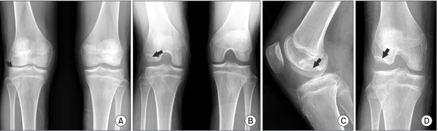
Eventually, the patient was transferred to our clinic at 4 months after the first surgery. Physical examination revealed tenderness on the lateral joint line of the right knee and positive McMurray test on internal rotation of the tibia. Plain radiographs of the right knee showed an obvious OCD lesion with a large bony defect in the LFC and narrowing of the lateral joint space (Fig. 1). Stand- ing alignment of the lower legs demonstrated about 2 degrees of slight valgus of the right knee compared to the left knee (Fig.
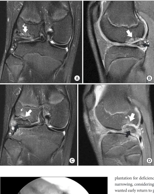
2). Magnetic resonance imaging (MRI; Intera Achieva, Philips, Amsterdam, The Netherlands) of the right knee revealed a 2.5 cm×3 cm sized lesion mainly in the posterolateral area of the LFC (Fig. 3). The lesion progressed more compared with the pre- operative initial MRI obtained 5 months ago (Fig. 3). The MRI
A B C D
Fig. 1. (A) Standing anteroposterior view showing lateral joint space narrowing (arrow) of the right knee. (B) Rosenberg view showing the osteochon- dral lesion (arrow) in the posterolateral aspect of the right lateral femoral condyle. (C) Lateral view of the right knee showing the extent of the osteo- chondral lesion (arrow) in the posterior aspect of the lateral femoral condyle. (D) Tunnel view of the right knee showing the osteochondral lesion with defect (arrow) in the posterolateral aspect of the lateral femoral condyle.
Fig. 2. Standing lower extremity radiograph showing slight valgus of the right knee (about 2 degrees compared to the left knee).
plantation for deficiency of the lateral meniscus with joint space narrowing, considering the patient was a high-level gymnast and wanted early return to previous level of sports activities.
1. Surgical technique
At one month after the first arthroscopic surgery performed at our hospital (5.5 months after the surgery performed at another hospital), she underwent osteoperiosteal autologous iliac crest graft implantation and lateral meniscus allograft transplantation as a one-stage procedure.
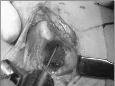
An anterolateral incision was used to expose the lateral com- partment of the knee joint. The OCD lesion was identified and completely debrided, then multiple drilling using a fine 1.2–1.5- mm diameter K-wire was performed toward the lesion to break down the sclerotic avascular zone (Fig. 5). In the next step, the size and depth of the defect was measured as 1.6 cm×2.4 cm and 0.8 cm, respectively using various-sized osteotomes. The ante- rior iliac crest was then accessed by making an incision 2–3 cm Fig. 4. The osteochondritis dissecans lesion treated with multiple drilling
at another hospital showed the large osteochondral defect without any regeneration.
A B
C D
Fig. 3. Initial preoperative magnetic reso- nance imaging (MRI) performed at an- other hospital showing the osteochondral lesion with subchondral cysts (white arrow) in the posterior aspect of the right lateral femoral condyle (LFC) in coronal plane (A) and the osteochondral lesion (white arrow) with posteriorly displaced lateral discoid meniscus (black arrow) in the posterolat- eral aspect in sagittal plane (B). Follow-up (5 months after initial MRI) preoperative MRI performed at our hospital showing the more progressed osteochondral lesion (white arrow) in coronal plane (C) and the massive defect of the LFC (white arrow) with retorn lateral meniscus adhering to the LFC (black arrow) in sagittal plane (D).
Knee joint motion was performed to check the stability of the inserted iliac crest plug and the transplanted lateral meniscus al- lograft. Finally, the integrity and congruency of the grafts were
months after surgery and return to sports at 6 months after sur- gery, although strenuous contact sports were prohibited.
3. follow-up evaluation
Plain radiography and 3-dimensional computed tomography (3D CT; Aquilion 64, Toshiba, Tokyo, Japan) were carried out on the first postoperative day, which showed a well integrated bone plug in the LFC and an allograft in the lateral tibial plateau (Fig. 7). At 24 months after surgery, the Lysholm score improved from 47 to 85, the International Knee Documentation Commit- tee subjective knee form improved from 57 to 75, and the Tegner activity score improved from 4 to 8. The visual analogue scale was 0 point. There was no donor-site complication such as pain and sensory loss as well as growth deformity. Isokinetic muscle strength test, follow-up MRI, and second-look arthroscopy were performed at 30 months after surgery. The deficit of isokinetic muscle strength (flexor peak torque/body weight) of the involved side was 9.2% and the hamstring-quadriceps ratio was 59%. On follow-up MRI and second-look arthroscopy, the iliac crest plug was well incorporated with the LFC, and the OCD lesion healed with fibrous cartilage regeneration as well as stabilized lateral me- Fig. 5. Photograph of the exposed right lateral femoral condyle. The os-
teochondritis dissecans lesion was debrided and multiple drilling using K-wire was performed toward the lesion.
A B C
Fig. 6. Bone plug harvest and transplantation. (A) A bone plug matching the size of the osteochondritis dissecans lesion was harvested without dam- aging the periosteum. (B) The harvested iliac crest plug with periosteum preservation was inserted into the lesion of the lateral femoral condyle and impacted. (C) The press-fitting mortise wider and deeper at the articular surface was used during graft insertion.
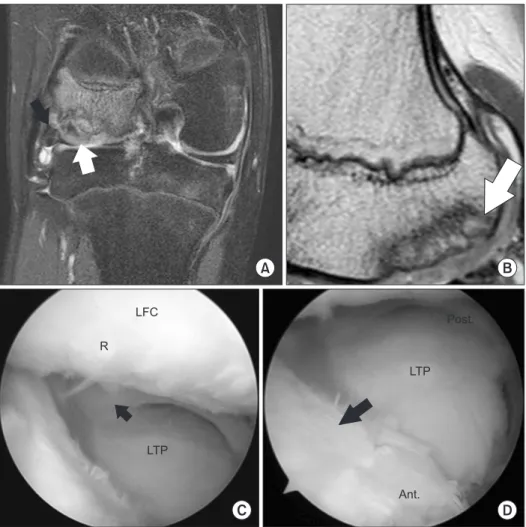
niscus allograft (Fig. 8). The surrounding cartilage revealed no progression of degeneration or formation of a kissing lesion. She could return to previous level of gymnastic activities without any swelling or pain at final evaluation at 24 months after surgery.
discussion
This case report describes osteoperiosteal autologous transplan- tation for massive OCD combined with lateral meniscus allograft transplantation for deficiency of the lateral meniscus as a “one- step” technique. The patient could return to high-level sports activities as well as full daily activities without symptoms. In ad- dition, a good morphological restoration with fibrous cartilage covering of the massive OCD lesion including osseous defect and well-maintained lateral meniscus allograft were shown on the follow-up MRI and second-look arthroscopy.
A discoid lateral meniscus sometimes results in OCD of the LFC, which has been reported in co-presence with discoid me- niscus in up to 19%1). The discoid meniscus, regardless of tear, could be an important risk factor for OCD, because it might
LFC
Fig. 7. Three-dimensional reconstructed computed tomography of the right knee revealing the stabilized periosteal plug (arrow) at the lateral femoral condyle (LFC).
LFC
A B
C D
R
LTP
Post.
LTP
Ant.
Fig. 8. Follow-up magnetic resonance im- aging showed well incorporated periosteal plug (white arrow) and lateral meniscus allograft (black arrow) in the posterior as- pect of the right LFC in coronal plane (A) and well integrated periosteal plug with surrounding cartilage of the LFC (white arrow) in sagittal plane (B). Second-look arthroscopy showed well covering (R) previous osteochondritis disseccans lesion without degeneration (C) and well main- tained lateral meniscus allograft without displacement (black arrow) (C, D). LFC:
lateral femoral condyle, R: regenerated cartilage, LTP: lateral tibial plateau, Post:
posterior, Ant: anterior.
young patients with use of improved techniques of meniscus al- lograft transplantation3). Andrish3) noted that the best candidate for meniscus transplantation could be young patients who had minimal degenerative joint disease. We could perform lateral me- niscus transplantation with a bone block technique without any violation of the physis of the tibia by making a shallow bone hole after failure of meniscectomy with repair at another hospital.
Several techniques such as multiple drilling, osteochondral autograft transplantation, and osteochondral allograft transplan- tation for OCD of the knees have been evolved. Osteochondral autograft transplantation can be used to treat osteochondral lesions with a large defect providing stability between cartilage and osseous levels2). However, obtaining large plugs in the osteo- chondral autograft transplantation, so called mosaicplasty, could result in significant donor-site morbidity4). For this reason, there have been several attempts to obtain autologus osteochondral grafts from other sites. Espregueira-Mendes et al.5) developed autologous osteochondral grafting from the upper tibio-fibular joint for the treatment of knee cartilage lesions. Even though the upper tibiofibular joint does not play a significant role, there are still problems of donor site morbidity such as disruption of the proximal joint or risk of peroneal nerve injury through the lateral approach. This technique was not suitable for high-level gymnast. Leumann et al.2) designed a modified mosaicplasty for severe OCD of the talus using bony periosteum-covered iliac crest plug transplantation and showed good radiological results in 82% at a mean follow-up of 25 months. The anterior part of the iliac crest has been widely used for autologous bone grafting including cancellous bone and structured bone2). In addition, the periosteum of membrane bone was identified to have abundant stem cells with the chondrogenic capacity and to provide me- chanical stability and biological interaction6). Matsushima et al.6) reported that the ileum represented relatively high gene expres- sion of type I collagen and type II collagen compared with cranial or mandibular bone. O’Driscoll and Fitzsimmons7) demonstrated
subchondral bone, and autologous cultured chondrocyte trans- plantation was performed using a double-layered “sandwich tech- nique”. This technique could be considered as a treatment option in our current patient who had a large bony defect with cartilage loss. However, autologous chondrocyte implantation requires two or three operative procedures and it takes long time for one stage operation with lateral meniscus allograft transplantation. Hence, we determined to perform “one-step” osteoperiosteal autologous iliac crest graft and lateral meniscus allograft transplantation.
Contrary to the osteochondral autograft transplantation, the periosteum reproduces fibrous cartilage2,7). Although hyaline car- tilage has been known to be superior to fibrous cartilage in terms of biomechanical property and longevity, there are some reports showing the regenerated fibrous cartilage is comparable to the grafted hyaline cartilage. Knutsen et al.9) performed a comparison study of clinical outcomes between the fibrous cartilage and the hyaline cartilage after microfracture and ACI, respectively and concluded that there were no significant differences in clinical and radiological results in the mid-term follow-up. Gobbi et al.10) also showed no significant differences in terms of clinical results between them in the ankle joint.
It is critical to provide a good congruency of articular cartilage in osteochondral autograft transplantation avoiding step off, which strongly related to better clinical outcomes in the long- term follow-up. In our patient, we used press-fitting mortise, which is wider and deeper at the articular surface, assuming that it could have stronger pullout strength (Fig. 6).
Although the short-term follow-up and lack of cases were the most significant limitations of this report, the modified osteo- chondral autograft transplantation demonstrated several advan- tages such as reproducibility, lower risk of donor site morbidity, and provision of fibrous cartilage coverage as well as filling of the bone defect.
In conclusion, this osteoperiosteal autologous iliac crest graft transplantation could be a strategy for a joint salvage procedure
with restoration of the osteochondral defect, reproduction of the fibrous cartilage, and mechanical stability as well as biological healing for massive OCD lesions. Additional aggressive treat- ments for combined torn discoid lateral meniscus, especially for deficiency of the meniscus, should be considered to prevent fail- ure of the treatment of OCD. The successful results of our “one- step” technique should be validated in further studies with long- term evaluation.
conflict of interest
No potential conflict of interest relevant to this article was re- ported.
references
1. Deie M, Ochi M, Sumen Y, Kawasaki K, Adachi N, Yasunaga Y, Ishida O. Relationship between osteochondritis dissecans of the lateral femoral condyle and lateral menisci types. J Pe- diatr Orthop. 2006;26:79-82.
2. Leumann A, Valderrabano V, Wiewiorski M, Barg A, Hin- termann B, Pagenstert G. Bony periosteum-covered iliac crest plug transplantation for severe osteochondral lesions of the talus: a modified mosaicplasty procedure. Knee Surg Sports Traumatol Arthrosc. 2014;22:1304-10.
3. Andrish JT. Meniscal injuries in children and adolescents:
diagnosis and management. J Am Acad Orthop Surg. 1996;
4:231-7.
4. Valderrabano V, Leumann A, Rasch H, Egelhof T, Hinter-
mann B, Pagenstert G. Knee-to-ankle mosaicplasty for the treatment of osteochondral lesions of the ankle joint. Am J Sports Med. 2009;37 Suppl 1:105S-111S.
5. Espregueira-Mendes J, Pereira H, Sevivas N, Varanda P, da Silva MV, Monteiro A, Oliveira JM, Reis RL. Osteochondral transplantation using autografts from the upper tibio-fibular joint for the treatment of knee cartilage lesions. Knee Surg Sports Traumatol Arthrosc. 2012;20:1136-42.
6. Matsushima S, Isogai N, Jacquet R, Lowder E, Tokui T, Lan- dis WJ. The nature and role of periosteum in bone and carti- lage regeneration. Cells Tissues Organs. 2011;194:320-5.
7. O’Driscoll SW, Fitzsimmons JS. The role of periosteum in cartilage repair. Clin Orthop Relat Res. 2001;(391 Suppl):
S190-207.
8. Peterson L, Minas T, Brittberg M, Nilsson A, Sjogren-Jans- son E, Lindahl A. Two- to 9-year outcome after autologous chondrocyte transplantation of the knee. Clin Orthop Relat Res. 2000;(374):212-34.
9. Knutsen G, Drogset JO, Engebretsen L, Grontvedt T, Isaksen V, Ludvigsen TC, Roberts S, Solheim E, Strand T, Johansen O.
A randomized trial comparing autologous chondrocyte im- plantation with microfracture: findings at five years. J Bone Joint Surg Am. 2007;89:2105-12.
10. Gobbi A, Francisco RA, Lubowitz JH, Allegra F, Canata G.
Osteochondral lesions of the talus: randomized controlled trial comparing chondroplasty, microfracture, and osteo- chondral autograft transplantation. Arthroscopy. 2006;22:
1085-92.