ⓒ 2018 Korean Association of Physical Anthropologists
This is an Open Access article distributed under the terms of the Creative Commons Attribution Non-Commercial License(http://creativecommons.org/ licenses/by-nc/3.0) which permits unrestricted non-commercial use, distribution, and reproduction in any medium, provided the original work is properly cited.
ISSN 2287-626X (Online)·ISSN 1225-150X (Print)
서 론
뇌병변을 포함하는 퇴행성질환에 대한 재생의학적 치료 방법 중 하나는 신경줄기/전구세포(neural stem/progenitor cell)를 이용한 손상된 신경조직의 재생과 신경기능회복이 다[1,2]. 신경전구세포는 뇌병변에 대한 매력적인 재생 신 경세포에 대한 공급원으로, 특히 성체 신경조직발생(neu- rogenesis)의 주된 부위인 가쪽뇌실의 뇌실아래층(subven- tricular zone of the lateral ventricle)과 해마의 치아이랑 과립아래층(subgranular zone of the hippocampal dentate
gyrus)에 대한 지속적인 신경전구세포의 증식과 분화에 주 목하고 있다[1,3]. 뇌병변과 더불어 증식분화가 활성화되 는 내재성 신경전구세포에 의존하는 줄기세포치료는 매우 한정된 세포만이 분화되어 중추신경계로 기능적 수렴됨으 로써 손상된 신경 기능의 회복은 매우 미미하다[4,5]. 성체 신경조직발생의 활성에는 다양한 내인성 및 외인성 인자 가 작용하는데, 외인성 인자의 예로 curcumin, resveratrol, blueberry polyphenol 등과 같은 영양인자들이 있다[6].
석창포(Acorus gramineus Solander)는 한국, 중국, 일본 등에서 자생하는 식물로 주로 뿌리줄기를 약용으로 사용 하고 있다. 한국 약전에 따르면 석창포는 진정작용, 소화 작용, 진통작용, 이뇨작용 및 항진균작용에 효과가 있다고 알려져 있다[7-9]. 뿐만 아니라 석창포의 추출물은 학습성 과의 향상과 노화 방지 효과가 있어 동아시아에서 오랫동 안 노인성 인지장애에 대한 약물로 사용되었다[8,10]. 석
신경전구세포의 증식과 분화에 미치는 α-asarone 의 영향
이홍주
1,2, 최병태
1,2,31부산대학교 한의학전문대학원 한의과학과, 2BK21 플러스 건강노화한의전문인력양성팀, 3건강노화한의과학연구센터
(2018년 3월 1일 접수, 2018년 3월 22일 수정접수, 2018년 4월 2일 게재승인)
간추림: 노인성 인지장애 등에 사용되어져 온 석창포(Acorus gramineus Solander)의 뿌리줄기에 존재하는 주요 성분 중 하나가 α-asarone이다. 본 연구는 신경전구세포와 해마조직절편배양을 이용하여 α-asarone이 신경전구세포의 증
식과 분화에 미치는 영향을 살펴보았다.
신경전구세포와 해마조직에서의 증식과 분화 정도는 cell counting kit-8 분석과 증식과 분화의 바이오마크를 이용
한 면역세포조직화학적 염색으로 확인하였다. 신경전구세포에 α-asarone을 처리하였을 때 특히 3 μM에서 현저한 세
포증식을 보였다. 분화 정도를 보면 nestin과 함께 별아교세포와 희소돌기아교세포의 분화마크인 GFAP와 PDGFR-α
이 일부 관찰되었으나 신경세포의 분화마크인 Dcx의 발현이 가장 현저하였다. 배양한 해마조직절편에 α-asarone을
처리했을 때 해마의 치아이랑에서 현저한 BrdU 및 Ki67 양성세포가 유의성 있게 증가하여 세포증식을 보였다. 이는
특히 30 μM/mL 농도에서 가장 높게 나타났으며 새로이 형성 분화된 신경모세포인 BrdU/Dcx 이중양성세포의 증가
도 현저하였다.
α-asarone은 신경전구세포와 해마조직절편배양에서 현저한 신경전구세포의 증식과 분화를 촉진하는 것을 알 수
있으며, 이는 뇌병변 질환에 대한 신경전구세포의 활성 치료에 대한 약물로서 가능성을 보여 준다.
찾아보기 낱말: α-asarone, 신경전구세포, 해마, 증식, 분화
*이 논문은 부산대학교 기본연구지원사업(2년)에 의하여 연구되었음.
저자 (들)는 ‘의학논문 출판윤리 가이드라인’을 준수합니다.
저자 (들)는 이 연구와 관련하여 이해관계가 없음을 밝힙니다.
교신저자 : 최병태(부산대학교 한의학전문대학원, 한의과학과, 해부학교실) 전자우편 : [email protected]
창포의 주요 활성 성분 중 하나인 α- 및 β-asarone은 석창 포 휘발성 오일의 약 95%를 차지하며, 치매, 파킨슨병을 비롯한 다양한 신경변성 질환에 대한 신경보호 효능을 가 진다[11-14]. 더욱이 asarone은 혈관-뇌 장벽(blood-brain barrier)을 통과할 수 있으며[15], 특히 α-asarone은 해마와 관자엽 피질에서 과산화질소 과다생성을 억제하여 β-am- yloid에 의한 신경독성 및 공간기억장애에 보호 효과를 보 인다[12].
본 연구진은 전통의학의 원전에 대한 텍스트 마이닝을 통해 신경전구세포의 증식과 분화에 미치는 외인성 영양 인자를 탐색하는 중 석창포를 도출하였으며 이의 주성분 중 하나인 α-asarone에 주목하였다[16]. α-asarone은 PC12 세포에서 신경돌기(neurite) 성장의 바이오마크인 neuro- filaments의 발현을 촉진한다는 보고만이 있으며 신경전 구세포의 증식과 분화에 대한 연구는 없다[17]. 따라서 본 연구는 신경전구세포의 증식과 분화에 대한 외부 영양인 자 도출의 일환으로 신경전구세포 및 해마조직절편배양계 (hippocampal slice culture)를 이용하여 α-asarone이 신경 전구세포의 증식 및 분화에 미치는 영향을 살펴보았다.
재료 및 방법
1. 신경전구세포의 분리 및 배양
임신 13주령의 암컷 C57/BL 마우스를 ㈜두열바이오텍 (서울, 한국)에서 구입하였다(n=3). 모든 실험과정은 부산 대학교 동물실험윤리위원회로부터 승인을 얻은 후 시행하 였다(승인번호: PNU-2017-1460). 임신 마우스를 2~3일 간 안정시킨 후, 8% chloral hydrate(Sigma-Aldrich Corpo- ration, St. Louis, MO, USA)를 복강주사하여 태아(E15)를
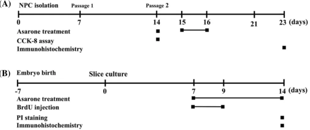
모체로부터 분리하였다. 태자의 뇌를 분리를 한 다음 다시 대뇌피질만을 취하였다. 이를 잘게 분쇄하여 원심분리기 와 70μM cell strainer(Corning Inc., One Riverfront Plaza, Corning, NY, USA)를 이용하여 신경전구세포만 분리 배 양하였다. 신경전구세포는 DMEM/F12(Gibco, Carlsbad, CA, USA)에 N2 supplement(Gibco), 1 M HEPES(Gibco), D-(+)-glucose(Sigma-Aldrich Corporation), bFGF(R&D system, Inc., Minneapolis, MN, USA), EGF(R&D sys- tem), Heparin(Sigma-Aldrich Corporation), amphotericin B(Sigma-Aldrich Corporation), 1% penicillin/streptomy- cin(Gibco)을 첨가하여 37℃ 5% CO2의 배양기에서 배양 하였다. 계대배양을 할 때 세포의 분화를 유도하기 위해 DMEM/F12에 10% FBS, N2 supplement, 1 M HEPES, D- (+)-glucose, amphotericin B, 1% penicillin/streptomycin 이 첨가된 배지에서 배양하였다. 신경전구세포에 대한 모 든 실험의 과정은 Fig. 1A에서 보는 바와 같다.
2. 해마조직절편의 제작 및 배양
출생 후 7일째 되는 신생 생쥐를 8% chloral hydrate (Sigma-Aldrich Corporation)를 이용해서 마취를 시킨 후, 뇌를 분리한 다음 뇌 부분 중 해마(hippocampus)만 을 따로 분리하였다. MEM(Gibco), 1 X HBSS(Hyclone, Logan, UT, USA), horse serum(Hyclone), 1 M HEPES (Gibco), D-(+)-glucose(Sigma-Aldrich Corporation)를 포함한 배지에서 vibratome(Campden Instruments LTD., Loughborough, Leics, England)을 이용해서 450μm의 해 마조직절편을 얻었다. 해마조직절편은 배지가 들어있는 insert(Merk-Millipore, Bredford, MA, USA)에 올려 37℃
5% CO2의 배양기에서 배양하였다. 해마조직절편배양에 대한 모든 실험의 과정은 Fig. 1B에서 보는 바와 같다.
(A)
(B)
Fig. 1. Schematic diagram for experimental procedures. All neural progenitor cells (A) and hippocampal slice culture (B) experiments were performed at the indicated time.
구세포의 세포증식률을 위해서 신경전구세포를 분리한지 14일부터 24시간 동안 α-asarone을 배지에 처리하였다.
α-asarone이 분화에 미치는 영향을 확인하기 위해서는 분 화를 유도한 신경전구세포에 1μM, 3μM 및 10μM 농도 로 배지에 3일간 처리하였다. Vehicle 그룹은 α-asarone과 동일한 기간 동안 10μM의 DMSO를 배지에 처리하였다.
실험결과는 일주일 후에 결과를 확인하였다. 해마조직절 편배양에 대한 처리는 배양 후 7일부터 7일 동안 10μM, 30μM 및 100μM 농도로 배지에 처리하였다. 이와 더불어 α-asarone 처리 첫날부터 3일 동안 5-Bromo-2ʹ-deoxyuri- dine(BrdU, Sigma-Aldrich Corporation)를 함께 처리하였 다.
4. Cell Counting Kit-8(CCK-8) 분석
세포의 증식률은 CCK-8(Dojindo, Rockville, MD, USA)을 사용하여 확인하였다. 96 well에 2×10⁴개의 세포 를 넣은 뒤 각 well 당 10μL씩 시약을 넣었다. 37℃ 5%
CO2의 배양기에서 1시간 동안 반응시켰다. 이후 spectro- photometer(Molecular Perices, Sunnyvale, CA, USA)를 이용하여 450nm 흡광도에서 세포 증식률을 측정하였다.
5. Propidium iodide(PI) 염색
배양한 해마조직절편은 α-asarone을 처리한 후 조직 내 손상을 확인하기 위해서 propidium iodide(PI, BD Biosci- ences, San Jose, CA, USA)를 처리하였다. 배지에 PI용액 을 처리한 후 빛을 차단하여 30분간 37℃ 5% CO2에 배양 하였다. 반응이 끝나면 PBS로 세척 한 후 형광현미경(Carl Zeiss Imager M1, Carl Zeiss Inc., Gottingen, Germany)으 로 확인하였다.
6. 면역세포조직화학적 염색
신경전구세포는 4% parafomaldehyde(PFA, Wako, Osa- ka, Japan)로 고정하고 PBS로 세척한 뒤 blocking serum (0.3% triton X-100, 1% BSA, 10% donkey serum in PBS) 을 처리 하였다. 1차 항체 nestin(R&D system), double- cortin(Dcx, Santa Cruz Biotechnology, Santa Cruz, CA, USA), glial fibrillary acidic protein(GFAP, Dako, Glostrup, Denmark), platelet-derived growth factor receptor-α(PDG- FR-α, Santa Cruz Biotechnology)를 사용하여 4℃에서 12 시간 처리하였다. 1차 항체 반응이 끝난 후 PBS로 씻어준
cein-conjugated goat anti-mouse IgG(Invitrogen corpo- ration, Carlsbad, CA, USA)와 Texas red-conjugated goat anti-rabbit 또는 mouse IgG(Life technology, Eugene, OR, USA)를 사용하였다. 반응이 끝나면 PBS로 세척 후 4℃
에 보관하고 confocal microscope(Carl Zeiss observer Z1, Carl Zeiss Inc)을 이용하여 형광염색을 확인하였다. α-asa- rone에 의한 치아이랑의 신경전구세포의 증식과 분화 를 알아보기 위해서 4% PFA로 고정하고 PBS로 세척 후 blocking serum을 처리하였다. 1차 항체인 BrdU(Bio-Rad, Hercules, CA, USA), Ki67(Abcam, Cambridge, UK) 및 Dcx(Santa Cruz Biotechnology)를 4℃에서 12시간 처리 하고, 반응 후 PBS로 세척한 뒤 2차 항체를 상온에서 2시 간 반응을 시켰다. 2차 항체는 fluorescein-conjugated goat anti-mouse IgG(Invitrogen corporation)와 Texas red-con- jugated goat anti-rabbit IgG(Life technology)를 사용하였 다. 반응이 끝나면 PBS로 세척한 후 봉입하여 confocal microscope(Carl Zeiss observer Z1)을 이용하여 형광염색 을 확인하였다.
7. 통계처리
실험 자료 분석은 SigmaPlot Statistical program version 11.2(Systat Sortware, San Jose, CA, USA)을 사용하였다.
각 실험데이터는 평균±표준오차로 나타내었고 모든 측정 값은 one-way analysis of variance(ANOVA)를 실시하여 F값을 구하였다. Turkey’s test를 이용하여 유의성을 검증 하였다. 통계적 유의성 판정을 위한 유의 수준은 P<0.05 로 하였다.
결 과
1. 신경전구세포의 세포증식률에 미치는 영향
CCK-8 분석을 통해 α-asarone이 신경전구세포의 증식 률에 미치는 영향을 알아보았다. Fig. 2에서 보는 바와 같 이, 대조군과 vehicle군에 비해 모든 α-asarone 처리군은 높은 세포증식률을 보여 주었다. 특히 3μM의 α-asarone 농도에서 가장 현저하였으나, 10μM 농도에서는 다소 감 소하는 경향을 보여 주었다. 이로 보아 α-asarone은 적정 농도에서 세포 증식을 보여주나 고농도에서는 세포 증식 이 저해되는 것을 확인하였다.
2. 신경전구세포의 분화에 미치는 영향
면역세포화학적 염색을 통해 α-asarone이 신경전구세포 의 분화에 미치는 영향을 알아보았다. 신경전구세포는 증 식을 거치며 분리 후 7일째에는 neurosphere를 형성하였 다. Fig. 3에서 보는 바와 같이, α-asarone처리군은 대조군
과 vehicle군에 비해 현저한 신경전구세포마크인 nestin의 발현을 관찰되었다. 이와 더불어 신경세포, 별아교세포, 희 소돌기아교세포의 분화 마크인 Dcx, GFAP, PDGFR-α를 nestin과 함께 이중염색으로 확인한 결과 모든 세포의 분 화마크가 확인되었다. 그러나 별아교세포, 희소돌기아교 세포의 분화마크인 GFAP, PDGFR-α와 nestin의 이중염색 이 미약한데 비해 신경세포 분화 마크인 Dcx와 nestin의 이중염색은 가장 현저하였다. 이러한 면역반응도 3μM의 α-asarone 농도에서 가장 높게 나타났다. 이로 보아 α-asa- rone은 신경전구세포의 분화에 영향을 주며 특히 신경세 포로의 분화를 촉진함을 알 수 있었다.
3. 해마조직절편의 손상에 대한 영향
배양한 해마조직절편에 대한 PI염색을 통해 α-asarone이 해마조직절편에 대한 손상을 확인하였다. Fig. 4에서 보는 바와 같이, 대부분의 α-asarone처리군은 대조군과 vehicle 군과 유사한 반응을 보여 주었다. 고농도 100μM의 α-asa- rone처리군은 다소 높은 PI 반응을 확인할 수 있었으나 조 직전반에 걸친 세포죽음과 손상은 관찰되지 않았다.
4. 해마 신경전구세포의 증식과 분화에 미치는 영향 해마의 치아이랑 과립아래층은 지속적인 줄기세포의 증
Fig. 3. Effect of α-asarone on the expression of differentiation markers in neural progenitor cells. Neural progenitor cells, isolated from embryonic mice(E15) brains, formed neurospheres. Immunofluorescent staining of neural progenitor cell marker nestin merged with neuro- blast marker Dcx, astrocyte marker GFAP and oligodenocyte marker PDGFR-α are presented. Scale bar=100μm.
Fig. 2. Effect of α-asarone on the proliferation of neural proge- nitor cells. α-asarone, especially at the 3μM concentration, sig- nificantly promoted the proliferation of neural progenitor cells.
All data are expressed as mean±SEM. *P<0.05 compared with control; #P<0.05 compared with vehicle.
식과 분화를 동반하므로 이에 대한 α-asarone의 영향을 알 아보았다. Fig. 5에서 보는 바와 같이, 분열하는 세포의 표
지인 Ki67은 BrdU와 함께 30μM 및 100μM의 α-asarone 처리군에서 증가하며 특히 30μM 처리군에서 현저하였 Fig. 4. Effects of α-asarone on the damage of hippocampal slice culture. A photomicrograph for PI-positive cells in the hippocampus.
Slight toxicity of α-asarone was detected at a concentration of 100μM. Scale bar=500μm.
Fig. 5. Effect of α-asarone on the proliferation of neural progenitor cells in the hippocampal slice culture system. A photomicrograph (A) and histogram (B) for BrdU and Ki67 positive cells. α-asarone promoted significantly proliferation of neural progenitor cells in the denate gyrus of the hippocampus at 30μM of α-asarone. Mean±SEM. *P<0.05 and **P<0.01 compared with control; #P<0.05 and ##P<0.01 compared with vehicle; $P<0.05 and $$$P<0.001 compared with 10μM/mL, &P<0.05 and &&&P<0.001 compared with 30μM. Scale bar=100μm.
(A) (B)
Fig. 6. Effect of α-asarone on the differentiation of neural progenitor cells in the hippocampal slice culture system. A photomicrograph (A) and histogram (B) for BrdU and Dcx positive cells. α-asarone at 30μM concentration significantly promoted neural progenitor cells differ- entiation into neuroblasts. Mean±SEM. **P<0.01 compared with control; ###P<0.001 compared with vehicle; $P<0.05 and $$P<0.01 compared with 100μM, &&&P<0.001 compared with 30μM/mL. Scale bar=20μm
(A) (B)
다. 이를 신생세포의 표지인 BrdU와 신경세포 분화마크인 Dcx를 복합염색 했을 때도 Fig. 6에서 보는 바와 같이 유 사한 결과를 보여 주었다. 또한 새로 증식분화된 신경아교 세포(BrdU와 Dcx 이중염색세포)가 30μM의 α-asarone처 리군에서 가장 높게 나타났다. 이로 보아, α-asarone은 해 마조직에서 신경전구세포로의 증식은 물론 신경세포로의 분화를 촉진함을 알 수 있었다.
고 찰
Asarone은 주로 Acorus 종의 뿌리줄기나 껍질에서 발견 되는 주요 식물화학물질(phytochemical)로서 많이 연구된 물질 중 하나이다[18]. α- 및 β-asarone 모두 항스트레스, 항울증, 항알츠하이머 및 항파킨슨 등 다양한 약리학적 효 능과 더불어 독성에 대한 보고가 있다[19,20]. 특히 예로부 터 치매 등 인지장애를 치료하는데 사용되어 온 석창포의 주요 활성성분인 α-asarone은 항경련제, 항불안제는 물론 신경보호작용 등의 다양한 신경생리학적 활성을 나타낸다 [12,20,21].
신경전구세포는 뇌병변 질환에서 급속히 증가하며 분화 됨으로서 신경기능회복에 관여한다[1,4,5]. 이들 세포는 손 상된 부위로 이동하여 세포에서 분비되는 다양한 성장인 자와 내인성 영양인자의 조절에 의해 혈관신생과 신경조 직발생에 영향을 준다[22,23]. 이러한 신경전구세포의 증 식과 분화는 만성 중추신경계질환과 더불어 노화나 만성 스트레스 등에 의해 감소한다[24-26]. 따라서 뇌병변 질환 에 대한 신경기능회복을 위해 이들 신경전구세포의 분화 와 증식을 위한 외부적 영양인자에 대한 연구는 이들 질환 의 예방과 치료에 좋은 전략적 타겟이 된다[27].
신경전구세포는 많은 신경변성질환의 기능회복에 관련 되어 있으므로, 우선 본 연구는 α-asarone이 신경전구세 포의 증식과 분화에 미치는 영향을 살펴보았다. 그 결과, α-asarone은 신경전구세포의 증식과 분화를 현저히 증가 하였다. 특히 세포의 분화마크를 이용한 검색에서 α-asa- rone은 별아교세포나 희소돌기아교세포보다 신경세포로의 분화를 더욱 촉진시키는 것을 확인할 수 있었다.
기관형 해마조직절편배양(organotypic hippocampal slice culture)은 in vitro 모델이나 in vivo 실험계를 잘 반영하고 있어 뇌병변 질환의 치료 기전과 전략에 좋은 모델이 된다 [28-30]. 해마조직절편의 배양은 조직특이적인 세포연결과 더불어 신경세포분포의 패턴과 특정 부분과의 연결회로 보존성이 뛰어나다[31]. 또한 생체와 유사하게 조직의 3차 원적 구조를 유지하면서 in vitro의 장점인 접근성의 용의
함을 가진다[32,33].
신경전구세포의 결과를 재확인하기 위하여, 배양한 해마 조직절편에 α-asarone을 처리하였다. α-asarone의 독성은 잘 알려져 있으나[18], 처리한 농도에서는 일부 100μM 농 도를 제외하고 조직손상을 보여 주지 않았다. 신경전구세 포의 결과와 유사하게 α-asarone은 해마의 신경전구세포 의 현저한 증식과 더불어 신경아교세포로의 분화를 촉진 하였다. 이는 α-asarone이 단일세포 뿐 아니라 기관형 조 직배양에서도 유사한 결과를 보여 줌을 알 수 있었다.
신경전구세포는 다양한 세포내외 인자에 의해 조절된 다[19]. 신경성장인자(neurotrophin)를 포함한 성장인자 (growth factor)는 물론 형태형성인자(morphogen) 등을 통 해 세포내 extracellular signal-regulated kinase(ERK), Akt 등의 특정 kinase cascade를 활성화함으로서 신경전구세포 의 증식과 분화를 촉진할 수 있다[34]. 최근에는 제한된 신 경전구세포의 증식과 분화를 조절하고 촉진할 수 있는 천 연물유래(herbal medicine) 외인성 인자에 대한 연구가 많 이 이루어지고 있다[19]. 본 연구에서 확인된 α-asarone의 신경전구세포와 해마조직절편의 세포증식과 분화에 대한 영향은 뇌병변에 대한 신경줄기세포치료에 적용할 가능성 을 보여 주고 있다.
결론적으로, α-asarone은 신경전구세포와 해마조직절편 에서 신경전구세포의 증식을 촉진시키며 신경세포로 분화 를 유도하는지 알 수 있었다. 따라서 α-asarone의 신경전 구세포에 대한 증식과 분화 효능은 알츠하이머, 파킨슨병, 뇌졸중 등과 같은 뇌병변 질환에 대한 줄기세포기반치료 에 적용할 수 있는 후보 약물이 될 수 있을 것이다.
REFERENCES
1. Geraerts M, Krylyshkina O, Debyser Z, Baekelandt V.
Concise review: therapeutic strategies for Parkinson dis- ease based on the modulation of adult neurogenesis, Stem Cells. 2007; 25:263-70.
2. Volkman R, Offen D. Concise Review: Mesenchymal stem cells in neurodegenerative diseases. Stem Cells. 2017;
35:1867-80.
3. Gage FH. Mammalian neural stem cells. Science. 2000;
287:1433-8.
4. Arvidsson A, Collin T, Kirik D, Kokaia Z, Lindvall O. Neu- ronal replacement from endogenous precursors in the adult brain after stroke. Nat Med. 2002; 8:963-70.
5. Parent JM, Vexler ZS, Gong C, Derugin N, Ferriero DM.
Rat forebrain neurogenesis and striatal neuron replacement after focal stroke. Ann Neurol. 2002; 52:802-13.
function. Adv Nutr. 2017; 8:804-11.
7. Han W, Xiong Y, Li Y, Fang W, Ma Y, Liu L, et al. Anti-ar- thritic effects of clematichinenoside(AR-6) on PI3K/Akt signaling pathway and TNF-alpha associated with colla- gen-induced arthritis. Pharm Biol. 2013; 51:13-22.
8. Rajput SB, Tonge MB, Karuppayil SM. An overview on traditional uses and pharmacological profile of Acorus cal- amus Linn.(Sweet flag) and other Acorus species. Phyto- medicine. 2014; 21:268-76.
9. Li M, Chen H. Antidepressant effect of water decoction of Rhizoma acori tatarinowii in the behavioural despair ani- mal models of depression. Zhong Yao Cai. 2001; 24:40-1.
10. Zhang H, Han T, Yu CH, Rahman K, Qin LP, Peng C.
Ameliorating effects of essential oil from Acori graminei rhizoma on learning and memory in aged rats and mice. J Pharm Pharmacol. 2007; 59:301-9.
11. Jiang J, Kim JJ, Kim DY, Kim MK, Oh NH, Koppula S, et al. Acorus gramineus inhibits microglia mediated neuroin- flammation and prevents neurotoxicity in 1-methyl-4-phe- nyl-1,2,3,6-tetrahydropyridine(MPTP)-induced mouse model of Parkinson’s disease. J Ethnopharmacol. 2012;
144:506-13.
12. Limon ID, Mendieta L, Diaz A, Chamorro G, Espinosa B, Zenteno E, et al. Neuroprotective effect of alpha-asarone on spatial memory and nitric oxide levels in rats injected with amyloid-beta(25-35). Neurosci Lett. 2009; 453:98- 13. Ma Y, Tian S, Sun L, Yao S, Liang Z, Li S, et al. The effect 103.
of acori graminei rhizoma and extract fractions on spatial memory and hippocampal neurogenesis in amyloid beta 1-42 injected mice. CNS Neurol Disord Drug Targets 2015;
14:411-20.
14. Manikandan S, Devi RS. Antioxidant property of al- pha-asarone against noise-stress-induced changes in differ- ent regions of rat brain. Pharmacol Res. 2005; 52:467-74.
15. Fang YQ, Shi C, Liu L, Fang RM. Pharmacokinetics of beta-asarone in rabbit blood, hippocampus, cortex, brain stem, thalamus and cerebellum. Pharmazie 2012; 67:120-3.
16. Pak ME, Kim YR, Kim HN, Ahn SM, Shin HK, Baek JU, et al. Studies on medicinal herbs for cognitive enhancement based on the text mining of Dongeuibogam and preliminary evaluation of its effects. J Ethnopharmacol. 2016; 179:383- 17. Lam KY, Chen J, Lam CT, Wu Q, Yao P, Dong TT, et al. 90.
Asarone from Acori Tatarinowii Rhizoma Potentiates the Nerve Growth Factor-Induced Neuronal Differentiation in Cultured PC12 Cells: A Signaling Mediated by Protein Ki- nase A. PLoS One 2016; 11:e0163337.
evidence. Phytomedicine. 2017; 32:41-58.
19. Mao J, Huang S, Liu S, Feng XL, Yu M, Liu J. A herbal medicine for Alzheimer’s disease and its active constituents promote neural progenitor proliferation. Aging Cell. 2015;
14:784-96.
20. Huang C, Li WG, Zhang XB, Wang L, Xu TL, Wu D. al- pha-asarone from Acorus gramineus alleviates epilepsy by modulating A-type GABA receptors. Neuropharmacology 2013; 65:1-11.
21. Kumar H, Kim BW, Song SY, Kim JS, Kim IS, Kwon YS.
Cognitive enhancing effects of alpha asarone in amnesic mice by influencing cholinergic and antioxidant defense mechanisms. Biosci Biotechnol Biochem. 2012; 76:1518- 22. Erlandsson A, Lin CH, Yu F, Morshead CM. Immunosup-22.
pression promotes endogenous neural stem and progenitor cell migration and tissue regeneration after ischemic injury.
Exp Neurol. 2011; 230:48-57.
23. Kolb B, Morshead C, Gonzalez C, Kim M, Gregg C, Shin- go T, et al. Growth factor-stimulated generation of new cor- tical tissue and functional recovery after stroke damage to the motor cortex of rats. J Cereb Blood Flow Metab. 2007;
27:983-97.
24. Donovan MH, Yazdani U, Norris RD, Games D, German DC, Eisch AJ. Decreased adult hippocampal neurogene- sis in the PDAPP mouse model of Alzheimer’s disease. J Comp Neurol. 2006; 495:70-83.
25. Drapeau E, Nora Abrous D. Stem cell review series: role of neurogenesis in age-related memory disorders. Aging Cell.
2008; 7:569-89.
26. Haughey NJ, Nath A, Chan SL, Borchard AC, Rao MS, Mattson MP. Disruption of neurogenesis by amyloid be- ta-peptide, and perturbed neural progenitor cell homeosta- sis, in models of Alzheimer’s disease. J Neurochem. 2002;
83:1509-24.
27. Lie DC, Song H, Colamarino SA, Ming GL, Gage FH.
Neurogenesis in the adult brain: new strategies for central nervous system diseases. Annu Rev Pharmacol Toxicol.
2004; 44:399-421.
28. Prager I, Patties I, Himmelbach K, Kendzia E, Merz F, Muller K, et al. Dose-dependent short- and long-term ef- fects of ionizing irradiation on neural stem cells in murine hippocampal tissue cultures: neuroprotective potential of resveratrol. Brain Behav. 2016; 6:e00548.
29. Wang X, Yu X, Xie C, Tan Z, Tian Q, Zhu D, et al. Rescue of brain function using tunneling nanotubes between neural stem cells and brain microvascular endothelial cells. Mol Neurobiol. 2016; 53:2480-8.
30. Ziemka-Nalecz M, Stanaszek L, Zalewska T. Oxygen-glu- cose deprivation promotes gliogenesis and microglia acti- vation in organotypic hippocampal slice culture: involve- ment of metalloproteinases. Acta Neurobiol Exp(Wars).
2013; 73:130-42.
31. Buchs PA, Stoppini L, Muller D. Structural modifications associated with synaptic development in area CA1 of rat hippocampal organotypic cultures. Brain Res Dev Brain Res. 1993; 71:81-91.
32. Mellentin C, Moller M, Jahnsen H. Properties of long-
term synaptic plasticity and metaplasticity in organotypic slice cultures of rat hippocampus. Exp Brain Res. 2006;
170:522-31.
33. Stoppini L, Buchs PA, Muller D. A simple method for or- ganotypic cultures of nervous tissue. J Neurosci Methods.
1991; 37:173-82.
34. Zhao Y, Li J, Liu Y, Yu KQ, Zhang J, Chen XG. Gu Ling Pian, a traditional Chinese medicine, regulates function and OPG/RANKL synthesis of osteoblasts via the p38 MAPK pathway. J Pharm Pharmacol. 2007; 59:1167-73.
Progenitor Cells
Hong Ju Lee
1,2, Byung Tae Choi
1,2,31Department of Korean Medical Science, School of Korean Medicine, Pusan National University
2BK21 Graduate Training Program of Korean Medicine for Healthy-Aging, School of Korean Medicine, Pusan National University
3Korean Medical Science Research Center for Healthy-Aging, Pusan National University
Abstract: This study investigated whether α-asarone could promote proliferation and differentiation of neural
progenitor cells into the neuronal cell types in in vitro and ex vivo studies.For in vitro assay, neural progenitor cells were isolated from fetal cerebral cortex(E15) and checked cell proliferation rate and neural progenitor cell marker in neurospheres. Treatment of α-asarone, particularly at a concentration of 3μM, promoted the proliferation of neural progenitor cells and effectively differentiated neural progenitor cells into neurons.
For ex vivo assay, a hippocampi slice culture system from 7 day postnatal rat fetuses was used. Although slight tissue damage was observed in the hippocampus after the high concentration(100μM) of α-asarone, however, α-asarone enhanced the proliferation of neural progenitor cells in dentate gyrus region and also effectively differentiated into neuroblast at concentration of 30μM.
Consequently, α-asarone promotes the proliferation of neural progenitor cells and effectively differentiates neural progenitor cells into neurons. Therefore, our results support the therapeutic benefits of α-asarone for treating neurodegenerative diseases.
Keywords: α-asarone, Neural progenitor cell, Hippocampus, Proliferation, Differentiation
Correspondence to : Byung Tae Choi(Department of Korean Medical Science, School of Korean Medicine, Pusan National University) E-mail : [email protected]