INTRODUCTION
The bronchial epithelium has traditionally been recognized as a physical barrier protecting the host from its environment. How- ever, epithelial cells play a central role in the regulation of airway immunity, affecting inflammation and host defenses in diseas- es of the airway. Epithelial cells release a wide range of proin- flammatory mediators and multifunctional cytokines in re- sponse to exposure to inhaled environmental factors or micro- organisms. The precise mechanisms are not fully understood, but epithelial cells are thought to play a major part in the regu- lation of host inflammatory status as well as airway structure and function.1,2
Lipid rafts are subdomains of the epithelial cell membrane that contain high concentrations of cholesterol and glycosphingo- lipids. They interact with one another and pack tightly together to form cell membrane structures. Thus, lipid rafts provide a platform for multiple signaling pathways and act as key modu- lators of certain disease pathways.3 Lipid entities render lipid rafts insoluble in nonionic detergents and cause them to sepa-
Effect of Cholesterol Depletion on Interleukin-8 Production in Human Respiratory Epithelial Cells
Min Jung Kim, Jung Yeon Hong, Kyung Eun Lee, Kyung Won Kim, Myung Hyun Sohn, Kyu-Earn Kim*
Department of Pediatrics and Institute of Allergy, Severance Medical Research Institute, Brain Korea 21 Project for Medical Science, Yonsei University College of Medicine, Seoul, Korea
rate from their surroundings. Cholesterol, the most abundant lipid component of animal cell membranes, regulates mem- brane fluidity and plays a crucial role in the formation and sta- bilization of membrane microdomains. It is also an important contributor to cell-cell adhesion, migration, and even endocy- tosis.4-7 However, despite increasing interest in the bronchial epithelium, the possible role of cholesterol in inflammation of the airway or the development of asthma has not been investi- gated.
Among the numerous cytokines and chemokines released from human airways, interleukin-8 (IL-8) is a representative Allergy Asthma Immunol Res. 2013 November;5(6):402-408.
http://dx.doi.org/10.4168/aair.2013.5.6.402 pISSN 2092-7355 • eISSN 2092-7363
Purpose: The lipid entities of cell membranes are components of the immune system and important mediators of inflammation. Despite increasing interest in the function of epithelial cells in inflammation, the role of cholesterol in this process has not been described. Here, we investigated the effect of cholesterol depletion on the inflammatory process in airway epithelial cells via the expression of interleukin (IL)-8 as a marker of inflamma- tion. Methods: A 549 cells were treated with 0.5% methyl-β-cyclodextrin as a selective cholesterol extractor. The IL-8 level was assessed by en- zyme-linked immunosorbent assay and reassessed after cholesterol repletion. Mitogen-activated protein kinase (MAPK) inhibitors were used to de- termine the upstream signaling pathway for IL-8 production in cholesterol-depleted cells. Results: We found a relationship between the amount of cholesterol in A 549 cells and inflammation of the airway. IL-8 production was increased in cholesterol-depleted A 549 cells and restored by cho- lesterol repletion. IL-8 production was decreased by pretreatment with the extracellular signal-regulated kinase (ERK) inhibitor U0126 but not with JNK inhibitor II or the p38 MAPK inhibitor SB202190. Conclusions: Our findings suggest that inflammatory responses are increased in cholester- ol-depleted epithelial cells via the MAPK signaling system, predominantly by the ERK pathway. We conclude that the lipid components of airwayep- ithelial cells may play a role in the inflammatory process.
Key Words: Cholesterol; epithelial cell; inflammation; interleukin-8; MAP kinase signaling system
This is an Open Access article distributed under the terms of the Creative Commons Attribution Non-Commercial License (http://creativecommons.org/licenses/by-nc/3.0/) which permits unrestricted non-commercial use, distribution, and reproduction in any medium, provided the original work is properly cited.
Correspondence to: Kyu-Earn Kim, MD, PhD, Department of Pediatrics and Institute of Allergy, Brain Korea 21 Project for Medical Science, Yonsei University College of Medicine, 50 Yonsei-ro, Seodaemun-gu, Seoul 120-752, Korea.
Tel: +82-2-2019-3353; Fax: +82-2-3461-9473; E-mail: kekim@yuhs.ac Received: September 14, 2012; Revised: November 26, 2012 Accepted: December 26, 2012
•Min Jung Kim and Jung Yeon Hong contributed equally to this work.
•There are no financial or other issues that might lead to conflict of interest.
chemokine expressed by bronchial epithelial cells. IL-8 medi- ates cell migration during inflammation of the airway.8,9 In ad- dition, patients with severe asthma have increased levels of IL-8 in their BAL fluids. In addition, various stimuli, including house dust mites, cockroaches, and microbes, induce IL-8 production in bronchial epithelial cells and promote inflammation.10-13
Here, we investigated the effect of cholesterol depletion in air- way epithelial cells on the production of IL-8 and its association with inflammation of the airway.
MATERIALS AND METHODS Cell culture
The human epithelial-like lung carcinoma cell line A 549 was obtained from the American Type Culture Collection (Manas- sas, VA, USA). Cells were cultured in F12K medium (Sigma, St.
Louis, MO, USA) supplemented with 10% fetal bovine serum containing 100 U/mL penicillin and streptomycin (GibcoBRL, Grand Island, NY, USA). At all stages of culture, the cells were maintained in an incubator at 37°C with 5% CO2.
Cholesterol depletion and repletion
Methyl-β-cyclodextrin (MβCD; Sigma) binds specifically to cholesterol to disturb the association of proteins with lipid rafts.14 It is therefore presumed to change the structure and function of the cell membrane by disrupting lipid rafts.15-17 A stock solution of 10% MβCD in phosphate-buffered saline (PBS) was stored at 4°C. This solution was used at concentrations of 0.5, 1, and 2%
(v/v). After serum starvation for 24 h, cells were incubated with the indicated concentrations of MβCD for 1 h at 37°C for cho- lesterol depletion. The culture medium was replaced with fresh serum-starved medium at the indicated times, and the cells were maintained at 37°C in an incubator with 5% CO2. For cholester- ol repletion, MβCD-treated cells were incubated for 1 h in the presence of 70 μg/mL cholesterol and 0.2% MβCD. The cells were then further incubated in fresh serum-free medium in an incubator.
Cell viability
A 549 cell viability at various concentrations of MβCD was measured with a Cell Counting Kit-8 (Dojindo, Kumamoto, Ja- pan). The day before the experiment, 100 μL cells were seeded into 96-well microplates at a density of 1×10⁴ cells per well. Af- ter 24 h of incubation, 10 μL cells per well were treated with var- ious concentrations of MβCD for 1 h, followed by incubation with an additional 10 μL Cell Counting Kit-8 solution for 1 h. The absorbance was then measured at 450 nm with an enzyme-linked immunosorbent assay (ELISA) reader; the compensated absor- bance was 590 nm.
Lipid extraction and cholesterol assay
A 549 cells pretreated with MβCD to deplete cholesterol were
washed in cold PBS buffer and extracted with Folch solution containing chloroform and methanol at a 2:1 ratio. The choles- terol released by the Folch method was solubilized in the organ- ic phase, and the amount of cholesterol was measured with a cholesterol probe following vaporization of the organic phase.
The organic phase was dried under a speed vacuum (Savant In- struments Inc., New York, NY, USA) for 30 min, then solubilized with 2-propanol and 10% Triton X-100. Membrane cholesterol was assayed using a Cholesterol/Cholesteryl Ester Quantitation Kit (Calbiochem-Novabiochem, La Jolla, CA, USA), according to the manufacturer’s instructions, Samples and standards were loaded and reacted by the addition of a reaction mixture con- taining cholesterol buffer, probe, and enzyme without light for 1 h. The absorbance at 570 nm was measured with an ELISA reader; the compensated absorbance was 590 nm. A BCA Pro- tein Assay Kit (Pierce, Rockford, IL, USA) was used to quantify the extracted proteins, which were collected with Mammalian Protein Extraction Reagent (Pierce) and normalized to the vol- ume.
Measurement of human IL-8 and MAP kinase (MAPK) assay After treatment with 0.5% MβCD for 1 h, cells were treated with fresh serum-free medium and incubated for 2, 5, 10, and 24 h at 37°C. They were then stored at -70°C until they were assayed.
The IL-8 levels in the culture supernatants were determined us- ing a specific ELISA against human IL-8 (R&D Systems, Minne- apolis, MN, USA) according to the manufacturer’s instructions.
To investigate the effect of changes in transcription factor activ- ity on IL-8 expression in human lung epithelial cells, the selec- tive MAP/extracellular signal-regulated kinase (ERK) [MEK] in- hibitor PD98059 or U0126, c-JUN NH2-terminal kinase (JNK) inhibitor JNK II Inhibitor, or p38 MAPK inhibitor SB203580 or SB202190 (Calbiochem-Novabiochem) was added 1 h before stimulation with MβCD and was present throughout the incu- bation period.
RNA preparation and real-time polymerase chain reaction (PCR)
Reverse transcription-PCR (RT-PCR) was used to examine the mRNA expression of IL-6, IL-8, and TNF-α. Total RNA was iso- lated from cells using Trizol reagent (Invitrogen, Carlsbad, CA, USA) prior to cDNA synthesis. PCR amplification was carried out using specific primer pairs. The primer sequences used were obtained from Primer Bank (http://pga.mgh.harvard.edu/prim- erbank). Fold changes were calculated using the comparative ΔΔCt method. The gene encoding glyceraldehyde-3-phosphate dehydrogenase (GADPH) was analyzed as an internal control in each sample.
Western blot analysis
MβCD-treated cells were lysed with lysis buffer (50 mM HEPES, pH 7.5; 150 mM NaCl; 1 mM EDTA; 10% glycerol; and 0.5% NP-
40). Protein samples were mixed with 5× SDS-PAGE buffer con- taining β-mercaptoethanol and heated at 95°C for 5 min. Whole- cell lysates (30 μg) were separated by 10% SDS-PAGE, then elec- trotransferred to nitrocellulose membranes (GE Healthcare, Stockholm, Sweden). The membranes were blocked for 1 h with 5% skim milk in TBST buffer (10 mM Tris-HCl, pH 7.5; 150 mM NaCl; and 0.2% Tween 20), then washed three times with TBST at room temperature. The blots were incubated overnight with specific antibodies (1:1,000) in 5% skim milk in TBST buffer at 4°C. After incubation with secondary antibodies (1:2,000), the signal was detected using the ECL Plus (GE Healthcare) and CL- XPosureTM film (Thermo Scientific, Waltham, MA, USA) or an ImageQuantTM LAS 4,000 Mini Biomolecular Imager (GE Health- care).
Statistical analysis
All data are expressed as the mean±SEM of at least 3 individ- ual experiments. A statistical analysis comparing the treatment and control groups was carried out using Student’s t-test.
P<0.05 was considered significant.
RESULTS
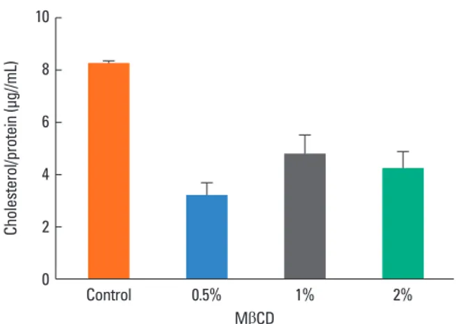
Induction of cholesterol depletion by MβCD in A 549 cells We estimated the amount of cholesterol in the growth media of control and MβCD-treated cultures. After treatment with 0.5, 1, or 2% MβCD for 1 h at 37°C, the cells showed a decreased amount of cholesterol (3.15±0.52, 4.73±0.75, and 4.17±0.68 μg/mL, respectively) compared to untreated cells (8.22±0.11 μg/mL). However, there was no significant difference in the amount of cholesterol among the MβCD-treated groups. In ad- dition, the amount of cholesterol in the cell was at its lowest fol- lowing treatment with 0.5% MβCD, showing an approximately 60% decrease compared to the control (Fig. 1). Subsequent ex-
periments were thus carried out using epithelial cells treated with 0.5% MβCD.
Alteration of cell viability by MβCD
To determine whether cholesterol depletion by MβCD had any effect on cell viability, we compared the untreated cells to the A 549 cell cultures treated with 0.5, 1, or 2% of MβCD for 1 h.
There were no dramatic changes in overall cell viability com- pared to the control (96.8, 95.3, and 94.2%, respectively, Fig. 2) but statistical differences showed in 1 and 2% of MβCD-treated cells. We concluded that 0.5% MβCD should be used in our subsequent experiments.
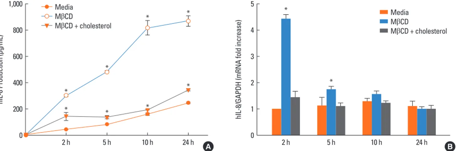
Effect of cholesterol depletion on human IL-8 production The effect of cholesterol depletion on IL-8 production was ex- amined in airway epithelial cells treated with 0.5% MβCD. Cho- lesterol-depleted cells produced more IL-8 than control cells at different time points (2, 5, 10, and 24 h). IL-8 production in- creased in a time-dependent manner, with maximum produc- tion at 24 h. Cells treated with 0.5% MβCD produced 300.4±7.9 pg/mL at 2 h, 477.7±1.5 pg/mL at 5 h, 816.7±55.2 pg/mL at 10 h, and 867.6±38.9 pg/mL at 24 h compared to 40.8±2.3 pg/mL at 2 h, 80.4±0.2 pg/mL at 5 h, 158.2±8.3 pg/mL at 10 h, and 244.6±6.0 pg/mL at 24 h for control cells. We verified that the IL-8 level decreased after treatment with both 0.5% MβCD and 70 μg/mL cholesterol, similar to in the control cells (Fig. 3A).
When tested by RT-PCR with GAPDH as an internal control, the A 549 cells depleted of cholesterol with 0.5% MβCD showed greater IL-8 mRNA expression. IL-8 expression peaked at 2 h and returned to baseline 24 h after MβCD stimulation (Fig. 3B).
Role of MAPK signaling in IL-8 production
To investigate the link between IL-8 production in cholester- ol-depleted cells and the MAPK signaling pathway, which is in-
Cholesterol/protein (μg//mL)
10
8
6
4
2
0 Control 0.5% 1% 2%
MβCD
Fig. 1. Determination of the cholesterol level in untreated and MβCD-treated A 549 cells. The amount of cholesterol in the cell decreased after MβCD treatment.
Each bar represents the mean±SEM of 3 independent experiments. Cholester- ol in the cell was at its lowest level following treatment with 0.5% MβCD.
* *
Cell viability (% of control)
10
8
6
4
2
0 Control 0.5% 1% 2%
MβCD
Fig. 2. Cholesterol depletion by 0.5% MβCD does not significantly change A 549 cell viability. A 549 cells preincubated with the indicated concentrations of 0.5% MβCD for 1 h did not show any significant change in viability compared to control cells. The data are expressed as the mean±SEM showing 96.8, 95.3, and 94.2% cell viability. *P<0.05 vs. the control alone.
volved in airway inflammatory diseases, we examined the ef- fect of MAPK inhibitors on IL-8 production. IL-8 production was decreased by pretreatment with the ERK inhibitor U0126 (50 μM), but JNK inhibitor II (100 μM) and the p38 MAPK in- hibitor SB202190 (50 μM) did not significantly reduce IL-8 pro- duction (Fig. 4A). The level of IL-8 was decreased by almost 80%
by U0126 but by only 10.9 and 31.4% by JNK inhibitor II and SB202190, respectively, compared to MβCD treatment alone.
To demonstrate the intracellular activation of MAPK signaling, we examined the phosphorylation of ERK, p38 MAPK, and JNK in MβCD-treated cells from 0 to 60 min. As shown in Fig. 4B, we detected small amounts of phosphorylated ERK 1/2 MAPK in untreated A 549 cells, whereas treatment with MβCD induced ERK 1/2 MAPK phosphorylation. ERK 1/2 MAPK activation was maximized at 30 min, but neither p38 MAPK nor JNK activation was detected.
Effect of cholesterol depletion on the production of other cytokines
To identify the effect of cholesterol depletion on inflammato- ry responses in the airway, we examined the production of oth- er cytokines released from A 549 cells. When tested by RT-PCR with GAPDH as an internal control, cholesterol depletion by MβCD resulted in elevated IL-6 and TNF-α mRNA expression in the A 549 cells. IL-6 and TNF-α expression peaked 2 h after MβCD stimulation (Fig. 5A and B).
DISCUSSION
The epithelium, which represents the first contact surface with the external environment, forms a tight and impermeable bar- rier for the maintenance of tissue homeostasis under normal circumstances.1 The structural integrity of the epithelium is also
Fig. 3. Effects of cholesterol depletion on IL-8 production in airway epithelial cells. (A) A 549 cells were treated with 0.5% MβCD and the supernatants were assessed by ELISA after 2, 5, 10, and 24 h. IL-8 production was increased in cholesterol-depleted cells and recovered after cholesterol repletion. (B) The induction of IL-8 mRNA expression in human respiratory epithelial cells. Total RNA was isolated and reverse-transcribed, and the resulting cDNA was amplified by real-time PCR. Cells stim- ulated with 0.5% MβCD showed enhanced IL-8 mRNA expression. *P<0.05 vs. the control.
hIL-8 Production (pg/mL)
1,000
800
600
400
200
0 2 h 5 h 10 h 24 h
Media MβCD
MβCD + cholesterol *
*
*
*
*
*
*
*
A
hIL-8/GAPDH (mRNA fold increase)
5
4
3
2
1
0
B
*
*
2 h 5 h 10 h 24 h
Media MβCD
MβCD + cholesterol
Fig. 4. Effects of pretreatment with MAPK inhibitors on MβCD-induced IL-8 production. (A) IL-8 production was decreased by pretreatment with the ERK inhibitor U0126 (50 μM) but not by JNK inhibitor II (100 μM) or the p38 MAPK inhibitor SB202190 (50 μM). **P<0.01 vs. MβCD-treated cells. (B) Phosphorylation assays of MAPK activity. Cells treated with MβCD were harvested at the indicated time points and then lysed. Equal amounts of the cell extracts were resolved on 10% acryl- amide gels and then subjected to Western blot analysis.
IL-8 production (pg/mL)
800
600
400
200
0
*
Control 0.5% MβCD
Media U0126 JNKII SB202190
A
Time (min) P-ERK 1/2 ERK 1/2 P-p38 p38 P-JNK JNK β-actin
0 15 30 45 60
B
maintained through cell-cell and cell-extracellular matrix inter- actions. However, air pollutants or infectious particles can in- crease the permeability of the epithelium and penetrate the air- way, inducing epithelial cell damage or death. Allergens such as house dust mites and pollen can also disrupt the integrity of the mucosa through the direct extracellular cleavage of tight junctions.2 Such allergens also enhance inflammatory pathways that make the epithelium interact with other cells.18,19 In addi- tion, epithelial cells can recruit and modulate immune cells to cause persistent inflammation through adhesive interactions, selective cytokine release, and cell-specific receptor expression.
Thus, epithelial cells can modulate the structure and function of the airway by contributing to host defenses and innate im- munity.1,2
A recent innovation in our understanding of the role of epi- thelial cells in inflammation of the airway has to do with the in- fluence of impaired innate immunity and epithelial cell injury on the progression of inflammation, especially in the case of asthma.18-20 The airway epithelium in asthma patients is dam- aged, with the disruption of tight junctions accompanied by a loss of junctional proteins in response to environmental factors such as dust mite allergens, fungal allergens, viral infections, and smoke.21,22 The damaged epithelium results in barrier dys- function, epithelial fragility, and abnormal repair responses in the asthmatic airway, creating a condition that is similar to in- flammatory conditions of the skin such as atopic dermatitis or those of the gut in the case of food allergy.18,19 Many studies have focused on compromised tight junctions and epithelial cell protein complexes to determine the relationship between asth- ma and the airway epithelium.18-22 However, the role of lipid components of the epithelial cell membrane in inflammation of the airway has not been comprehensively studied.
Lipid rafts are dynamic nanoscale membrane microdomains that contain large amounts of cholesterol and sphingolipids in- volved in signaling platforms.3 Cholesterol, an essential compo-
nent of the plasma membrane, has been implicated in the reg- ulation of membrane fluidity, permeability, and cell surface re- ceptor function.4-6 Because cholesterol contributes to the estab- lishment of a cornified barrier, it plays a crucial role in keratino- cytes. Atopic dermatitis has even been shown to occur in paral- lel with keratinocytes disrupted by lipid rafts.17 We hypothesize that inflammatory diseases of the airway are influenced by air- way epithelial cells with disrupted lipid rafts, similar to kerati- nocytes in atopic dermatitis.
To clarify the relationship between cholesterol and inflamma- tion of the airway, we used MβCD to damage lipid rafts in airway epithelial cells. We identified a concentration of MβCD capable of inducing significant changes in cholesterol levels in the cell.
We then demonstrated that the amount of cholesterol in the cell was significantly decreased after incubation with 0.5% MβCD, without effect on cell viability.
IL-8 is a major chemokine of the CXC chemokine subfamily that acts as a chemoattractant for neutrophils released from the bronchial epithelium.8 IL-8 plays a key role in triggering and perpetuating inflammation in patients with severe asthma. In addition, the expression and release of IL-8 increase in response to other stimuli such as allergens and infectious particles.10-13 Hence, we used IL-8 as an inflammatory marker in this study, and IL-8 production rose in cholesterol-depleted epithelial cells. Based on these results, we recognize that IL-8 production in human respiratory epithelial cells could be affected by the cholesterol content in the membrane through the depletion of cholesterol in the cell in the absence of any other allergen.
MAPK regulates IL-8 expression and release in response to various allergens and viruses in airway epithelial cells, resulting in an influence on cell signaling pathways in diseases of the air- way. Among ERK, JNK, and p38 MAPK, ERK-regulated IL-8 production has been suggested as a distinct signaling process in chronic diseases of the airway.10-13 We investigated the effect of MAPK inhibitors on IL-8 production using U0126, JNK inhib- Fig. 5. Effect of cholesterol depletion on IL-6 and TNF-α mRNA expression. (A) A 549 cells were treated with 0.5% MβCD and/or 70 μg/mL cholesterol for the indicat- ed time. Total RNA was isolated and reverse-transcribed, and the resulting cDNA was amplified by real-time PCR. The cholesterol-depleted cells showed enhanced proinflammatory cytokine release. The mRNA expression of IL-6 (A) and TNF-α (B) increased and peaked at 2 h following MβCD treatment. *P<0.05 vs. the control.
hIL-6/GAPDH (mRNA fold increase)
4
3
2
1
0
A
*
2 h 5 h 10 h 24 h
Media MβCD
MβCD + cholesterol
hTNF-
α/GAPDH (mRNA fold increase) 5
4
3
2
1
0
B
*
*
2 h 5 h 10 h 24 h
Media MβCD
MβCD + cholesterol
itor II, and SB202190 and sought to determine whether IL-8 production in cholesterol-depleted cells was affected by the MAPK signaling pathway. Our results suggest that activation of the ERK pathway is a relatively strong contributor to IL-8 pro- duction in cholesterol-depleted cells, in agreement with previ- ous studies of allergen-induced inflammation of the airway.10-13 In addition, we investigated the expression of other cytokines generated by airway epithelial cells. IL-6 can stimulate T cells and contribute to the development of Th2-mediated inflamma- tion. It was previously demonstrated that epithelial IL-6 expres- sion increased after exposure to fungi, house dust mites, or com- mon respiratory viruses.1 In addition, airway epithelial cells pro- duce pleiotropic cytokines, including TNF-α, which plays an im- portant role in severe refractory asthma. Targeting TNF-α may be a novel therapeutic approach to treating asthma.23 We found that the mRNA expression of IL-6 and TNF-α was increased in cholesterol-depleted epithelial cells. Cholesterol depletion in the airway epithelium may be related to inflammatory respons- es through the production of several inflammatory products.
Genome expression profiling has been performed in choles- terol-depleted keratinocytes from patients with atopic dermati- tis.17 What is interesting is that patients with genetic differences tend to be susceptible to both this condition and other related allergic diseases.24,25 Such large-scale genome profiling and ge- netic experiments have confirmed that the disruption of lipid rafts can lead to inflammatory disease under certain circum- stances.
In conclusion, our findings suggest that the disruption of lipid rafts plays an important role in inflammation of the airway.
Bronchial epithelial cells with cholesterol depletion released a large amount of IL-8, and IL-8 production was regulated by the MAPK signaling pathway, predominantly through ERK. In ad- dition, cholesterol depletion in airway epithelial cells induced the elevation of other proinflammatory cytokines generated by the airway epithelium. Considering that the bronchial epitheli- um may be a novel target for new therapies for inflammatory diseases of the airway, additional studies of cholesterol and lip- id rafts in human airway epithelial cells are needed.
ACKNOWLEDGMENTS
This study was supported by a faculty research grant (6-2007- 0098) from the Yonsei University College of Medicine for 2007.
REFERENCES
1. Proud D, Leigh R. Epithelial cells and airway diseases. Immunol Rev 2011;242:186-204.
2. Holgate ST. The sentinel role of the airway epithelium in asthma pathogenesis. Immunol Rev 2011;242:205-19.
3. Pike LJ. Lipid rafts: bringing order to chaos. J Lipid Res 2003;44:655- 67.
4. Chang TH, Segovia J, Sabbah A, Mgbemena V, Bose S. Cholesterol-
rich lipid rafts are required for release of infectious human respira- tory syncytial virus particles. Virology 2012;422:205-13.
5. Saeki K, Fukuyama S, Ayada T, Nakaya M, Aki D, Takaesu G, Hana- da T, Matsumura Y, Kobayashi T, Nakagawa R, Yoshimura A. A ma- jor lipid raft protein raftlin modulates T cell receptor signaling and enhances Th17-mediated autoimmune responses. J Immunol 2009;
182:5929-37.
6. San-Juan-Vergara H, Sampayo-Escobar V, Reyes N, Cha B, Pacheco- Lugo L, Wong T, Peeples ME, Collins PL, Castaño ME, Mohapatra SS. Cholesterol-rich microdomains as docking platforms for respi- ratory syncytial virus in normal human bronchial epithelial cells. J Virol 2012;86:1832-43.
7. Abraham SN, Duncan MJ, Li G, Zaas D. Bacterial penetration of the mucosal barrier by targeting lipid rafts. J Investig Med 2005;53:318- 21.
8. Strieter RM. Interleukin-8: a very important chemokine of the hu- man airway epithelium. Am J Physiol Lung Cell Mol Physiol 2002;
283:L688-9.
9. Mukaida N, Harada A, Matsushima K. Interleukin-8 (IL-8) and monocyte chemotactic and activating factor (MCAF/MCP-1), che- mokines essentially involved in inflammatory and immune reac- tions. Cytokine Growth Factor Rev 1998;9:9-23.
10. Hong JY, Lee KE, Kim KW, Sohn MH, Kim KE. Chitinase induce the release of IL-8 in human airway epithelial cells, via Ca2+-dependent PKC and ERK pathways. Scand J Immunol 2010;72:15-21.
11. Lee KE, Kim JW, Jeong KY, Kim KE, Yong TS, Sohn MH. Regulation of German cockroach extract-induced IL-8 expression in human airway epithelial cells. Clin Exp Allergy 2007;37:1364-73.
12. Sohn MH, Lee KE, Kim KW, Kim ES, Park JY, Kim KE. Calcium- calmodulin mediates house dust mite-induced ERK activation and IL-8 production in human respiratory epithelial cells. Respiration 2007;74:447-53.
13. Sohn MH, Lee KE, Choi SY, Kwon BC, Chang MW, Kim KE. Effect of Mycoplasma pneumoniae lysate on interleukin-8 gene expres- sion in human respiratory epithelial cells. Chest 2005;128:322-6.
14. Pontes Soares C, Portilho DM, da Silva Sampaio L, Einicker-Lamas M, Morales MM, Costa ML, Dos Santos Mermelstein C. Membrane cholesterol depletion by methyl-beta-cyclodextrin enhances the expression of cardiac differentiation markers. Cells Tissues Organs 2010;192:187-99.
15. Kabouridis PS, Janzen J, Magee AL, Ley SC. Cholesterol depletion disrupts lipid rafts and modulates the activity of multiple signaling pathways in T lymphocytes. Eur J Immunol 2000;30:954-63.
16. Chen X, Resh MD. Cholesterol depletion from the plasma mem- brane triggers ligand-independent activation of the epidermal growth factor receptor. J Biol Chem 2002;277:49631-7.
17. Mathay C, Pierre M, Pittelkow MR, Depiereux E, Nikkels AF, Colige A, Poumay Y. Transcriptional profiling after lipid raft disruption in keratinocytes identifies critical mediators of atopic dermatitis pathways. J Invest Dermatol 2011;131:46-58.
18. Hackett TL, Knight DA. The role of epithelial injury and repair in the origins of asthma. Curr Opin Allergy Clin Immunol 2007;7:63-8.
19. Holgate ST. Epithelium dysfunction in asthma. J Allergy Clin Im- munol 2007;120:1233-44.
20. Davies DE. The role of the epithelium in airway remodeling in asthma. Proc Am Thorac Soc 2009;6:678-82.
21. Holgate ST, Davies DE, Lackie PM, Wilson SJ, Puddicombe SM, Lor- dan JL. Epithelial-mesenchymal interactions in the pathogenesis of asthma. J Allergy Clin Immunol 2000;105:193-204.
22. Swindle EJ, Collins JE, Davies DE. Breakdown in epithelial barrier function in patients with asthma: identification of novel therapeu- tic approaches. J Allergy Clin Immunol 2009;124:23-34.
23. Brightling C, Berry M, Amrani Y. Targeting TNF-alpha: a novel therapeutic approach for asthma. J Allergy Clin Immunol 2008;121:
5-10.
24. Henderson J, Northstone K, Lee SP, Liao H, Zhao Y, Pembrey M, Mukhopadhyay S, Smith GD, Palmer CN, McLean WH, Irvine AD.
The burden of disease associated with filaggrin mutations: a popu- lation-based, longitudinal birth cohort study. J Allergy Clin Immu- nol 2008;121:872-7.e9.
25. Marenholz I, Nickel R, Rüschendorf F, Schulz F, Esparza-Gordillo J, Kerscher T, Grüber C, Lau S, Worm M, Keil T, Kurek M, Zaluga E, Wahn U, Lee YA. Filaggrin loss-of-function mutations predispose to phenotypes involved in the atopic march. J Allergy Clin Immu- nol 2006;118:866-71.