23
마우스 혈장과 조직에서의 doxorubicin 측정 HPLC-MS/MS 방법
박정선a#·김혜경b·이혜원b·이미현b·김현기a·채수완a,b·채한정a,b
a전북대학교, 의과대학 약리학교실 및 심혈관연구소,
b전북대학교병원 기능성식품 임상시험지원센타
Validation of a HPLC MS/MS Method for Determination of Doxorubicin in Mouse Serum and its Small Tissues
Jung-Sun Park
a#, Hye-Kyung Kim
b#, Hye-Won Lee
b, Mi-Hyun Lee
b, Hyun-Gi Kim
a, Soo-Wan Chae
a,b, Han-Jung Chae
a,ba
Department of Pharmacology and Cardiovascular Research Center, Medical School, Chonbuk National University,
b
Clinical Trial Center for Functional Foods, Chonbuk Hospital, Jeonju, Chonbuk, Korea
(
# equally contributed Jung-Sun Park
1#, Hye-Kyung Kim
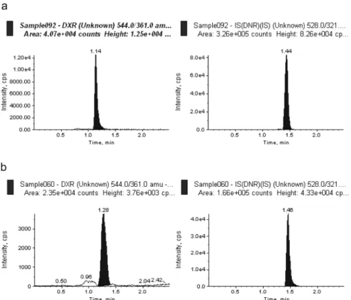
2#)Doxorubicin (DXR) is a type of anti-cancer drug called an “anthracycline glycoside”. It works by impairing DNA synthesis, a crucial feature of cell division, and thus is able to target rapidly dividing cells. Doxorubicin is a very serious anti-cancer medication with definite potential to do great harm as well as great good. A liq- uid chromatography-tandem mass spectroscopy (LC–MS/MS) method was developed to identify and quantify DXR in small-volume biological samples. After the addition of internal standard (IS, 5
µL of 1
µM/ml daunorubicin methanol solution) into the serum sample, the drug and IS were extracted by methanol. Following vortex for a 1 min and centrifugation at 15,000g for 10 min the organic phase was transferred and evaporated under a vac- uum. The residue was reconstituted with 350
µL of mobile phase and 10
µL was injected into C18 column with mobile phase composed of 0.05 M ammonium acetate (0.1 M acetic acid adjusted to pH 3.5) and aceto- nitrile (40:60, v/v). The flow rate was kept constant at 350
µL/min. The ions were quantified in the multiple reaction mode (MRM), using positive ions, on a triple quadrupole mass spectrometer. The lower limits of quantification for Doxorubicin in plasma and small tissues were approximately 0.5 ng/mL and 0.5 ng/mL respectively. Intra- and inter-assay accuracy (% of nominal concentration) and precision (% CV) for all ana- lytes were within 15%, respectively.
□ Key words - doxorubicin, daunorubicin, HPLC-MS/MS, validation
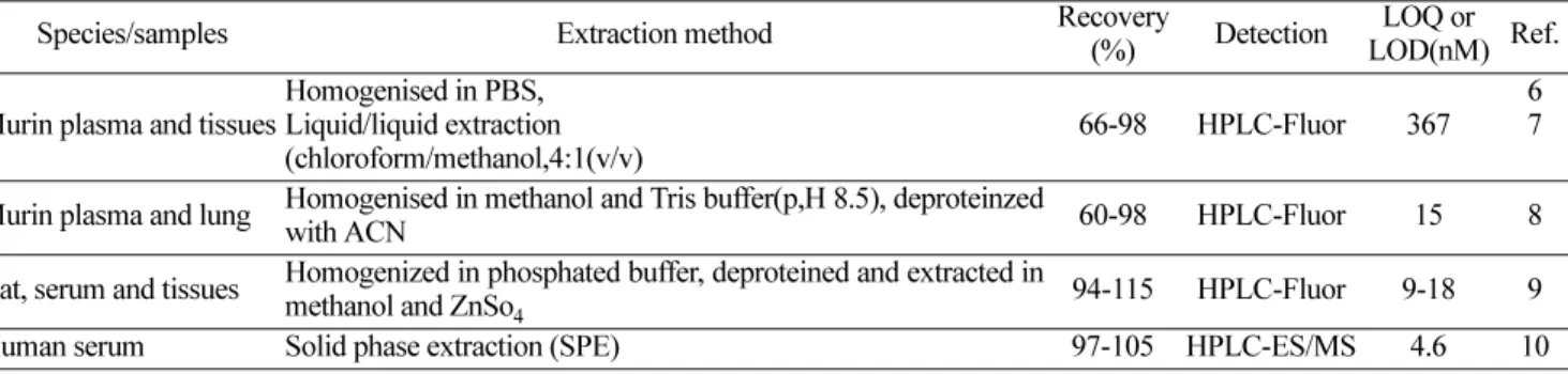
DXR is an anthracycline antibiotic that possesses broad spectrum antineoplastic activity, and is one of the most important anticancer agents in use
1-5). However, clinical utility is hampered by cumulative, dose-limiting cardiotoxicity, myelosuppression, and the development of drug resistance.
DXR is composed of an aglycone backbone linked to a daunosamine sugar through an O -glycosidic bond at carbon 7. A variety of procedures to extract, resolve,
and quantify DXR has been published
6-10)(Table 1).
Many of these techniques are laborious and necessitate long analytical run times to achieve sufficient peak res- olution, owing to the spectral and structural similarities of DXR
11). Among the techniques, LC-MS/MS can per- mit and exact and efficient determination of doxorubi- cin. In this study, a simplified, rapid extraction procedure and a liquid chromatography-tandem mass spectroscopy (LC-MS/MS) method were developed to identify and quantify DXR and metabolites in plasma and tissue extracts. Tandem mass spectroscopy can permit the determination of DXR analysis and quantification in complex biological samples.
Correspondence to : Han-Jung Chae
전북대학교의과대학약리학교실 전주시덕진구금암동