Nephrotic syndrome is a disease characterized by albuminuria, hypoalbuminemia, edema, and hy- perlipidemia, and thrombus and embolism are ma- jor complications of nephrotic syndrome. The loca- tion of the thrombus varies among reports, but it mostly occurs in the vein, with renal vein throm- bosis being most common; it also occurs in the deep vein or artery of the pelvic limbs.1,2 Thrombosis is a complication of adult nephrotic syndrome, which mainly occurs in membranous glomerulonephritis, and its occurrence is relatively lower in minimal change disease.1
Minimal change disease shows good response to steroid treatment, and 85% – 90% of patients
show complete remission of albuminuria.3 However, there is relapse in 56% - 76% of adult patients that show a response to steroids, while 10% – 25% show frequent relapse.4 Treatment of recurrent minimal change disease is achieved by first using an alkylating agent (cyclophosphamide), as well as other drugs such as calcineurin inhibitors (cyclosporine or tacrolimus), mycophenolate mo- fetil (MMF), and rituximab, but there is still no clear- ly established treatment protocol.3
The author experienced a case in which a patient with frequently relapsing minimal change disease, who also showed steroid resistance and the clinical features of hepatic portal vein thrombosis, went
Case Report
A Case of Steroid Resistant Minimal Change Disease Associated with Portal Vein Thrombosis Treated by Combined Immunosuppressive Agents
Hyo Jin Jung1, Su Mi Lee1, Seo Hee Rha2, Seong Eun Kim1, Young Ki Son1, Ki Seung Kim1, Won Suk An1
1Department of Internal Medicine, Dong-A University College of Medicine, Busan, Korea
2Department of Pathology, Dong-A University College of Medicine, Busan, Korea
Minimal change disease (MCD) is a common cause of nephrotic syndrome and relatively well responds with steroid treatment. However, nearly half of patients with MCD experience recurrence of nephrotic syndrome.
Thromboembolic events including renal vein thrombosis may occur in patients with MCD, but portal vein thrombosis rarely occurs. We experienced a case of frequent relapse/steroid dependent MCD with nephrotic syndrome progressed to steroid resistance associated with portal vein thrombosis. This patient showed complete remission of MCD and resolution of portal vein thrombosis after treatment with corticosteroid, cyclosporine, mycophenolate mofetil, and anticoagulant.
Key Words: Minimal change disease, Nephrotic syndrome, Recurrence, Steroid, Thrombosis
Corresponding Author: Won Suk An, Department of Internal Medicine, Dong-A University, 26, Daesingongwon-ro, Seo-gu, Busan 49201, Republic of Korea
Tel: +82-51-240-2811 Fax: +82-51-242-5852 E-mail: [email protected]
Received:
Revised:
Accepted:
Aug. 12, 2015 Sep. 22, 2015 Sep. 24, 2015
1. Patient: Age, 28 Years; Male.
Chief Complaint: Edema and Hydrops Abdominis.
History of presenting illness: The patient was hos- pitalized in October 2003 with chief complaints of albuminuria, hypoalbuminemia, edema, and hy- perlipidemia, and he was diagnosed with minimal change disease following renal biopsy (Fig. 1A).
He showed complete remission at 4 weeks after using 60 ㎎ (1 ㎎/㎏/day) of prednisolone (Solondo®;
Yuhan Corporation, Seoul, Korea); he showed side effects during dose reduction such as blushing.
Thus, the drug was switched to deflazacort (Calcort®; Handok Inc., Seoul, Korea), which was continuously taken. After 6 months of treatment, the patient showed complete remission of albu- minuria, and thus he discontinued his intake of deflazacort. Two months later in June 2004, the first relapse occurred with at least 2 g/g for the spot urine protein/creatinine ratio, and thus the patient took 60 ㎎ of deflazacort and then reached complete remission. Later, the patient’s steroid in- take was randomly discontinued and the patient showed a second relapse with a spot urine pro- tein/creatinine ratio of 5.97 g/g in April 2005. The
other day. The spot urine protein/creatinine ratio in December 2006 and October 2007 was 12.42 g/g and 5.24 g/g, respectively, which were thus indicative of the third and fourth relapse episodes.
Therefore, the combination therapy of deflazacort and cyclosporine was maintained. Most of the re- lapses were caused by the patient’s arbitrary dis- continuity of medication. After the fifth relapse in April 2010, with a spot urine protein/creatinine ratio of 11.51 g/g, another relapse occurred multi- ple times at 3–6-month intervals; thus, a renal biop- sy was performed again in December 2012. The results showed no focal segmental glomerulo- sclerosis aside from minimal change disease (Fig.
1B, 1C, and 1D). Later, while observing the patient’s progress on an outpatient basis, he was hospitalized due to hydrops abdominis and severe edema in November 2013.
2. Medical History: Minimal Change Disease 12 Years Ago.
Family History: No Significant Findings.
Findings and laboratory opinions: At the time the patient was hospitalized, his height was 159
㎝ and his weight was 68 ㎏, which was 8 ㎏ more
Fig.1. Renal histologic finding.
A. Light microscopy. Χ400, PAS (October, 2003). A glomerulus is normal in size and shows normal cellularity.
B. Light microscopy. X40, PAS (December, 2012). There is no focal and segmental glomerulosclerosis on lots of glomerulus.
C. Light microscopy. Χ400, PAS (December, 2012). Compared to the previous biopsy (October, 2003), there is no interval change.
D. Electron microscopy. (December, 2012). The electron microscopic examination shows effaced foot process of epithelial cell (arrow).
had no oppressive pain in either of his kidneys, and he exhibited pitting edema that was more se- vere than moderate in both pelvic limbs.
On peripheral blood examination, the patient’s hemoglobin was 15.9 g/dL, hematocrit 45.2%, and leucocytes 8,780/㎣. During the serum electrolyte test, the patient’s sodium was 136 mmol/L, potas- sium 4.6 mmol/L, chlorine 107 mmol/L, calcium 7.0 ㎎/dL, and phosphorous 5.2 ㎎/dL. During the serum biochemistry test, the patient’s blood urea nitrogen was 17 ㎎/dL, creatinine 0.8 ㎎/dL, glomer- ular filtration rate 124.2 mL/min/1.73㎡, aspartate aminotransferase 30 U/L, alanine aminotransferase 22 U/L, alkaline phosphatase 250 U/L, and total bilirubin 0.5 ㎎/dL. The total cholesterol increased to 427 ㎎/dL and the albumin decreased to 1.6 g/dL.
Antithrombin Ⅲ was 49.3% (normal range: 70%–
130%), protein C was 158% (normal range: 70%–
140%), and protein S was 102% (normal range: 60%–
130%). With respect to the urinalysis, protein 3+, the erythrocyte count was 10–14 high-power fields (HPF), and the spot urine protein/creatinine ratio was 8.05 g/g. Chest radiograph showed normal results.
Treatment and clinical course: A diuretic was
abdominis, hepatic portal vein thrombosis was dis- covered, and thus the patient began to receive treat- ment using heparin and warfarin, which was ad- justed within the prothrombin time range of 2–3 INR (International Normalized Range) (Fig. 2A). Due to thrombosis, prednisolone was reduced to 0.5
㎎/㎏/day, and the patient began to take 100 ㎎ of cyclosporine twice a day. When using the steroid exclusively, the edema continued even after taking a high-dose furosemide intravenous injection, but it was somewhat controlled after adding cyclo- sporine; thus, the patient was discharged. Severe albuminuria continued and the albumin index fell to below 2.0 g/dL, even after using cyclosporine for 4 weeks; thus, 500 ㎎ of MMF (Cellcept®; Roche Korea, Seoul, Korea) was added twice a day while gradually reducing the steroid. After using MMF for 4 weeks, the spot urine protein/creatinine ratio decreased to 2.25 g/g, so the steroid use was further reduced; 2 weeks later, the patient reached com- plete remission. Since then, he has been receiving combination therapy consisting of three drugs; he started taking 4.5 ㎎ of deflazacort 1 time/day, 100
㎎ of cyclosporine 2 times/day, and 250 ㎎ of MMF 2 times/day. He has been showing stable progress
without relapse for at least a year thus far (Fig.
3A and 3B). The hepatic portal vein thrombus was completely gone as per the abdominal ultrasound taken in March 2015, and warfarin was discontinued a year and a half after beginning the thrombosis treatment (Fig. 2B).
DISCUSSION
Minimal change disease responds excellently to steroids as a key factor of nephrotic syndrome in both adults and children. Adults show slower re- sponses to steroids than do children, and thus 85%
of children show remission 8 weeks after beginning treatment, while only approximately 50% of adults show remission.5 Approximately 50%–60% of pa- tients with minimal change disease that reach com- plete remission end up experiencing relapse.
Relapse is related to the patient’s age and the period of steroid intake, and recurrence rates are com-
parably lower in adults than in children.6 In this case, the patient had low compliance with his medi- cation and he discontinued treatment by himself, thereby showing frequent relapse. He also showed resistance to the drugs in 2013 when he was hospi- talized, showing no response even after using a steroid and cyclosporine for more than 8 weeks.
Therefore, it is important to warn those patients with minimal change disease to always comply with their prescribed medication.
Steroid is increased, or cyclophosphamide, cy- closporine, MMF, and rituximab are administered, when the first relapse occurs, but there are still not enough clinical studies to support this.
Cyclophosphamide has a longer maintenance peri- od of remission than cyclosporine, and it was shown to be effective for the patient who had trouble enduring the steroid; however, it may cause severe cytotoxicity such as sterility, myelosuppression, in- fection, and malignant tumors.7 When treating ste- roid-dependent minimal change disease, cyclo- Fig. 2. Abdominal ultrasonography image.
A. Internal hyperechoic material suggesting a portal vein thrombosis was shown on main portal vein (December, 2013).
B. Regression of portal vein thrombosis with normal portal venous flow was shown on doppler ultrasonography (March, 2015).
presented as an alternative that reduces the side new occurrence of focal segmental glomerulo-
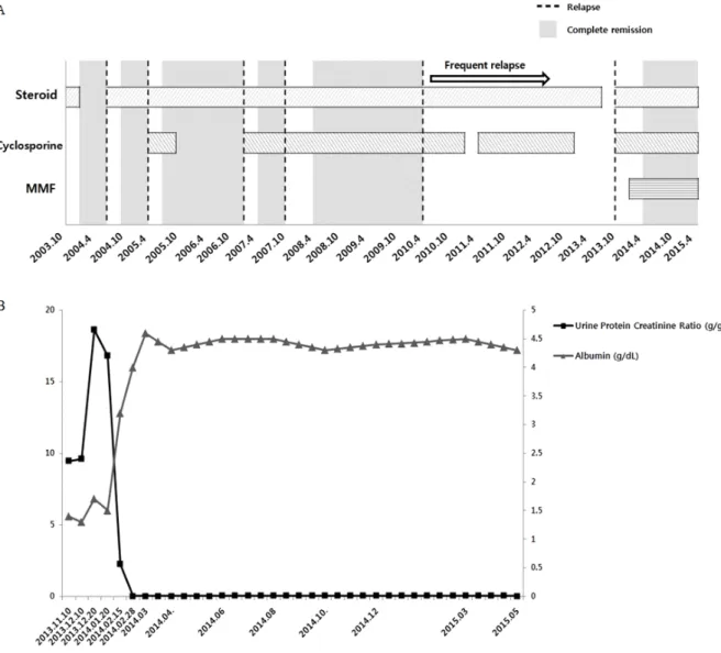
Fig. 3. Long-term treatment course of steroid resistant minimal change disease.
A. Treatment with steroid, cyclosporine, and mycophenolate mofetil (MMF) resulted in complete remission.
B. Changes of urine protein to creatinine ratio and serum albumin levels. The left side Y-axis shows urine
sclerosis was present rather than minimal change disease. However, the biopsy findings showed that the patient presented with minimal change disease.
As such, it is necessary to assess whether focal segmental glomerulosclerosis has newly occurred in cases where patients show frequent relapse of minimal change disease, or if they are showing secondary resistance to steroid treatment after re- peated relapses.10
Even though this case showed no other findings aside from minimal change disease during renal biopsy, severe hypoalbuminosis and albuminuria continued during the combination therapy that in- cluded cyclosporine; thus, MMF was added. MMF is a drug that has been used since the 1990s to prevent rejection following kidney transplantation, and many recent studies have shown the effects of MMF on glomerulonephritis. However, most of these investigations are small-scale retrospective studies that do not clearly detail the role of MMF in treating glomerulus diseases.11 Clara et al. re- vealed that MMF is useful for patients with con- stantly relapsing minimal change disease despite the use of steroids, cyclophosphamide, and cyclo- sporine, but the researchers reported that it is un- clear whether this combination reduces relapse af- ter treatment termination.12 Zhao et al. further dis- covered that MMF has fewer side effects than cyclo- phosphamide and cyclosporine; it also enables the successful reduction of steroids in steroid-depend- ent glomerulus diseases, reduces this dependency, and also plays a significant role in reducing the side effects associated with the long-term use of
steroids.7 Moreover, if MMF is administered to those pediatric patients with steroid-dependent neph- rotic syndrome who are under treatment with a steroid and cyclosporine, then the amount of ste- roid and cyclosporine can be reduced, and the re- currence rate also decreases.13 This case also used cyclosporine and steroids along with MMF, thereby inducing quick remission and reducing the use of steroids at an early stage.
The mechanism that causes thrombosis, which is a major complication of nephrotic syndrome, is not clear, but various factors seem to be asso- ciated with its development. The genetic factors that cause thrombus embolism are accompanied by risk factors such as inflammation, drugs, and central vein catheterization, thereby causing thrombus. Moreover, antithrombin and protein S, which are important proteins involved in coagu- lation control due to a deficiency of glomerulus, are lost, and the hemostasis protein can be gen- erated in the liver to maintain a balance between this protein loss and synthesis, ultimately affecting thrombus development.14 The platelet count and activity increased in pediatric patients with neph- rotic syndrome, and there is also the excessive co- agulation of blood platelets, the secretion of active materials, and an increased expression of activity signs on the surface of the blood platelets.
Accordingly, it is assumed that the blood platelets may be involved in the occurrence of a thrombus in the vein and artery that occurs in nephrotic syndrome. However, the correlation between blood platelet variation and thrombosis occurrence in
there are clinical findings of nephrotic syndrome.
The patient in this case took warfarin for a year and a half after the occurrence of hepatic portal vein thrombosis in November 2013, and he was only able to stop taking warfarin after verifying that there were no additional clinical symptoms and that the hepatic portal vein thrombus improved on ultrasound. It is important to suspect that those high-risk patients that show severe nephrotic syn- drome, and those that are resistant to treatment, may have phlebothrombosis.16
The patient showed resistance to the steroid, and thus a combination therapy of steroid and cyclo- sporine was used as a secondary drug, but the pa- tient showed no response. Therefore, MMF was add- ed as an alternative option, and the phlebo- thrombosis was completely gone following com- plete remission. The results of this study were re- ported in association with a literature review.
REFERENCES
1. Singhal R, Brimble KS. Thromboembolic complica- tions in the nephrotic syndrome: pathophysiology
3. Hogan J, Radhakrishnan J. The treatment of minimal change disease in adults. J Am Soc Nephrol 2013;24:702-11.
4. Nakayama M, Katafuchi R, Yanase T, Ikeda K, Tanaka H, Fujimi S. Steroid responsiveness and frequency of relapse in adult-onset minimal change nephrotic syndrome. Am J Kidney Dis 2002;39:503-12.
5. Waldman M, Crew RJ, Valeri A, Busch J, Stokes B, Markowitz G, et al. Adult minimal-change disease:
clinical characteristics, treatment, and outcomes.
Clin J Am Soc Nephrol 2007;2:445-53.
6. Korbert SM, Schwartz MM, Lewis EJ. Minimal-change glomerulopathy of adulthood. Am J Nephrol 1988;8:291-7.
7. Zhao M, Chen X, Chen Y, Liu Z, Liu Y, Lu F, et al. Clinical observations of mycophenolate mofetil therapy in refractory primary nephrotic syndrome.
Nephrology (Carlton) 2003;8:105-9.
8. Ponticelli C, Edefonti A, Ghio L, Rizzoni G, Rinaldi S, Gusmano R, et al. Cyclosporine versus cyclo- phosphamide for patients with steroid-dependent and frequently relapsing idiopathic nephrotic syn- drome: a multicenter randomized controlled trial.
Nephrol Dial Transplant 1993;8:1326-32.
9. Eguchi A, Takei T, Yoshida T, Tsuchiya K, Nitta
K. Combined cyclosporine and prednisolone ther- apy in adult patients with the first relapse of mini- mal-change nephrotic syndrome. Nephrol Dial Transplant 2010;25:124-9.
10. Mak SK, Short CD, Mallick NP. Long-term outcome of adult-onset minimal-change nephropathy.
Nephrol Dial Transplant 1996;11:2192-202.
11. Shigidi MM. The treatment of relapse in adults with minimal change nephrotic syndrome: myths and facts. Saudi J Kidney Dis Transpl 2011;22:10-7.
12. Day CJ, Cockwell P, Lipkin GW, Savage CO, Howie AJ, Adu D. Mycophenolate mofetil in the treatment of resistant idiopathic nephrotic syndrome. Nephrol Dial Transplant 2002;17:2011-3.
13. Fujinaga S, Ohtomo Y, Umino D, Takemoto M,
Shimizu T, Yamashiro Y, et al. A prospective study on the use of mycophenolate mofetil in children with cyclosporine-dependent nephrotic syndrome.
Pediatr Nephrol 2007;22:71-6.
14. Kerlin BA, Ayoob R, Smoyer WE. Epidemiology and pathophysiology of nephrotic syndrome-associated thromboembolic disease. Clin J Am Soc Nephrol 2012;7:513-20.
15. Eneman B, Levtchenko E, van den Heuvel B, Van Geet C, Freson K. Platelet abnormalities in nephrotic syndrome. Pediatr Nephrol 2016;31:1267-79.
16. Suri D, Ahluwalia J, Saxena AK, Sodhi KS, Singh P, Mittal BR, et al. Thromboembolic complications in childhood nephrotic syndrome: a clinical profile.
Clin Exp Nephrol 2014;18:803-13.