227
II. Case Report
A 52-year-old man presented to the Department of Oral and Maxillofacial Surgery, Gachon University Gil Medi- cal Center (Incheon, Korea) with reports of pain in the right TMJ, acute malocclusion with posterior open bite of the ipsi- lateral side (Fig. 1), deviation of the lower dental midline to left side, and limited mouth opening. The symptoms began 10 days prior to presentation and progressively worsened. At the onset of symptoms, he was diagnosed with temporoman- dibular disorder from another dental clinic and prescribed a muscle relaxant and analgesics for 7 days; however, his pain did not improve. He did not have any systemic predisposing factors. Clinical examination showed maximum mouth open- ing limited to 25 mm, posterior open bite on the right side, and preauricular swelling. Joint space widening on the right TMJ was identified on plain film.(Fig. 2) Serum leukocyte and C-reactive protein were tested prior to arthrocentesis, which showed a normal serum leukocyte and increased C-re- active protein level. We suspected a systemic infection from primary TMJ infectious disease.
After clinical evaluation with laboratory tests and plain films, a provisional diagnosis was made of septic arthritis of the right TMJ. The patient underwent arthrocentesis on the right TMJ under local anesthesia. A traditional two-needle technique was used for arthrocentesis: an imaginary line was marked from the tragus of the ear to the lateral canthus of the eye. At first, one needle was placed approximately 10 mm
I. Introduction
Septic arthritis of the temporomandibular joint (TMJ) pres- ents as a severe infection with fever, pain, swelling, redness, and hypofunction in the affected joint. It is also characterized by preauricular edema and trismus1,2. Septic arthritis of the TMJ occurs from either local dissemination or hematogenous spread from a distant primary infectious site3.
A mortality rate of 12% has been reported for cases of sep- tic arthritis in whole body joints, and significant hypofunction of the affected joint is observed in up to 75% of survivors1. Septic arthritis of the TMJ is known to result in significant morbidity if diagnosis is delayed4,5. Therefore, we describe the importance of early diagnosis and appropriate treatment of TMJ septic arthritis.
CASE REPORT
Jin-Yong Cho
Department of Oral and Maxillofacial Surgery, Gachon University Gil Medical Center, 21 Namdong-daero 774beon-gil, Namdong-gu, Incheon 21565, Korea TEL: +82-32-460-3372 FAX: +82-32-460-3101
E-mail: [email protected]
ORCID: http://orcid.org/0000-0001-7945-3923
This is an open-access article distributed under the terms of the Creative Commons Attribution Non-Commercial License (http://creativecommons.org/licenses/by-nc/4.0/), which permits unrestricted non-commercial use, distribution, and reproduction in any medium, provided the original work is properly cited.
CC
Septic arthritis of the temporomandibular joint: a case report
Sung-Won Yang, Jin-Yong Cho, Hyeon-Min Kim
Department of Oral and Maxillofacial Surgery, Gachon University Gil Medical Center, Incheon, Korea
Abstract(J Korean Assoc Oral Maxillofac Surg 2016;42:227-230)
Septic arthritis of the temporomandibular joint (TMJ) is a rare disease. The most common symptoms of this disease are acute malocclusion, limited mouth opening, swelling, and tenderness of affected TMJ. These symptoms are often confused with internal derangement of the articular disc, rheu- matoid arthritis, retrodiscitis, or osteoarthritis. Therefore, differential diagnosis by image examination is required. Usually, antimicrobial treatment and surgical drainage by needle aspiration, arthroscopy, or arthrotomy are effective treatment approaches. In this study, a patient who was diagnosed with septic arthritis was treated with arthrocentesis and antibiotics without significant complications. We present a case report with a review of the literature.
Key words: Infectious arthritis, Temporomandibular joint arthrocentesis, Temporomandibular joint aspiration
[paper submitted 2016. 5. 21 / revised 2016. 6. 29 / accepted 2016. 8. 3]
Copyright Ⓒ 2016 The Korean Association of Oral and Maxillofacial Surgeons. All rights reserved.
http://dx.doi.org/10.5125/jkaoms.2016.42.4.227 pISSN 2234-7550·eISSN 2234-5930
J Korean Assoc Oral Maxillofac Surg 2016;42:227-230
228
observed on contrast enhanced CT, so the patient was pre- scribed an additional 7 day course of both medications; we also recommended mouth opening exercises.
forward along this line and 2 mm below it. Joint fluid was obtained from the upper joint cavity and was sent for micro- biological examination. We observed turbid fluid (Fig. 3) and whitish debris macroscopically. One more needle was insert- ed in the upper joint cavity and the joint was thoroughly irri- gated with 500 mL of 0.9% normal saline. A broad-spectrum antibiotic (amoxicillin 250 mg/clavulanate 125 mg, Cramo- tin; Dong-A ST, Seoul, Korea) and analgesic (naproxen sodi- um 275 mg, Anaprox; Chong Kun Dang, Seoul, Korea) were prescribed for 7 days. Staphylococcus aureus was founded in the microbiological examination.
At follow-up 7 days later, the patient’s symptoms were slightly improved. However, preauricular swelling, TMJ pain during opening and mastication, and posterior open bite mal- occlusion were still present.(Fig. 4) Computed tomography (CT) was performed to evaluate the residual exudate in the right TMJ space and the need for additional arthrocentesis.
A widening of the joint space and bony change were not
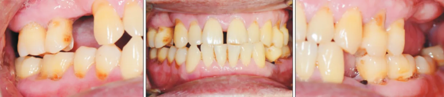
Fig. 1. Clinical photos presenting acute malocclusion.
Sung-Won Yang et al: Septic arthritis of the temporomandibular joint: a case report. J Korean Assoc Oral Maxillofac Surg 2016
Fig. 2. Transcranial projection of both temporomandibular joint (TMJ) show- ing enlarged joint space and limitation of translation on right TMJ.
Sung-Won Yang et al: Septic arthritis of the temporomandibular joint: a case report. J Korean Assoc Oral Maxillofac Surg 2016
Fig. 3. Turbid fluid with white debris was aspirated from right tem- poromandibular joint space.
Sung-Won Yang et al: Septic arthritis of the temporomandibular joint: a case report. J Korean Assoc Oral Maxillofac Surg 2016
Septic arthritis of the temporomandibular joint
229 its severe complications. Therefore, early diagnosis and treat- ment of TMJ septic arthritis are required to obtain better clin- ical outcomes. Unfortunately, the primary symptoms of sep- tic TMJ arthritis may be confused with other TMJ disorders such as internal derangement, synovial chondromatosis, or rheumatoid arthritis6. Therefore, the diagnostic criteria should include imaging findings, joint fluid analysis, and laboratory examination as well as clinical appearance1.
Imaging studies are useful for accurate diagnosis. Increased intracapsular fluid caused by accumulation of pus and inflam- matory exudate results in restriction of condyle movement and joint space widening1. These pathologic changes in the TMJ can be observed on plain film. Contrast-enhanced CT is used to identify pathologic changes such as joint effusions, condy- lar surface change, ankylosis, and osteomyelitis of the TMJ.
Magnetic resonance imaging (MRI) is useful for earlier de- tection of joint effusions and evaluation of joint surfaces and periarticular soft tissue7. Therefore, MRI is considered a very useful method in the diagnosis of septic arthritis. T2 scan on MRI is highly recommended to evaluate joint effusions. How- ever, in our case, septic arthritis of the TMJ was already con- firmed after arthrocentesis and microbiological examination of the aspirated joint fluid prior to additional imaging. Therefore, MRI was not deemed useful after proceeding with arthrocen- tesis because the infused saline and remaining exudate could At the 1-month follow-up, pain with opening and clench-
ing were absent. Occlusion returned to almost normal (Fig.
5), and maximum mouth opening was increased to 35 mm without pain. We recommended aggressive range of motion exercises to minimize the risk of long-term restricted mouth opening6.
III. Discussion
TMJ arthritis is caused by either hematogenous dissemina- tion of microorganisms into the well-vascularized synovial membrane or the direct spread of an adjacent contiguous infection1. The most common cause is hematogenous spread from a distant primary infection4. In adults, the spread of lo- cal infection to the TMJ has occurred in cases of infection after molar extraction, intra-articular injection, facial burns, and otitis externa. Blunt trauma with resulting capsular injury may also facilitate the hematogenous spread of infection into the joint space5.
Complications of septic arthritis of the TMJ include a high incidence of local spread, recurrence, destruction of joint articular surfaces, and bony ankyloses or fibrotic changes of the TMJ1,4. Symptoms of acute malocclusion, limited mouth opening, swelling and tenderness may also occur.
Septic arthritis of the TMJ is a medical emergency due to
Fig. 4. Occlusion discrepancy of the right side was improved but open bite of right molars was remained at 7 days after treatment.
Sung-Won Yang et al: Septic arthritis of the temporomandibular joint: a case report. J Korean Assoc Oral Maxillofac Surg 2016
Fig. 5. Normal occlusion was observed at 1 month after treatment.
Sung-Won Yang et al: Septic arthritis of the temporomandibular joint: a case report. J Korean Assoc Oral Maxillofac Surg 2016
J Korean Assoc Oral Maxillofac Surg 2016;42:227-230
230
can occur after acute inflammation, so exercises are highly recommended to recover the length of mouth opening. In this patient, joint drainage with arthrocentesis and a 14-day course of antibiotics taken for 14 days were prescribed. Phys- ical therapy and active mouth opening exercises improved mouth opening to 35 mm.
In conclusion, septic arthritis may be confused with other temporomandibular disorders. Therefore, when a patient presents with pain, fever, swelling, preauricular edema, trismus, and acute malocclusion, septic arthritis should be considered depending on imaging findings, joint aspiration, aspirated fluid analysis, and laboratory examination. Septic arthritis can be managed with antibiotics, surgical drainage, and joint immobilization.
Conflict of Interest
No potential conflict of interest relevant to this article was reported.
ORCID
Sung-Won Yang, http://orcid.org/0000-0002-7265-800X Jin-Yong Cho, http://orcid.org/0000-0001-7945-3923 Hyeon-Min Kim, http://orcid.org/0000-0002-5963-6543
References
1. Cai XY, Yang C, Zhang ZY, Qiu WL, Chen MJ, Zhang SY. Septic arthritis of the temporomandibular joint: a retrospective review of 40 cases. J Oral Maxillofac Surg 2010;68:731-8.
2. Klüppel LE, Bernabé FB, Primo BT, Stringhini DJ, da Costa DJ, Rebellato NL, et al. Septic arthritis of the temporomandibular joint.
J Craniofac Surg 2012;23:1752-4.
3. Parmar J. Case report: septic arthritis of the temporomandibular joint in a neonate. Br J Oral Maxillofac Surg 2008;46:505-6.
4. Leighty SM, Spach DH, Myall RW, Burns JL. Septic arthritis of the temporomandibular joint: review of the literature and report of two cases in children. Int J Oral Maxillofac Surg 1993;22:292-7.
5. Bounds GA, Hopkins R, Sugar A. Septic arthritis of the temporo- mandibular joint--a problematic diagnosis. Br J Oral Maxillofac Surg 1987;25:61-7.
6. Gayle EA, Young SM, McKenna SJ, McNaughton CD. Septic ar- thritis of the temporomandibular joint: case reports and review of the literature. J Emerg Med 2013;45:674-8.
7. Trimble LD, Schoenaers JA, Stoelinga PJ. Acute suppurative ar- thritis of the temporomandibular joint in a patient with rheumatoid arthritis. J Maxillofac Surg 1983;11:92-5.
8. Sembronio S, Albiero AM, Robiony M, Costa F, Toro C, Politi M.
Septic arthritis of the temporomandibular joint successfully treated with arthroscopic lysis and lavage: case report and review of the literature. Oral Surg Oral Med Oral Pathol Oral Radiol Endod 2007;103:e1-6.
not be distinguished on MRI. At the patient’s second visit, his symptoms were considerably improved. Therefore, MRI was not felt to be necessary. Joint fluid aspiration is recommended for additional investigation when septic arthritis has been diagnosed. The joint aspirate should be examined grossly for color and turbidity as indicators of infection, microscopically for joint fluid analysis, and then submitted for culture, Gram stain, and sensitivity analysis7. Joint fluid will be light yellow, transparent, and glistening in the patient with a temporoman- dibular disorder or rheumatoid arthritis. In contrast, it will be turbid or semitransparent in an infected joint1. The most common pathogen isolated in septic arthritis of the TMJ is Staphylococcus aureus, followed by Neisseria, Haemophilus influenzae, and Streptococcus4.
Several laboratory parameters are also useful for diagnosis, such as white blood count with differential and C-reactive protein testing1. The serum leukocyte count can be normal or increased due to antibiotic use.
C-reactive protein level indicates the severity of disease quite accurately. It may be used to identify disease resolution and may be increased at the initial stage8. In this case, we suspected septic arthritis based on clinical and radiographic examinations. Therefore, we performed arthrocentesis for di- agnosis and management, and aspirated turbid and semitrans- parent fluid. Staphylococcus aureus was found on culture of the aspirated fluid, which is known to be common in septic arthritis.
In terms of treatment, no definitive concurrence on the management of TMJ septic arthritis has been reached2. In general, appropriate joint drainage, antimicrobial treatment, and joint immobilization are recommended. However, surgi- cal drainage methods are controversial as no single diagnostic criterion has been proven4,8.
Empiric medication with antibiotics should be started im- mediately when acute septic arthritis has been diagnosed.
Broad-spectrum antibiotics such as intravenous penicillins or cephalosporins are often used and can be changed to an oral regimen after the joint infection is under control1. Antibiotics should be tailored according to culture and sensitivity tests.
In the acute period, septic arthritis may be managed by aspiration alone with the addition of arthrotomy and washout for pediatric patients5. Aspiration of the infected synovial fluid in addition to antibiotic administration are used for the treatment of TMJ. Within 48 to 72 hours after drainage, the infected joints become sterile. Different surgical drain- age methods such as needle aspiration, arthroscopy, and arthrotomy have been described1. Mouth opening restriction