http://dx.doi.org/10.12671/jkfs.2015.28.1.82
82
Copyright ⓒ 2015 The Korean Fracture Society. All rights reserved.
This is an Open Access article distributed under the terms of the Creative Commons Attribution Non-Commercial License (http://creativecommons.org/licenses/
by-nc/3.0) which permits unrestricted non-commercial use, distribution, and reproduction in any medium, provided the original work is properly cited.
Address reprint requests to: Jae-Ang Sim, M.D., Ph.D.
Department of Orthopaedic Surgery, Gachon University Gil Hospital, 21 Namdong-daero 774beon-gil, Namdong-gu, Incheon 405-760, Korea
Tel: 82-32-460-2645ㆍFax: 82-32-423-3384 E-mail: [email protected]
Financial support: None. Conflict of interest: None.
외상 환자에서의 정맥 혈전 색전증의 예방 및 치료
윤용철⋅심재앙*
가천대학교 길병원 권역외상센터, 가천대학교 길병원 정형외과*
Prophylaxis and Management of Deep Vein Thrombosis in Trauma Patients
Yong-Cheol Yoon, M.D., Jae-Ang Sim, M.D., Ph.D.*
Regional Trauma Center, Gachon University Gil Hospital,
Department of Orthopaedic Surgery, Gachon University Gil Hospital*, Incheon, Korea
서 론
정맥 혈전 색전증(venous thromboembolism, VTE)으로 알려져 있는 심부 정맥 혈전증(deep vein thrombosis, DVT) 및 폐 색전증(pulmonary embolism, PE)은 매년 미 국 인구 중 약 900,000명에서 발병하여 300,000명이 죽음 에 이르게 되며,1) 일반적으로 약 2/3는 DVT로, 1/3은 PE 로 진단된다.2,3)
골절을 동반한 중증 외상 환자의 경우 VTE가 나타날 수 있는데 이는 생명을 위협하는 잠재적 합병증이다.4-9) VTE 예방을 하지 않은 암 절제 수술이나 일반 외과 수술 환자 에서 DVT 발병률은 30%이며 이로 인한 사망률은 1%로 알려져 있다.10) 그러나 중증 외상 환자는 일반적인 수술 환자보다 더욱 높은 VTE의 발생 가능성을 내재하고 있으 며, DVT로 진단받은 환자의 6%에서, PE로 진단받은 환자 중 12%에서 1개월 안에 사망한다고 알려져 있다.11) 외상
환자에서 DVT 발병률은 환자의 위험 요인, 예방, 양상 및 감별 방법에 따라 5%에서 65%까지 다양하게 나타난
다.12,13) 또한 외상 환자 중 25%에서는 혈액 응고 장애
(coagulopathy)가 수상 당시부터 발생하며 혈액 응고 장애로 인한 사망률이 5배 증가하게 된다.14) Sevitt와 Gallagher15)가 중증 외상으로 사망한 125명의 환자를 대상으로 수행한 부 검 연구에서 DVT 발병률이 65%, PE 발병률이 16%로 밝 혀진 바 있고, Dunbar와 Chandler13)는 7.6년간 VTE가 발 생한 21,680명을 추적한 연구에서 예방을 통해 중증 외상 환자에서 VTE의 빈도를 1,300명(6%)으로 낮출 수 있었다 고 보고하였다. 그러므로 외상 환자들에 있어 VTE의 예방 은 반드시 필요하며, 만약 적절한 항 응고 요법(thrombo- prophylaxis) 치료를 하지 않으면 DVT 및 그에 따른 PE가 환자의 목숨을 위협할 수 있다.16)
본 종설에서는 외상 환자들에서의 VTE의 원인, 발생 기 전 및 예방을 위한 다양한 접근법에 대한 연구들을 요약하 고 외상 환자들에게 가장 적합한 VTE 예방에 대해 알아보 고자 한다.
외상 환자에서 심부 정맥 혈전증의 위험 인자
외상 후 DVT 발병 위험 인자로는 골반 및 하지 골절, 두부 손상, 그리고 장기간의 부동 상태(prolonged immobi-
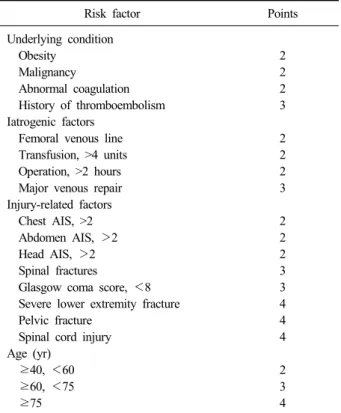
Table 1. Individual Risk Factors and the Risk Assessment Profile Score
Risk factor Points
Underlying condition Obesity
Malignancy
Abnormal coagulation History of thromboembolism Iatrogenic factors
Femoral venous line Transfusion, >4 units Operation, >2 hours Major venous repair Injury-related factors Chest AIS, >2 Abdomen AIS, >2 Head AIS, >2 Spinal fractures
Glasgow coma score, <8 Severe lower extremity fracture Pelvic fracture
Spinal cord injury Age (yr)
≥40, <60 ≥60, <75 ≥75
2 2 2 3
2 2 2 3
2 2 2 3 3 4 4 4
2 3 4
AIS: Abbreviated injury scale. Data from Greenfield LJ et al. J Trauma 1997;42:100-3.24)
lization) 등이 거론되어 왔지만, 최근의 전향적 무작위 연구 를 통하여 위험 인자에 대한 수정 보완이 있어 왔다.2,6,12) 뇌에서 자주 발견되는 조직 인자(tissue factor)는 두부 외상 후 혈액 응고 장애의 발생에 있어서 중요한 역할을 한다.6) 외상성 두부 손상 후 초기에 발생하는 혈액 응고 장애의 경우 혈액 응고의 외재성 경로(extrinsic pathway) 를 활성화시키는 조직 인자의 손상-매개성 국소적 분비 (injury-mediated local release)로 인한 것이라 생각되어 왔 다.17) 그러나 Geerts 등8)은 전향적 연구를 통하여 주요 두 부 손상이 DVT와 유의한 연관성을 갖지 못하며, 이외에도 성별, 손상 중증도 계수(injury severity score, ISS), 골반 골절도 유의한 연관성을 갖지 못한다고 하였다. 또한 독립 적인 DVT 예측 인자들로서는 연령, 수혈, 수술, 대퇴골 및 경골 골절, 그리고 척수 손상이라고 주장하였다. 다만 수혈 여부는 DVT와 관련이 있으나, 수혈량은 관련이 적은 것으 로 보고하였다. Kudsk 등18)도 ISS의 증가에 따른 DVT 증 가를 발견하지 못한 반면 연령의 증가에 따른 DVT가 증가 되었음을 발견하였다. 수혈에 있어서 Spinella 등19)은 5 unit 이상의 적혈구 농축액을 수혈 받은 외상 환자의 후향 적 코호트 연구에서 장기간 보관된 적혈구 농축액을 수혈 받는 경우 DVT 및 병원 내 사망률이 증가했다고 하였다.
Knudson 등3)은 미국외과학회(American College of Surgeons, ACS)의 국가 외상 자료 은행(National Trauma Data Bank, NTDB) 450,375명 환자의 광범위한 자료 분석 으로 외상 환자에서 VTE 발병에 있어 독립적인 유의성을 갖는 여섯 개의 위험 인자를 제시했다. 위험 인자로는 40 세 이상의 연령, 약식 손상 척도(abbreviated injury scale, AIS) 3점 이상의 하지 골절, 인공 호흡기 사용 일수가 3일 초과인 경우, AIS 3점 이상의 두부 손상, 정맥 손상, 그리 고 주요 수술적 치료(major operative procedure)가 필요했 던 경우였다. 일반적으로 연령이 높을수록 위험도가 증가 하지만 정확히 어느 연령에서 위험도가 증가하는지는 명확 하지 않다.2,10) Selby 등9)은 전향적 코호트 연구를 통해 외 상 환자에 있어 연령의 증가가 VTE 발병의 가장 중요한 임상적 예측 인자라고 보고하였다. 연령에 따른 혈전증 발 병 위험 증가의 원인에 대해서는 명확하지 않으나 혈전 발 병 성향을 가진 다른 질환의 증가나 혈액 응고 잠재성의 증가, 또는 이러한 요인들이 복합되어 발생할 수 있으리라 생각된다. 급성 척수 손상 및 마비 환자들에서도 DVT의 발병 위험이 매우 높은 것으로 알려져 있다.20-23) Fujii 등21) 은 방사성 섬유소원(radiofibrinogen) 흡수 검사를 통해 척 수 손상 환자들에서 DVT의 전체 발생률이 57% 정도임을 보고하였다.
Greenfield 등24)은 외상 환자에 있어서 문헌 고찰을 통 해 위험 인자들을 정리하여 점수화하는 위험도 평가 프로
파일(risk assessment profile, RAP)을 제안하였다(Table 1).
이들은 RAP에서 최소 두 개의 인자를 갖는 5점 이상이 되 는 경우를 고 위험군으로 정의하였다. Gearhart 등25)의 후 속 연구에서도 RAP 점수가 5점 이상인 환자들의 VTE 발 병 확률이 5점 미만인 환자들에 비해 3배 이상 높음을 밝 혀 RAP의 유용성을 뒷받침하였다.
외상 환자에서 심부 정맥 혈전증 발병 기전
혈전 생성에 기여하는 세 가지 주요 인자는 혈류(blood flow), 혈액 구성 요소(blood component), 혈관(blood ves- sels)이다.2,26) 외상은 종종 혈액 응고 항진(hypercoagul- ability), 내피 손상(endothelial injury), 정맥 울혈(venous stasis)이라는 Virchow의 삼징후(Virchow’s triad)의 위험 인 자들 중 하나 혹은 여러 인자들의 발생을 야기한다.27) 직 접적인 혈관 손상은 혈관 내막 손상과 혈전증을 일으킬 수 있으며 동반된 다른 손상으로 인해 장기간 침상 휴식, 부동 상태, 관류 저하(hypoperfusion) 및 마비로 인하여 정맥 울 혈이 촉진된다.2,7) 항트롬빈(antithrombin) III 수치의 감소 및 섬유소 용해(fibrinolysis) 현상의 억제로 인해 외상 환자
들에서 응고 항진 상태를 초래할 수 있다.28-31) Okamura 등32)은 고관절 골절 환자들의 혈장 내 D-이합체(D-dimer) 및 수용성 섬유소 단위 복합체(soluble fibrin monomer complex) 농도가 정상 수준보다 높았음을 보고하였다.
Peetz 등33)도 정형외과 수술을 받은 환자에서 저 위험군과 비교하여 고 위험군의 D-이합체가 상승되어 있음을 보고하 였다.
외상 후 트롬보플라스틴(thromboplastin, tissue factor) 및 트롬빈(thrombin) 생성 표지자가 증가했으며,34-36) 항트 롬빈, 단백질(protein) C, 단백질 S와 같은 자연 항 응고 물질(natural anticoagulants)이 저하된다고 여러 연구들을 통해 알려져 있다.29,35) Selby 등9)은 중증 외상이 트롬빈 조 절을 방해하여 혈중 트롬빈을 현저히 상승 및 유지시킨다 고 보고하였다. 관류 저하의 악화는 혈장 내 트롬보모듈린 (thrombomodulin)의 증가 및 단백질 C의 감소와 연관되어 있다. 이는 급성 혈액 응고 장애가 단백질 C 경로의 활성 화를 통한 전신성 항 응고 작용에 기인함을 시사한다.14) 응고 인자(Clotting factors) 소모 외에도 산증(acidosis) 및 저체온증(hypothermia)으로 인한 응고인자의 활성 및 작용 저하 그리고 정맥 내 수액 요법으로 인한 희석 및 적 혈구 투여 또한 외상성 혈액 응고 장애의 원인으로 받아들 여지고 있다.2,11,14,37)
하지만 Brohi 등14)은 외상에서 나타나 는 혈액 응고 장애 초기 단계의 급성 외상성 혈액 응고 장 애는 산증, 중증도의 저체온증, 혹은 희석으로 인한 응고 인자의 소모 혹은 장애에 기인하지 않으며, 쇼크(shock) 그 자체로 인한 항 응고성 및 섬유소 용해성 경로의 전신적 활성화에 기인하는 혈액 응고 장애로 이어진다고 주장하였 다.
부동성(immobility) 또한 VTE의 원인으로 알려져 있다.
마비로 인한 부동성은 척수에 외상을 입은 환자들에서 DVT를 일으키는 주요 기여 인자 중 하나이다.21) 근육의 수축 작용의 부재로 인해 혈류가 감소하고 종아리 근육 내 공간(intramuscular sinuses)에 혈액이 고이게 됨으로써 DVT를 초래한다.38) 적혈구 용적(hematocrit)의 증가, 섬유 소원(fibrinogen)과 폰 빌리브란트 인자(von Willebrand factor) 고분자 복합체(macromolecular complex) 상승으로 혈액의 점도가 높아지며 이로 인해 혈류에의 영향이 더욱 커질 수 있다.39,40) 혈류의 감소는 내피 손상, 혈액 응고 활 성 및 응고 억제 인자의 국소적 감소를 초래할 수 있으며, 이들 모두는 혈액의 응고성을 증가시키게 된다.21)
중증 외상 환자에서의 심부 정맥 혈전증 예방
다발성 손상 환자에서 혈전증을 초래하는 인자들이 수상 직후부터 발생하여 예방적 치료를 행하는 것이 어렵기 때
문에 비 외상성 환자처럼 효과적인 예방 조치를 대부분 적 절하게 사용할 수 없다.2,4,27,41) 또한 외상 환자에서는 때로 는 동반된 손상으로 인해 항 응고 약물의 투여를 할 수 없 는 경우도 있어 DVT 예방 조치가 힘들 수도 있다.24) 그러 나 항 응고 요법을 시행하지 않는다면 외상으로 인한 DVT 발병률이 50% 이상 되므로 반드시 어떠한 방식으로든 항 응고 요법을 시작하여야 한다.8,18)
지금까지 여러 연구들을 통해 중증 외상 환자에 관한 DVT 예방 및 치료 알고리즘을 확립해 왔다.3,25,41-43)
그러나 다발성 손상을 입은 외상 환자에서의 예방 조치에 대한 무 작위 통제 임상 연구(randomized controlled clinical study) 에는 제한이 따른다. 이는 ‘복합적인 손상을 입은 외상 환 자’라는 이질성을 띤 집단을 대상으로 하려면 모집 대상의 규모가 매우 커야 하는데 기존의 연구들에서는 대규모 임 상 연구가 이루어진 바 없기 때문이다.1,3) 따라서 외상 후 환자에서의 최적의 예방법에 대해서는 아직 논란이 존재하 지만, 외상 환자에서 이용 가능한 항 응고 요법들로는 약 물적 치료, 물리적 치료 및 하부 정맥 여과법(inferior vena cava filters) 등으로 분류될 수 있다.5,7,42,43)
1. 약물적 예방
1) 저용량 헤파린(low dose heparin)
Becker44)는 소용량의 헤파린을 이용 시 DVT 발병이 20%에서 40%까지 감소될 수 있다고 하였다. 과거에는 5,000 unit의 복용량으로 1일 2회 혹은 3회 피하 주사하는 저용량 헤파린(low dose heparin, LDH) 투여가 DVT 및 PE 예방을 위한 주된 약물 치료법으로 간주되어 왔다.45) 그러나 의학적 타당성에 대한 논의 없이 이러한 치료가 외 상 환자들에게 적용되어 왔음을 지적한 Ruiz 등27)은 LDH 의 효과를 파악하기 위해 복합적인 외상을 입은 100명의 환자들을 대상으로 한 연구에서 ISS가 10점을 초과하는 중 증 외상 환자들에서는 적절한 예방 조치가 되지 못함을 발 견하였다. 또한 Ganzer 등46)도 정형외과 및 외상외과라는 고 위험 분야에서 혈전 색전성 합병증의 예방에 있어 미분 획성 표준 헤파린(unfractioned standard heparin, UFH)은 그다지 효과적이지 않았으며 오히려 출혈 등의 부작용의 발생 위험이 높았다고 하였다. Geerts 등47)은 LDH 치료와 저 분자량 헤파린(low molecular weight heparin, LMWH) 치료를 비교한 전향적 무작위 이중 맹검 연구에서 DVT 예 방에 있어 LDH 치료가 현저히 불충분함을 밝혔다. 심지어 는 예방법을 적용하지 않은 경우와 LDH 치료를 비교한 다 른 몇몇 연구에서도 유의한 차이가 없었다.3,48-51) 그러나 대 부분 연구들에서 LDH 치료가 효과적이지 않음을 보고한 것과는 달리 최근 Arnold 등52)은 5,000 unit 헤파린을 1일
3회 처방 받은 외상 환자와 표준 용량인 30 mg 에녹사파 린(enoxaparin)을 1일 2회 처방 받은 외상 환자의 비교 연 구에서 유사한 수준의 낮은 DVT를 보고하여 비용 절감 효 과를 얻을 수 있다고 주장하였다.
2) 저분자량 헤파린(low molecular weight heparin, enoxaparin)
LMWH는 미분획성 헤파린(unfractioned heparin, UH)의 화학적 저 분자화(depolymerization)을 통해 생성된다. 이 같은 과정을 통해 크기, 전하 및 중량이 감소된다. UH와 비교하여 LMWH는 작은 크기와 더불어 Xa 인자에 대해 현저하게 큰 작용력을 가진다.53)
LMWH는 1990년대 초반에 VTE 예방 제제로서 명성을 얻었으며, 1990년대 후반에는 외상 환자에서 DVT를 예방 하는 가장 효과적인(혹은 유일한) 방법으로 부상했다.45) 여 러 연구들이 최적의 DVT 예방법을 조사해 왔으며 LMWH 로 치료받은 외상 환자에서 DVT 및 PE 발생률이 현저히 감소됨을 확인하였다.42,47,54-56)
Green 등56)은 척수 손상 환 자들을 대상으로 한 연구에서 LMWH가 완전 운동 신경 마 비(complete motor paralysis)에서 안전하고 효과적으로 VTE를 예방했으며 피하 주사된 헤파린보다 우월했다고 밝 혔다. Knudson 등54)은 고 위험 외상 환자에서의 DVT 예 방에 있어 LMWH 치료가 안전하고 효과적이라고 하였다.
1998년 미국 흉부의학회(American College of Chest Physcians, ACCP)는 복합적 외상 환자에서 LMWH를 이용 한 DVT 예방을 처음으로 권고하였다. Cothren 등42)은 전 향적 연구를 통해 고 위험 외상 환자에서 1일 1회의 예방 적 LMWH 치료가 시행하기 용이하고 안전하며 효과적이라 고 하였고, 전신적 예방에 골고루 작용(operate through)하 여 뇌 손상 및 다발성 외상 환자에서 있어서 시기 적절한 예방(timely prophylaxis)을 확보할 수 있었다고 하였다. 최 근에 Sems 등57)은 고 에너지 하지 외상 환자에서 일시적 가교 외고정술(temporary joint spanning external fixator placement)과 LMWH 투여를 동시에 이용함으로써 DVT의 비율을 2.1%로 낮출 수 있다고 하였다.
그러나 LMWH의 용량에 대해서는 논란이 분분하다. 일 부 저자들은 외상 환자에서의 DVT 예방에 있어 D-이합체 수준에 따른 용량 조절이 필요하다고 주장하였다.14,33) Warwick58)는 표준 용량의 LMWH를 투여 받은 환자들과 체중에 맞춘 복용량을 투여 받은 환자들 사이에서 DVT 발 병에는 차이가 없었다고 하였다. 그러나 Malinoski 등59)은 외과 ICU 환자의 절반에서 표준 용량의 LMWH가 DVT의 발생 위험을 현저하게 상승시키는 항 Xa 수치를 감소시킨 다고 보고하였다. 따라서 LMWH의 용량에 관한 연구가 더 필요한 실정이다.
3) 폰다파리녹스(fondaparinux)
폰다파리녹스는 최초로 합성된 Xa 인자를 선택적으로 억제하는 오당류(pentasaccharide)이다.60,61) 정형외과 수술 후 VTE 예방 및 고 위험 복부 수술 환자에서 폰다파리녹 스가 가지는 항 혈전적 효능과 더불어 안정성은 여러 임상 실험에서 입증되어 왔다.61) 고관절 골절 수술에서 에녹사 파린보다 폰다파리녹스를 투약한 경우 VTE 위험도가 56.4% 감소되었고,62) DVT의 발병률은 1.4%로 보고하였 다.63) 1등급 외상 센터(level 1 trauma center)에서 시행한 전체 외상 환자에서의 DVT 발병률은 4.6%였으나 폰다파 리녹스를 투약한 경우 발병률이 1.2%로 감소하였다.60) 또 한 폰다파리녹스로 인한 PE, 혈소판 감소증, 혹은 출혈이 발생한 예는 없었으며, 고 위험 외상 환자에서 이 제제가 VTE을 예방할 수 있다고 하였다. 폰다파리녹스는 1일 1회 복용법으로 복용이 쉽고 비용이 절감되었으며 헤파린 유도 성 혈소판 감소증(heparin induced thrombocytopenia)으로 부터 자유로울 수 있다. 하지만 아직은 대부분 소규모 연 구들이어서 향후 폰다파리녹스의 안정성 및 부작용에 관한 연구는 좀 더 필요한 실정이다.
2. 물리적 예방
부동 상태의 환자들이나 외상 환자에서 DVT 예방을 위 한 여러 종류의 외부 압박 기기들이 이용되는데, 이 기기 들은 정맥의 내강 지름을 감소시킴으로써 정맥 혈류의 속 도를 증가시키게 된다. 외부 압박 기기들의 장점은 이용이 쉽고, 출혈의 위험이 적으나,16) 단점은 환자의 순응도가 낮 으며, 하지의 골절이나 심한 창상이 있는 경우 착용하기가 어렵다.64)
1) 압박 스타킹(graduated compression stockings) 압박 스타킹(graduated compression stockings, GSC)은 비 외상성 환자에서 DVT의 예방과 치료에 널리 이용된
다.65,66) DVT에 있어서 GSC의 효과에 대해서는 이견이 있
으나, 최근 18개의 무작위 비교 연구(randomized clinical trials)에서 GSC가 입원 환자에서 DVT의 위험 감소에 효과 적이라고 하였다.67) 그러나 외상 환자에서 GSC의 효과에 대한 연구는 부족하다.
2) 공기 압박 기기(pneumatic compression devices) 외상 환자에서 DVT 예방을 위한 공기 압박 기기 (pneumatic compression devices, PCD)에 대한 연구가 현 재까지 진행되어 왔고, 이용이 증가하고 있는 추세이
다.68-70) 대조군과 비교하여 PCD가 DVT 발생률을 현저히
낮추는 데 상당한 효과가 있다고 밝혀졌고,53,55) 순차적 압
박 기기(sequential compression device), LDH, 혹은 이 둘 을 병합하여 예방 조치를 취한 환자들에서 낮은 수준의 DVT 발병률이 관찰되었다.45) Kurtoglu 등71)은 두부 및 척 추 외상 환자를 대상으로 한 PCD와 LMWH의 비교 연구에 서 DVT 및 PE의 발생 감소와 사망률이 차이가 없었다고 보고하여 PCD를 안전하게 적용할 수 있다고 주장하였다.
3) 족부 펌프(foot pumps)
1983년 Gardner와 Fox는 발이 단독적으로 갖는 생리적 펌핑 기전을 처음으로 관찰하였고, 1990년 Laverick은 동정 맥(arteriovenous, A-V) 족부 펌프가 슬와 정맥(popliteal vein)의 혈류를 250%까지 증가시킨다고 보고한 이후 여러 외상 환자군에서 A-V 족부 펌프에 대한 연구가 이루어졌 다.45) Knudson 등54)은 족부펌프, LMWH과 비교 연구에서 족부 펌프를 이용하는 경우 DVT 발생률이 높았다고 보고 하였고, Anglen 등72)도 PCD와 족부 펌프의 비교 연구에서 DVT 발생률이 각각 0%와 8%로 족부 펌프 치료에 회의적 이었다. 그러나 Spain 등73)은 PCD와 족부 펌프의 비교 연 구에서 DVT 발생률이 각각 7%와 3%로 유의적 차이가 없 었다고 보고하였고, Stannard 등74)은 외상 환자에 있어 에 녹사파린 단독 투여와 에녹사파린과 족부 펌프의 병용 치 료에서 DVT 유병률(prevalence)은 각각 13.4%와 8.7%, PE 유병률은 각각 2.1%와 0%로 보고하여 족부 펌프의 병용 치료가 유용함을 주장하였다.
3. 대정맥 필터(vena cava filters, VCFs)
근위부 DVT 환자에서 PE의 예방에 있어 대정맥 필터 (vena cava filters, VCFs)의 효과는 잘 알려져 있다.2,24,41) 급성 근위부 DVT 환자, 항 응고 요법에도 불구하고 PE가 발생한 환자, 직전에 PE가 발병한 환자로 헤파린을 이용할 수 없거나 헤파린 치료 시 출혈이 있었던 경우에 VCFs를 장착해 왔다.3) 따라서 약물적으로나 물리적 예방을 시행하 기 어려운 외상 환자들에서 VCFs는 VTE의 유병률 및 사 망률을 감소시키는 방법이 될 수 있다.
예전에는 VCFs 시술에는 위험이 따르며 대부분의 외상 환자에서 응고 항진 상태를 잠재적으로 지속시킬 수 있어 선호도가 낮았다.75) 그러나 외상 환자에서 이용 가능한 VTE 예방법이 없을 때나 PE, DVT이 발생하지 않은 고 위 험 환자에서 VCFs의 장착을 추천하기도 하였다.76-78) 이는 분명 논란의 여지가 있으나 Shackford 등79)이 ACS NTDB 의 자료를 후향적으로 분석한 결과에 의하면 1991년에서 2002년 사이 617,349명의 환자 중 6,282명(1%)이 VCFs 치 료를 받았으며 이들 중 86%는 예방적 차원에서 이 치료법 을 받았다고 보고하였다.
외상 환자에서 예방적 차원의 VCFs 치료법의 이용에 관 한 연구들 중 PE 발병률이 현저하게 저하됨을 보인 몇몇 연구들이 있다. Khansarinia 등80)은 예방적 차원의 VCFs를 장착한 고 위험 외상 환자들과 VCFs를 장착하지 않은 비 슷한 손상을 입은(inury-matched) 대조군을 비교하였을 때 PE 및 PE 관련 사망 모두에서 유의한 차이가 있음을 관찰 하였다. Velmahos 등81)도 메타 분석에서 VCFs 시술을 받 지 않은 환자들(1.5%)와 비교하여 예방적 차원의 VCFs를 시술 받은 환자들에서 폐색전증 발병률이 낮았다고(0.2%) 보고하였다. Toro 등37)은 VCFs가 PE 예방에 안전하고 효 과적이었으며 DVT 재발 위험 또한 낮았다고 하였다. 하지 만 일부 저자들은 영구적 VCFs가 장기적인 측면에서 DVT 의 위험을 상승시킬 수 있다고 보고하였다.82-84) Phelan 등85)은 영구적 VCFs 시술을 받은 외상 환자들을 장기간 추적 관찰한 연구에서 영구적 필터는 노령 환자들을 위한 방법이라고 주장하였으며 대부분의 외상 환자들은 비교적 젊기 때문에 다른 해결책이 필요하다고 하였다.
최근에는 회수 가능한 VCFs를 이용할 수 있어 VCFs에 대한 관심이 다시 생겼다. 회수 가능한 VCFs를 이용함으로 써 위험 기간 동안 PE를 방지할 수 있고, VCFs를 제거할 수 있어 뒤늦게 생길 수 있는 합병증을 피할 수 있어 VCFs의 단점을 보완할 수 있게 되었다.83) Gorman 등84)은 회수 가능한 VCFs를 고 위험 외상 환자에 이용하여 안전 하고 효과적으로 PE을 예방한다고 하였다. Rosenthal 등86) 도 회수 가능한 VCFs가 중환자실(intensive care unit, ICU) 침상에서 중증 외상 환자에 바로 시술 가능하며 환자를 ICU 밖으로 이동시킬 필요가 없는 안전하고도 간단한 기 법이라고 하였다. Cherry 등87)도 외상 환자들에서 회수 가 능한 VCFs를 예방적으로 시술하였고, PE 발생률은 1.6%, 회수율은 59%, 합병증 발생률은 0.1%라고 보고하였다.
외상 환자에서 심부 정맥 혈전증 예방에 대한 최신 권고 지침
본 종설에서는 일반적인 환자를 대상으로 한 DVT에 대 한 예방 지침은 제외하고 오로지 외상 환자에 대해 명확하 게 제시된 예방 권고 지침에 대해 소개하고자 한다. 미국 정형외과학회(American Academy of Orthopedic Surgeons) 의 경우 고관절 및 슬관절 치환술에 대한 VTE 예방 권고 지침 외에 외상 환자에 대한 예방 권고 지침은 제시하지 않아 언급하지 않기로 한다.
ACCP는 권고 지침(9th edition, 2012)을 통해 VTE 예방 의 진료 가이드라인(Table 2)을 발표해왔다.88) 중증 외상 환자는 항 응고 요법을 쓰지 않는 것보다 어떤 방법으로든 수상 즉시 항 응고 요법을 시행하는 것이 VTE 발병률과
Table 2. Ninth American College of Chest Physicians Guidelines for VTE Prevention (2012)
Recommendation Major trauma patients
At high risk for VTE Contraindications to chemoprophylaxis Isolated lower-leg injuries requiring leg immobilization
LMWH, LDUH, or IPC (>18 hours) LMWH or LDUH+IPC (>18 hours) IPC (>18 hours)
No prophylaxis
VTE: Venous thromboembolism, LMWH: Low molecular weight heparin, LDUH: Low-dose unfractionated heparin, IPC: Intermittent pneumatic compression. Data from Kearon C et al. Chest 2012;
141(2 Suppl):e419S-494S.88)
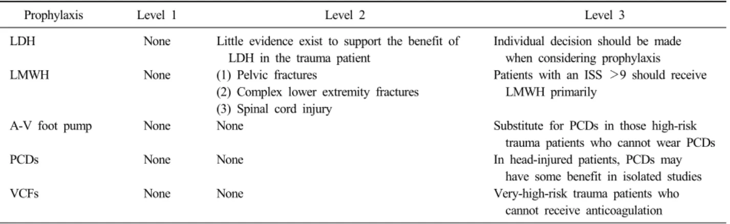
Table 3. Recommendations of the EAST Group for the Venous Thromboembolism Prophylaxis in Trauma Patients (2002)
Prophylaxis Level 1 Level 2 Level 3
LDH
LMWH
A-V foot pump
PCDs
VCFs
None
None
None
None
None
Little evidence exist to support the benefit of LDH in the trauma patient
(1) Pelvic fractures
(2) Complex lower extremity fractures (3) Spinal cord injury
None
None
None
Individual decision should be made when considering prophylaxis
Patients with an ISS >9 should receive LMWH primarily
Substitute for PCDs in those high-risk trauma patients who cannot wear PCDs In head-injured patients, PCDs may have some benefit in isolated studies Very-high-risk trauma patients who cannot receive anticoagulation
Level 1: recommendation is justifiable based on the available scientific evidence alone; recommendation is based on class I or a preponderance of class II evidence. Level 2: recommendation is reasonably justifiable based on the available scientific evidence and supported by expert opinion; recommendation is supported by class II evidence or a preponderance of class III evidence. Level 3:
recommendation is supported by available data, but inadequate scientific data are available; recommendation is supported by class III evidence. EAST: Eastern Association for the Surgery of Trauma, LDH: Low dose heparin, LMWH: Low molecular weight heparin, ISS: Injury severity score, A-V: Arteriovenous, PCDs: Pneumatic compression devices, VCFs: Vena cava filters. Data from Cushman JG et al. Trauma 2001;51:1016-1026.89)
사망률을 낮출 수 있다고 하였으며, LMWH를 다른 어떤 약물 치료보다도 우선적으로 고려해야 한다고 권고하였다.
또한 항 응고 약물 치료를 받고 있는 환자에 있어서 입원 기간 동안 PCD를 같이 사용할 것을 권장하였다. 적어도 항 응고 요법은 35일간 지속되어야 하며, 출혈 위험이 있 는 경우의 환자에게는 PCD를 사용할 것을 권장하였다. 약 물적 혹은 물리적 예방법을 이용할 수 없는 외상 환자들에 서 VCFs는 유병률 및 사망률을 감소시키는 방법이 될 수 있다. 퇴원 전에 초음파를 통해 VTE 선별(screening)이 필 요하며, 단독 하지 외상만 있는 경우에는 항 응고 요법을 권하지는 않는다.
외상 수술 동부 학회(Eastern Association for the Surgery
of Trauma, EAST)는 외상 환자를 위한 증거 기반 지침 (evidence based guideline)의 수립에 있어 선구적인 역 할을 해 왔고, VTE 예방을 위해 권장되는 진료 지침 (Table 3)도 발표하였다.89) EAST에서는 3등급(level 3) 권 고 사항으로 출혈이 손상을 더욱 악화시킬 수 있는 환자들 에서 LDH의 안전성이 아직 확립되지 않았으며, 예방법으 로 고려 시 환자에 따른 결정이 이루어져야 한다고 권고하 고 있다. 2등급(level 2) 권고 사항으로 LMWH는 (1) 골반 골절(수술적 혹은 장기간 동안의 침상 휴식), (2) 복합성 하지 골절(수술적 혹은 장기간 동안의 침상 휴식), (3) 척 수 손상 시 이용할 수 있다. 3등급 권고 사항으로 ISS가 9 를 초과하는 경우 기본적으로 LMWH 치료를 받아야 하며, 두부 손상 환자 중 일부에서 PCD를 이용하는 것이 어느 정도 이로울 수 있고, 항 응고 요법을 받을 수 없는 고 위 험 외상 환자들의 경우 출혈의 위험이 크고 장기간 동안 부동 상태여야 하기 때문에 예방 목적의 VCFs 삽입을 권고 하였다.
영국에서는 영국 국립 보건 임상 연구소(National Institute for Health and Clinical Excellence)를 통해서 VTE 예방을 위해 권장되는 지침을 발표해 왔으며, 중증 외상 환자 발생 시 GSC, PCD, 족부 펌프와 같은 물리적 예방법을 먼저 이 용하고, VTE의 위험 요인과 출혈 경향을 평가한 후 위험 요인이 출혈 경향보다 큰 경우 LMWH를 사용할 수 있으며, 심각한 신장 기능 부전일 경우 UFH을 쓸 수 있다고 권고 하였다. 또한 항 응고 요법의 기간은 VTE의 위험이 없다는 의학적 판단이 있기까지 사용해야 한다고 권고하였다.90-92)
결 론
VTE가 외상 환자에서 큰 문제 중 하나라는 점은 명백하 다. 외상 환자에서의 VTE 예방에 관한 문헌 고찰에서 살 펴 본 것과 같이 이용할 수 있는 확실한 예방법을 임상의 가 결정하는 데 도움이 되는 신뢰도 있는 임상 연구가 부 족한 실정이다. 앞서 언급된 예방법 중 어떠한 예방법도 VTE를 완벽히 예방하지 못하지만 그럼에도 불구하고 예방 법을 시행하지 않는다면 잠재성 및 비 잠재성 DVT의 발병 률이 증가되고 이로 인한 유병률 및 사망률도 증가하게 된 다.
가장 최근에 이루어진 임상 연구들이 외상 환자에서의 VTE 예방에 있어 LMWH 이용을 지지하고 있고, 두 가지 중요 지침인 ACCP 및 EAST 권고 지침 또한 외상 환자에 서 LMWH를 우선적으로 이용할 것을 권장하고 있다. 또 한, 여러 연구들과 ACCP 및 EAST 권고 지침에서 기계적 예방법을 지지했으나 대부분은 LMWH의 보조 요법으로서 혹은 LMWH의 이용이 금기시되는 경우에 이용해야 한다고 권고하고 있다. VCFs는 LMWH를 이용할 수 없는 경우 PE 예방에 주로 이용되어 왔는데 이를 중증 외상 환자에서 주 된 예방법으로 이용될 수도 있다는 것에 대해 널리 논의되 어 왔다. 현재 ACCP 및 EAST 권고 지침이 매우 큰 위험 을 갖고 있는 중증 외상 환자에서 LMWH를 이용할 수 없 는 경우 VCFs를 이용하도록 권장하고 있다.
결론적으로 외상 환자에서 VTE 예방은 필요하며 환자에 게 이로운 선택일 것으로 판단된다. 그러나 아쉽게도 현재 까지 알려진 예방 조치에 따른 VTE의 발병 감소가 유병률 과 사망률을 완전히 소거시키지는 못했다. 이러한 논란을 잠재우고 외상 환자에서 가장 이상적인 VTE 예방법을 규 명하기 위해서 대규모의 신뢰도 있는 무작위적이고 전향적 인 임상 시험이 필요하다.
References
1) Raskob GE, Silverstein R, Bratzler DW, Heit JA, White RH: Surveillance for deep vein thrombosis and pulmonary embolism: recommendations from a national workshop. Am J Prev Med, 38: S502-S509, 2010.
2) Cushman M: Epidemiology and risk factors for venous thrombosis. Semin Hematol, 44: 62-69, 2007.
3) Knudson MM, Ikossi DG, Khaw L, Morabito D, Speetzen LS: Thromboembolism after trauma: an analysis of 1602 episodes from the American College of Surgeons National Trauma Data Bank. Ann Surg, 240: 490-496, discussion 496-498, 2004.
4) Paffrath T, Wafaisade A, Lefering R, et al: Trauma Registry of DGU: Venous thromboembolism after severe trauma: incidence, risk factors and outcome. Injury, 41:
97-101, 2010.
5) Chiasson TC, Manns BJ, Stelfox HT: An economic evaluation of venous thromboembolism prophylaxis strat- egies in critically ill trauma patients at risk of bleeding.
PLoS Med, 6: e1000098, 2009.
6) Reiff DA, Haricharan RN, Bullington NM, Griffin RL, McGwin G Jr, Rue LW 3rd: Traumatic brain injury is associated with the development of deep vein thrombosis independent of pharmacological prophylaxis. J Trauma, 66: 1436-1440, 2009.
7) Feliciano DV, Mattox KL, Moore EE: Trauma. 6th ed.
New York, McGraw-Hill Medical: 1251-1270, 2008.
8) Geerts WH, Code KI, Jay RM, Chen E, Szalai JP: A prospective study of venous thromboembolism after major trauma. N Engl J Med, 331: 1601-1606, 1994.
9) Selby R, Geerts W, Ofosu FA, et al: Hypercoagulability after trauma: hemostatic changes and relationship to ve- nous thromboembolism. Thromb Res, 124: 281-287, 2009.
10) Eppsteiner RW, Shin JJ, Johnson J, van Dam RM:
Mechanical compression versus subcutaneous heparin ther- apy in postoperative and posttrauma patients: a systematic review and meta-analysis. World J Surg, 34: 10-19, 2010.
11) White RH: The epidemiology of venous thromboembolism.
Circulation, 107(23 Suppl 1): I4-I8, 2003.
12) Bendinelli C, Balogh Z: Postinjury thromboprophylaxis.
Curr Opin Crit Care, 14: 673-678, 2008.
13) Dunbar NM, Chandler WL: Thrombin generation in trauma patients. Transfusion, 49: 2652-2660, 2009.
14) Brohi K, Cohen MJ, Ganter MT, et al: Acute coagulop- athy of trauma: hypoperfusion induces systemic anti- coagulation and hyperfibrinolysis. J Trauma, 64: 1211-1217;
discussion 1217, 2008.
15) Sevitt S, Gallagher N: Venous thrombosis and pulmonary embolism. A clinico-pathological study in injured and burned patients. Br J Surg, 48: 475-489, 1961.
16) Datta I, Ball CG, Rudmik L, Hameed SM, Kortbeek JB: Complications related to deep venous thrombosis pro- phylaxis in trauma: a systematic review of the literature. J Trauma Manag Outcomes, 4: 1, 2010.
17) Sase T, Wada H, Kamikura Y, et al: Tissue factor mes- senger RNA levels in leukocytes compared with tissue factor antigens in plasma from patients in hypercoagulable
state caused by various diseases. Thromb Haemost, 92:
132-139, 2004.
18) Kudsk KA, Fabian TC, Baum S, Gold RE, Mangiante E, Voeller G: Silent deep vein thrombosis in immobilized multiple trauma patients. Am J Surg, 158: 515-519, 1989.
19) Spinella PC, Carroll CL, Staff I, et al: Duration of red blood cell storage is associated with increased incidence of deep vein thrombosis and in hospital mortality in pa- tients with traumatic injuries. Crit Care, 13: R151, 2009.
20) Agarwal NK, Mathur N: Deep vein thrombosis in acute spinal cord injury. Spinal Cord, 47: 769-772, 2009.
21) Fujii Y, Mammen EF, Farag A, Muz J, Salciccioli GG, Weingarden ST: Thrombosis in spinal cord injury.
Thromb Res, 68: 357-368, 1992.
22) Do JG, Kim du H, Sung DH: Incidence of deep vein thrombosis after spinal cord injury in Korean patients at acute rehabilitation unit. J Korean Med Sci, 28: 1382-1387, 2013.
23) Miranda AR, Hassouna HI: Mechanisms of thrombosis in spinal cord injury. Hematol Oncol Clin North Am, 14:
401-416, 2000.
24) Greenfield LJ, Proctor MC, Rodriguez JL, Luchette FA, Cipolle MD, Cho J: Posttrauma thromboembolism prophylaxis. J Trauma, 42: 100-103, 1997.
25) Gearhart MM, Luchette FA, Proctor MC, et al: The risk assessment profile score identifies trauma patients at risk for deep vein thrombosis. Surgery, 128: 631-640, 2000.
26) Kitagawa K, Sakoda S: Mechanism underlying thrombus formation in cerebral infarction. Rinsho Shinkeigaku, 49:
798-800, 2009.
27) Ruiz AJ, Hill SL, Berry RE: Heparin, deep venous thrombosis, and trauma patients. Am J Surg, 162: 159-162, 1991.
28) Seyfer AE, Seaber AV, Dombrose FA, Urbaniak JR:
Coagulation changes in elective surgery and trauma. Ann Surg, 193: 210-213, 1981.
29) Owings JT, Bagley M, Gosselin R, Romac D, Disbrow E: Effect of critical injury on plasma antithrombin activ- ity: low antithrombin levels are associated with throm- boembolic complications. J Trauma, 41: 396-405; dis- cussion 405-406, 1996.
30) Attar S, Boyd D, Layne E, McLaughlin J, Mansberger AR, Cowley RA: Alterations in coagulation and fi- brinolytic mechanisms in acute trauma. J Trauma, 9:
939-965, 1969.
31) Enderson BL, Chen JP, Robinson R, Maull KI:
Fibrinolysis in multisystem trauma patients. J Trauma, 31:
1240-1246, 1991.
32) Okamura K, Nakagawa I, Hidaka S, Okada Y, Kubo T, Kato T: Preoperative hypercoagulopathy in patients undergoing orthopedic lower extremity surgery. Masui, 57:
1207-1212, 2008.
33) Peetz D, Hafner G, Hansen M, et al: Dose-adjusted thrombosis prophylaxis in trauma surgery according to levels of D-Dimer. Thromb Res, 98: 473-483, 2000.
34) Meissner MH, Chandler WL, Elliott JS: Venous throm- boembolism in trauma: a local manifestation of systemic hypercoagulability? J Trauma, 54: 224-231, 2003.
35) Engelman DT, Gabram SG, Allen L, Ens GE, Jacobs LM: Hypercoagulability following multiple trauma. World J Surg, 20: 5-10, 1996.
36) Dries DJ: Activation of the clotting system and comple- ment after trauma. New Horiz, 4: 276-288, 1996.
37) Toro JB, Gardner MJ, Hierholzer C, et al: Long-term consequences of pelvic trauma patients with thromboem- bolic disease treated with inferior vena caval filters. J Trauma, 65: 25-29, 2008.
38) Nicolaides AN, Kakkar VV, Field ES, Fish P: Venous stasis and deep-vein thrombosis. Br J Surg, 59: 713-717, 1972.
39) Rossi EC, Green D, Rosen JS, Spies SM, Jao JS:
Sequential changes in factor VIII and platelets preceding deep vein thrombosis in patients with spinal cord injury.
Br J Haematol, 45: 143-151, 1980.
40) Myllynen P, Kammonen M, Rokkanen P, et al: The blood F VIII:Ag/F VIII:C ratio as an early indicator of deep venous thrombosis during post-traumatic immobilization. J Trauma, 27: 287-290, 1987.
41) Hak DJ: Prevention of venous thromboembolism in trau- ma and long bone fractures. Curr Opin Pulm Med, 7:
338-343, 2001.
42) Cothren CC, Smith WR, Moore EE, Morgan SJ:
Utility of once-daily dose of low-molecular-weight heparin to prevent venous thromboembolism in multisystem trau- ma patients. World J Surg, 31: 98-104, 2007.
43) Matthiasson SE, Lindblad B, Bergqvist D: Prevention of experimental venous thrombosis in rabbits with low mo- lecular weight heparin, dextran and their combinations, ad- ministered before or during induction of venous endothe- lial trauma. Thromb Res, 74: 655-663, 1994.
44) Becker DM: Venous thromboembolism: epidemiology, di- agnosis, prevention. J Gen Intern Med, 1: 402-411, 1986.
45) Rogers FB, Cipolle MD, Velmahos G, Rozycki G, Luchette FA: Practice management guidelines for the pre- vention of venous thromboembolism in trauma patients:
the EAST practice management guidelines work group. J Trauma, 53: 142-164, 2002.
46) Ganzer D, Gutezeit A, Mayer G: Potentials risks in drug prevention of thrombosis--low-molecular-weight heparin versus standard heparin. Z Orthop Ihre Grenzgeb, 137:
457-461, 1999.
47) Geerts WH, Jay RM, Code KI, et al: A comparison of low-dose heparin with low-molecular-weight heparin as prophylaxis against venous thromboembolism after major trauma. N Engl J Med, 335: 701-707, 1996.
48) Shackford SR, Davis JW, Hollingsworth-Fridlund P, Brewer NS, Hoyt DB, Mackersie RC: Venous throm- boembolism in patients with major trauma. Am J Surg, 159: 365-369, 1990.
49) Dennis JW, Menawat S, Von Thron J, et al: Efficacy of deep venous thrombosis prophylaxis in trauma patients and identification of high-risk groups. J Trauma, 35:
132-138; discussion 138-139, 1993.
50) Upchurch GR Jr, Demling RH, Davies J, Gates JD, Knox JB: Efficacy of subcutaneous heparin in prevention of venous thromboembolic events in trauma patients. Am Surg, 61: 749-755, 1995.
51) Napolitano LM, Garlapati VS, Heard SO, et al:
Asymptomatic deep venous thrombosis in the trauma pa- tient: is an aggressive screening protocol justified? J Trauma, 39: 651-657; discussion 657-659, 1995.
52) Arnold JD, Dart BW, Barker DE, et al: Gold Medal Forum Winner. Unfractionated heparin three times a day versus enoxaparin in the prevention of deep vein thrombo- sis in trauma patients. Am Surg, 76: 563-570, 2010.
53) Weitz JI: Low-molecular-weight heparins. N Engl J Med, 337: 688-698, 1997.
54) Knudson MM, Lewis FR, Clinton A, Atkinson K, Megerman J: Prevention of venous thromboembolism in trauma patients. J Trauma, 37: 480-487, 1994.
55) Schwarcz TH, Quick RC, Minion DJ, Kearney PA, Kwolek CJ, Endean ED: Enoxaparin treatment in high-risk trauma patients limits the utility of surveillance venous duplex scanning. J Vasc Surg, 34: 447-452, 2001.
56) Green D, Lee MY, Lim AC, et al: Prevention of throm-
boembolism after spinal cord injury using low-molec- ular-weight heparin. Ann Intern Med, 113: 571-574, 1990.
57) Sems SA, Levy BA, Dajani K, Herrera DA, Templeman DC: Incidence of deep venous thrombosis af- ter temporary joint spanning external fixation for complex lower extremity injuries. J Trauma, 66: 1164-1166, 2009.
58) Warwick D: Thromboembolic prophylaxis in orthopaedic trauma patients: a comparison between fixed dose and an individually adjusted dose of a low molecular weight heparin. Injury, 28: 233-234, 1997.
59) Malinoski D, Jafari F, Ewing T, et al: Standard prophy- lactic enoxaparin dosing leads to inadequate anti-Xa levels and increased deep venous thrombosis rates in critically ill trauma and surgical patients. J Trauma, 68: 874-880, 2010.
60) Lu JP, Knudson MM, Bir N, Kallet R, Atkinson K:
Fondaparinux for prevention of venous thromboembolism in high-risk trauma patients: a pilot study. J Am Coll Surg, 209: 589-594, 2009.
61) Turpie AG: The safety of fondaparinux for the prevention and treatment of venous thromboembolism. Expert Opin Drug Saf, 4: 707-721, 2005.
62) Eriksson BI, Bauer KA, Lassen MR, Turpie AG; Steering Committee of the Pentasaccharide in Hip-Fracture Surgery Study: Fondaparinux compared with enoxaparin for the prevention of venous thromboembolism after hip-fracture surgery. N Engl J Med, 345: 1298-1304, 2001.
63) Eriksson BI, Lassen MR, Colwell CW Jr: Efficacy of fondaparinux for thromboprophylaxis in hip fracture patients. J Arthroplasty, 19(7 Suppl 2): 78-81, 2004.
64) Rahman A, Colak MC, Ustünel L, Koc M, Kocakoc E, Colak C: A comparison of different treatment manage- ments in patients with acute deep vein thrombosis by the effects on enhancing venous outflow in the lower limb.
Med Sci Monit, 15: CR588-CR593, 2009.
65) Xu B: DVT in acute stroke: the use of graduated com- pression stockings. Aust Fam Physician, 39: 485-487, 2010.
66) Arpaia G, Carpenedo M, Pistelli R, Mastrogiacomo O, Cimminiello C, Agnelli G: Attitudes to prescribing com- pression stockings for patients with acute DVT: the MASTER registry. J Thromb Thrombolysis, 28: 389-393, 2009.
67) Sachdeva A, Dalton M, Amaragiri SV, Lees T: Elastic compression stockings for prevention of deep vein
thrombosis. Cochrane Database Syst Rev, (7): CD001484, 2010.
68) Velmahos GC, Nigro J, Tatevossian R, et al: Inability of an aggressive policy of thromboprophylaxis to prevent deep venous thrombosis (DVT) in critically injured pa- tients: are current methods of DVT prophylaxis in- sufficient? J Am Coll Surg, 187: 529-533, 1998.
69) Gersin K, Grindlinger GA, Lee V, Dennis RC, Wedel SK, Cachecho R: The efficacy of sequential compression devices in multiple trauma patients with severe head injury. J Trauma, 37: 205-208, 1994.
70) Fisher CG, Blachut PA, Salvian AJ, Meek RN, O'Brien PJ: Effectiveness of pneumatic leg compression devices for the prevention of thromboembolic disease in orthopaedic trauma patients: a prospective, randomized study of compression alone versus no prophylaxis. J Orthop Trauma, 9: 1-7, 1995.
71) Kurtoglu M, Yanar H, Bilsel Y, et al: Venous throm- boembolism prophylaxis after head and spinal trauma: in- termittent pneumatic compression devices versus low mo- lecular weight heparin. World J Surg, 28: 807-811, 2004.
72) Anglen JO, Bagby C, George R: A randomized compar- ison of sequential-gradient calf compression with inter- mittent plantar compression for prevention of venous thrombosis in orthopedic trauma patients: preliminary results. Am J Orthop (Belle Mead NJ), 27: 53-58, 1998.
73) Spain DA, Bergamini TM, Hoffmann JF, Carrillo EH, Richardson JD: Comparison of sequential compression devices and foot pumps for prophylaxis of deep venous thrombosis in high-risk trauma patients. Am Surg, 64:
522-525; discussion 525-526, 1998.
74) Stannard JP, Lopez-Ben RR, Volgas DA, et al:
Prophylaxis against deep-vein thrombosis following trau- ma: a prospective, randomized comparison of mechanical and pharmacologic prophylaxis. J Bone Joint Surg Am, 88: 261-266, 2006.
75) Winchell RJ, Hoyt DB, Walsh JC, Simons RK, Eastman AB: Risk factors associated with pulmonary em- bolism despite routine prophylaxis: implications for im- proved protection. J Trauma, 37: 600-606, 1994.
76) Greenfield LJ, Michna BA: Twelve-year clinical experi- ence with the Greenfield vena caval filter. Surgery, 104:
706-712, 1988.
77) Rogers FB, Shackford SR, Ricci MA, Wilson JT, Parsons S: Routine prophylactic vena cava filter insertion
in severely injured trauma patients decreases the incidence of pulmonary embolism. J Am Coll Surg, 180: 641-647, 1995.
78) Rodriguez JL, Lopez JM, Proctor MC, et al: Early placement of prophylactic vena caval filters in injured pa- tients at high risk for pulmonary embolism. J Trauma, 40:
797-802, 1996.
79) Shackford SR, Cook A, Rogers FB, Littenberg B, Osler T: The increasing use of vena cava filters in adult trauma victims: data from the American College of Surgeons National Trauma Data Bank. J Trauma, 63: 764-769, 2007.
80) Khansarinia S, Dennis JW, Veldenz HC, Butcher JL, Hartland L: Prophylactic Greenfield filter placement in se- lected high-risk trauma patients. J Vasc Surg, 22: 231-235, 1995.
81) Velmahos GC, Kern J, Chan LS, Oder D, Murray JA, Shekelle P: Prevention of venous thromboembolism after injury: an evidence-based report: part II: analysis of risk factors and evaluation of the role of vena caval filters. J Trauma, 49: 140-144, 2000.
82) Helling TS, Kaswan S, Miller SL, Tretter JF: Practice patterns in the use of retrievable inferior vena cava filters in a trauma population: a single-center experience. J Trauma, 67: 1293-1296, 2009.
83) McKenzie S, Gibbs H, Leggett D, et al: An Australian experience of retrievable inferior vena cava filters in pa- tients with increased risk of thromboembolic disease. Int Angiol, 29: 53-57, 2010.
84) Gorman PH, Qadri SF, Rao-Patel A: Prophylactic in- ferior vena cava (IVC) filter placement may increase the relative risk of deep venous thrombosis after acute spinal cord injury. J Trauma, 66: 707-712, 2009.
85) Phelan HA, Gonzalez RP, Scott WC, White CQ, McClure M, Minei JP: Long-term follow-up of trauma patients with permanent prophylactic vena cava filters. J Trauma, 67: 485-489, 2009.
86) Rosenthal D, Kochupura PV, Wellons ED, Burkett AB, Methodius-Rayford WC: Günther Tulip and Celect IVC filters in multiple-trauma patients. J Endovasc Ther, 16:
494-499, 2009.
87) Cherry RA, Nichols PA, Snavely TM, David MT, Lynch FC: Prophylactic inferior vena cava filters: do they make a difference in trauma patients? J Trauma, 65:
544-548, 2008.
88) Kearon C, Akl EA, Comerota AJ, et al; American
College of Chest Physicians: Antithrombotic therapy for VTE disease: Antithrombotic Therapy and Prevention of Thrombosis, 9th ed: American College of Chest Physicians Evidence-Based Clinical Practice Guidelines. Chest, 141:
e419S-e494S, 2012.
89) Cushman JG, Agarwal N, Fabian TC, et al; EAST Practice Management Guidelines Work Group: Practice management guidelines for the management of mild trau- matic brain injury: the EAST practice management guide- lines work group. J Trauma, 51: 1016-1026, 2001.
90) Treasure T, Hill J: NICE guidance on reducing the risk of venous thromboembolism in patients admitted to hospital. J R Soc Med, 103: 210-212, 2010.
91) Howard LS, Hughes RJ: NICE guideline: management of venous thromboembolic diseases and role of thrombo- philia testing. Thorax, 68: 391-393, 2013.
92) Langford Nj, Stansby G, Avital L: The management of venous thromboembolic diseases and the role of thrombo- philia testing: summary of NICE Guideline CG144. Acute Med, 11: 138-142, 2012.