Korean J Gastroenterol Vol. 61 No. 1, 54-57 http://dx.doi.org/10.4166/kjg.2013.61.1.54 pISSN 1598-9992 eISSN 2233-6869
IMAGE OF THE MONTH
Korean J Gastroenterol, Vol. 61 No. 1, January 2013 www.kjg.or.kr
유방암의 위 전이
윤소윤, 심기남
이화여자대학교 의학전문대학원 내과학교실
Gastric Metastasis from Breast Cancer
So Yoon Yoon and Ki-Nam Shim
Department of Internal Medicine, Ewha Womans University School of Medicine, Seoul, Korea
CC This is an open access article distributed under the terms of the Creative Commons Attribution Non-Commercial License (http://creativecommons.org/licenses/
by-nc/3.0) which permits unrestricted non-commercial use, distribution, and reproduction in any medium, provided the original work is properly cited.
교신저자: 심기남, 158-710, 서울시 양천구 목동 911-1, 이화여자대학교 의과대학 내과학교실
Correspondence to: Ki-Nam Shim, Department of Internal Medicine, Ewha Womans University School of Medicine, 911-1 Mok-dong, Yangcheon-gu, Seoul 158-710, Korea. Tel: +82-2-2650-2632, Fax: +82-2655-2076, E-mail: [email protected]
Financial support: None. Conflict of interest: None.
Fig. 1. Abdominal CT and PET-CT findings. (A) Diffuse wall thickening and enhancement (black arrow) were seen on the fundus. (B) It showed fluorodeoxyglucose uptake in the stomach and fundus (black circle), maximum standardized uptake value of 5.2.
증례: 33세 여자 환자가 내원 1주일 전부터 시작된 질 출혈 을 주소로 지역 병원을 내원하여 시행한 초음파 검사 결과 자궁 종괴가 의심되어 추가 검사 및 치료를 위해 전원되었다.
환자는 질 출혈 이외에 호소하는 증상은 없었으나, 신체검진 결과 양쪽 유방의 종괴가 촉지되어 유방 초음파와 흡인 생검 을 통하여 침윤성 소엽암을 진단받고 복부 CT, 골주사 검사 및 양전자방출 단층촬영을 추가로 시행하였다. 골주사 검사와 양전자방출 단층촬영에서 파종성 골전이 및 자궁내막과 위의 전이가 의심되었고 복부 CT 결과 위 기저부의 미만성 비후와
조영 증강 소견을 보여(Fig. 1) 위내시경 검사를 위하여 소화 기내과에 의뢰되었다. 내원 시 신체활력징후는 혈압 95/50 mmHg, 맥박 75회/분, 호흡수 18회/분, 체온 36.5oC였다. 검 사실 소견으로 말초혈액검사에서 백혈구 5,900/mm3, 혈색소 8.9 g/dL, 헤마토크리트 27.2%, 혈소판 395,000/mm3, BUN 7 mg/dL, 크레아티닌 0.8 mg/dL, AST 15 IU/L, ALT 7 IU/L였고, ALP는 587 IU/L로 상승되어 있었다. 종양표지자 검사에서 CEA 18.1 IU/mL, carcinoma antigen (CA15-3) 842.4 IU/mL로 증가된 소견을 보였다. 위내시경 검사에서는
Yoon SY, Shim KN. Gastric Metastasis from Breast Cancer
55
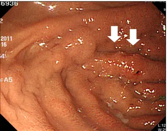
Vol. 61 No. 1, January 2013 Fig. 2. Initial esophagogastroduodenoscopic finding. Congestive
nodularity with hemorrhagic spot (white arrows) was seen on the fundus.
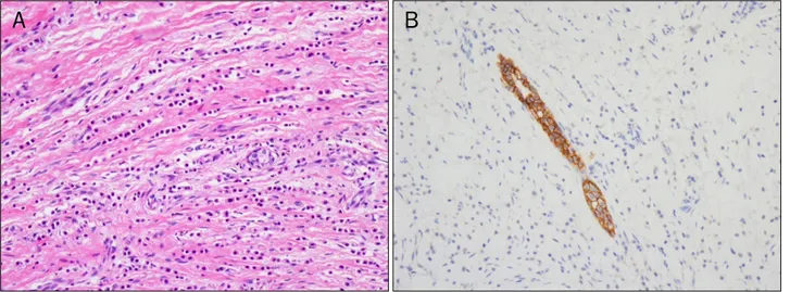
Fig. 3. Microscopic findings of the gastric biopsy. (A) It showed diffuse infiltrated adenocarcinoma with signet ring cell feature, consistent with metastatic lobular carcinoma with signet ring cell feature from the breast (H&E, ×200). (B) In additional estrogen receptor immunohistochemical stain, it revealed positive finding (Estrogen receptor, ×200).
위 기저부의 출혈 반점을 동반한 울혈성 결절이 관찰되었다 (Fig. 2). 병변에서 조직생검을 시행하였으며 병리검사 결과 인환세포의 형태를 가진 선암의 조직학적 소견을 보였다(Fig.
3A). 유방의 조직학적 소견은 소엽암종으로 진단되었으며 (Fig. 4), 위의 병변에 대하여 원발암과 유방암의 위전이의 감 별을 하기 위하여 에스트로겐 수용체와 프로게스테론 수용체 에 대한 면역조직화학염색을 하였고 유방암의 위 전이를 진단 하는 데 특이적인 면역조직화학염색법인 gross cystic disease fluid protein (GCDFP)-15에 대한 검사를 추가로 시행하였다.
그 결과 에스트로겐과 프로게스테론 수용체에 양성을 나타내 었고 GCDFP-15 검사에서 부분적으로 양성 소견을 보여 원발 부위가 유방인 전이성 위선암으로 확인되었다(Fig. 3B). 환자
는 다발성 전이를 동반한 양쪽 유방암 진단 하에 7개월 간 총 9차례의 선행화학요법(doxorubicin, docetaxel) 이후 양쪽 변형근치유방절제술을 시행받았다. 이후 방사선 치료를 하였 고 현재 고식적 항암화학요법(gemcitabine)을 유지하며 진단 시부터 13개월 간 추적 관찰 중이다. 1년 후 추적 관찰 시 시행한 복부 CT에서 전이가 의심되었던 위 기저부 점막의 비 후는 다소 호전되었고 양전자방출 단층촬영에서도 골 전이 외 에 동위원소 흡수가 뚜렷이 관찰되는 병변은 없었다. 위내시 경 검사는 선행화학요법이 종료된 후 1년 추적 관찰 시점에서 시행되었으며, 위 기저부의 주름 일부에 발적과 결절성 점막 변화가 관찰되었고 두 차례 검사 결과 내시경 소견에서 큰 변화는 보이지 않았다(Fig. 5). 추적관찰 시 시행한 조직 생검 결과 역시 인환세포를 포함하는 전이성 위선암으로 확인되었 다.
진단: 침윤성 소엽 유방암에 의한 전이성 위선암 복강 외 암종에 의한 위장관의 전이는 매우 드물고, 유방암 은 폐암 다음으로 두 번째로 흔히 전이하는 원발암으로 알려 져 있다.1-3 유방암의 위 전이는 0.1-8.0%의 빈도로 발견되며 부검에 의한 검사에서는 2.1-15.2%로 다양하게 보고되어 있 다.4-6 일반적으로 유방암은 관암종이 더 흔하지만(75%) 특징 적으로 유방암의 위 전이는 소엽암종에서 70-97%로 흔히 발 생한다.6,7 유방암에 의한 위 전이가 있는 환자에서 상부위장 관 증상은 대부분이 식욕부진, 구역, 구토, 상복부 동통과 같 은 비특이적인 증상으로 나타나며 체중 감소, 위장관 출혈, 빈혈이 동반되는 경우도 있다.8,9 유방암의 위 전이는 기저부 와 전정부에서 주로 발생하고 위내시경 소견은 정상부터 미만 성 위암침윤에 이르기까지 매우 다양하며 궤양이나 용종성의
56
윤소윤, 심기남. 유방암의 위 전이The Korean Journal of Gastroenterology
Fig. 5. One year follow-up esopha- gogastroduodenoscopic findings. (A) At the fundus, nodular elevated lesion with enlarged thichkened folds was seen without significant interval chan- ge. (B) It showed irregular nodularity in narrow band image.
Fig. 4. Microscopic findings of the breast biopsy. (A) It was composed of non-cohesive cells individually dispersed in single-file linear patterns,
‘Indian files’, in fibrous stroma (H&E, ×200). (B) There was no detection of E-cadherin expression, and it was compatible with invasive lobular carcinoma (E-cadherin, ×200).
종괴, 미란, 점막하 종양 형태, 발적과 주름 비후 등으로 광범 위하게 나타나 위염, 원발성 위암과 림프종과의 감별이 어렵
다.7,10,11 이 증례에서는 다발성 전이가 있는 유방의 소엽암종
환자에서 특이적 복부 증상은 없었으나 위내시경 검사에서 위 기저부의 출혈 반점을 동반한 울혈성 결절이 발견되었다. 이 러한 경우 위 전이를 감별하기 위하여 조직생검이 필수적이며 면역조직화학검사를 통하여 원발 부위의 확인이 가능하다. 이 번 증례에서 양성소견을 보인 에스트로겐 수용체, 프로게스테 론 수용체는 원발 위암에서 역시 23%가 양성으로 나타날 수 있어서 민감도와 특이도가 떨어지나, 유방암에서 높은 민감도 와 특이도를 갖는 GCDFP-15 면역염색에서 양성소견을 나타 내어 유방암에 의한 위 전이로 진단할 수 있었다.12-14 유방암 에서 위전이 발생 시 전이가 흔하다고 알려진 뼈, 간, 폐 등으 로 다발성 전이가 이미 확인된 경우가 57-94%로 보고되어 있 고,6,8 이번 증례에서도 다발성 전이가 있는 환자에서 위 전이 가 발견되었다. 유방암에서 위 전이를 포함한 다발성 전이가
확인된 경우 주된 치료법은 전신 화학항암요법과 호르몬 요법 이며 평균 생존율은 약 10개월 가량으로 알려져 있다.8 이번 증례의 환자는 전신 화학항암요법과 고식적 수술 및 방사선 요법으로 치료하였고 현재 13개월 간 추적 관찰 중으로 치료 에 일부 반응을 보이고 있으며 추가로 호르몬 요법을 고려 중이다.
유방암과 위암은 국내에서 그 발생률이 비교적 높은 암으 로 과거에 유방암을 진단받았거나 다발성 전이가 있는 유방암 환자에서 위내시경 검사 시 위암이 의심되는 경우 위 전이에 대한 감별이 매우 중요하다. 특히 복부 증상이 없거나 비전형 적인 증상을 호소하는 환자에서도 유방의 소엽암종이며 타 장 기로의 전이가 있는 경우에는 위내시경 검사를 반드시 시행해 야하고 추적 관찰이 필요하며, 위내시경 검사에서 위염, 미란, 궤양, 점막하 종양, 위용종과 위암 등 다양한 형태로 나타날 수 있으므로 단순히 위에서 기원한 병변으로 간과하지 말고 조직생검을 통하여 전이 여부에 대하여 확인해야 할 것이다.
Yoon SY, Shim KN. Gastric Metastasis from Breast Cancer
57
Vol. 61 No. 1, January 2013
REFERENCES
1. McLemore EC, Pockaj BA, Reynolds C, et al. Breast cancer: pre- sentation and intervention in women with gastrointestinal metastasis and carcinomatosis. Ann Surg Oncol 2005;12:
886-894.
2. Jones GE, Strauss DC, Forshaw MJ, Deere H, Mahedeva U, Mason RC. Breast cancer metastasis to the stomach may mim- ic primary gastric cancer: report of two cases and review of literature. World J Surg Oncol 2007;5:75.
3. Koike K, Kitahara K, Higaki M, Urata M, Yamazaki F, Noshiro H.
Clinicopathological features of gastric metastasis from breast cancer in three cases. Breast Cancer 2011. [Epub ahead of print]
4. Taal BG, den Hartog Jager FC, Steinmetz R, Peterse H. The spectrum of gastrointestinal metastases of breast carcinoma:
I. Stomach. Gastrointest Endosc 1992;38:130-135.
5. Jeon SH, Lee YS, Kwon TK, et al. A case of gastric metastasis from breast carcinoma manifested by upper gastrointestinal bleeding. Korean J Gastrointest Endosc 2002;24:220-224.
6. Almubarak MM, Laé M, Cacheux W,et al. Gastric metastasis of breast cancer: a single centre retrospective study. Dig Liver Dis 2011;43:823-827.
7. Cheoi KS, Lee WY, Eum YO, et al. A case of stomach metastasis from breast cancer. Korean J Med 2006;71:567-572.
8. Taal BG, Peterse H, Boot H. Clinical presentation, endoscopic features, and treatment of gastric metastases from breast carcinoma. Cancer 2000;89:2214-2221.
9. Cormier WJ, Gaffey TA, Welch JM, Welch JS, Edmonson JH.
Linitis plastica caused by metastatic lobular carcinoma of the breast. Mayo Clin Proc 1980;55:747-753.
10. Dumoulin FL, Sen Gupta R. Breast cancer metastasis to the stomach resembling small benign gastric polyps. Gastrointest Endosc 2009;69:174-175.
11. Yamamoto D, Yoshida H, Sumida K, et al. Gastric tumor from metastasis of breast cancer. Anticancer Res 2010;30:3705- 3708.
12. Kojima O, Takahashi T, Kawakami S, Uehara Y, Matsui M.
Localization of estrogen receptors in gastric cancer using im- munohistochemical staining of monoclonal antibody. Cancer 1991;67:2401-2406.
13. Wick MR, Lillemoe TJ, Copland GT, Swanson PE, Manivel JC, Kiang DT. Gross cystic disease fluid protein-15 as a marker for breast cancer: immunohistochemical analysis of 690 human neoplasms and comparison with alpha-lactalbumin. Hum Pathol 1989;20:281-287.
14. Park KW, Im YH, Lee J, et al. Use of GCDFP-15 (BRST-2) as a spe- cific immunocytochemical marker for diagnosis of gastric metastasis of breast carcinoma. Cancer Res Treat 2003;35:
460-464.