326
<Received:November 11, 2013, Revised (1st:February 7, 2014, 2nd:March 3, 2014, Accepted:March 7, 2014>
Corresponding to:Sung Jae Choi, Division of Rheumatology, Department of Internal Medicine, Korea University Ansan Hospital, 123, Jeokgeum-ro, Danwon-gu, Ansan 425-707, Korea. E-mail:[email protected]
pISSN: 2093-940X, eISSN: 2233-4718
Copyright ⓒ 2014 by The Korean College of Rheumatology
This is a Free Access article, which permits unrestricted non-commerical use, distribution, and reproduction in any medium, provided the original work is properly cited.
Leflunomide was licensed for the treatment of rheumatoid ar- thritis in 1998 and has been available in Korea since 2003.
Allergic cutaneous reactions (rash, purpura) are common (<10%) side effects of leflunomide, but severe cases such as Stevens-Johnson syndrome (SJS) or toxic epidermal necrolysis (TEN) are rarely reported. There has not been a report of SJS or TEN induced by leflunomide in Korea. Here we report
a case of leflunomide-induced TEN in a patient with rheuma- toid arthritis. Leflunomide was discontinued, and the TEN was treated with methylprednisolone, cholestyramine and immunoglobulin. The skin lesion eventually resolved over four weeks with residual post-inflammatory hyperpigmentation.
Key Words. Leflunomide, Toxic epidermal necrolysis, Rheumatoid arthritis
Introduction
Leflunomide is a disease-modifying anti-rheumatic drug (DMARD) that is effective in relieving symptoms of rheuma- toid arthritis and reducing radiological progression. It was first licensed for the treatment of rheumatoid arthritis in 1998 and has been available in Korea since 2003. Fifteen years after the initial licensing of leflunomide, its safety profile has been well documented in clinical trials, post-marketing surveillance, and epidemiological studies (1).
Common adverse effects are gastrointestinal (diarrhea, dys- pepsia, nausea/vomiting, abdominal pain, oral ulcers), abnor- mal liver function tests, drug eruptions, alopecia, infections, weight loss and hypertension (2,3). In clinical trials, le- flunomide-induced allergic cutaneous reactions (rash, purpura) were common (<10%) side effects. However, there are very few reports of severe side effects, such as Stevens-Johnson syndrome or toxic epidermal necrolysis. Only four cases worldwide have been reported to date (4-7).
Toxic epidermal necrolysis is a rare, acute and life-threat- ening disease characterized by mucocutaneous tenderness, er-
ythema, and extensive exfoliation. Stevens-Johnson syndrome and Toxic epidermal necrolysis are classified by the extent of skin detachment; >30% of the body surface area. The mortal- ity rate ranges from 25% to 50%.
A case of Stevens-Johnson syndrome or toxic epidermal nec- rolysis induced by leflunomide has not been reported in Korea. However, we recently encountered a case of toxic epi- dermal necrolysis induced by leflunomide in a patient with rheumatoid arthritis. Here we provide a description of the case along with a review of the relevant literature.
Case Report
A 31-year-old woman presented to the emergency room complaining of a painful, itchy maculopapular rash distributed over her entire body and oral cavity pain for the last two days.
She was diagnosed with seropositive rheumatoid arthritis four years previously, and she had been followed up as an outpatient. When rheumatoid arthritis was first diagnosed, she was treated with methotrexate monotherapy until she got pregnant. After giving birth, she was treated with pre-
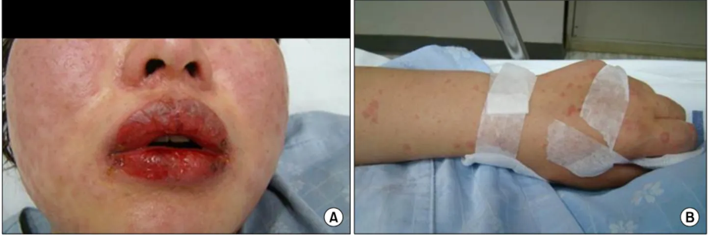
Figure 1. Day 1, The patient had diffuse erythema of the face, erosion of the lips and oral mucosa, and erythematous macules and multiple clear, small, fluid-filled vesicles on her extremities.
Figure 2. H&E stain, ×200, subepidermal blister.
dnisolone, 5 mg once a day, and hydroxychloroquine, 200 mg twice a day, combined with symptomatic agents (aceclofenac, 200 mg/day) for a year.
Seventeen days before coming to the emergency room, she visited the outpatient clinic for a routine checkup. On physical examination, she had joint swelling and pain in both wrists, and her PIP and MCP joints. Based on the CRP/ESR results and physical findings, leflunomide 10 mg once a day was add- ed to her regimen.
Upon presentation to the ER, the patient was conscious but had difficulty verbally communicating due to painful ulcers of the oral mucosa and throat. She also had an itchy burning rash around her lips and eyes and on her trunk and extremities. She indicated that the rash had first appeared on her lips and in her mouth, and subsequently spread to her face, trunk and extremities over the past two days.
On physical examination, the patient had temperature of 37.1oC, pulse of 88 beats/min, respiration rate of 18 breaths/min, and a blood pressure of 120/70 mm Hg. Her face was edematous, and she had erosions on her lips, oral mucosa, and genitalia. She also had a conjunctival injection.
Erythematous macules and multiple clear, small, fluid-filled vesicles appeared over her lips, face and extremities. Pruritus and bullous lesions were evident on her hands with few dis- rupted bullae (Figure 1). Evaluation by an ophthalmologist showed catarrhal conjunctivitis.
Laboratory examination revealed 11,510 WBCs/mg, LDH of 646 IU/L, and C-reactive protein of 4.71 mg/dL. Kidney and the liver function tests and urinalysis were within normal lim- its, and a chest radiograph was normal. A blood sample col- lected for surveillance culture in the emergency room revealed a methicillin-sensitive strain of Staphylococcus epidermidis.
Biopsy of a palm lesion showed subepidermal vesicular der-
matitis with scant inflammation (Figure 2). Direct fluorescent antibody staining was negative.
She was suspected of having overlapping Stevens-Johnson syndrome and toxic epidermal necrolysis, possibly related to the leflunomide that had been started seventeen days earlier. She was treated with massive hydration, antibiotics (glycopeptides), silver-coated dressings, and supportive care in the ICU. The leflunomide was discontinued at that time.
After two days, the vesicles and macules changed to bullae that spread rapidly to the rest of her body (abdomen, back, extremities, palms, and soles) and covered 64% of her total body surface area. Eventually, the patient was diagnosed with toxic epidermal necrolysis. The patient was treated with meth- ylprednisolone 24 mg/day and colestyramine 24 mg/day for leflunomide wash-out and immunoglobulin, 1 g/kg total dose over three days.
After five days, the lesions coalesced into large areas of epidermal detachment, and Nikolsky’s sign was present
Figure 3. Day 6, The vesicles and macules changed to bullae, and Nikolsky’s sign was present.
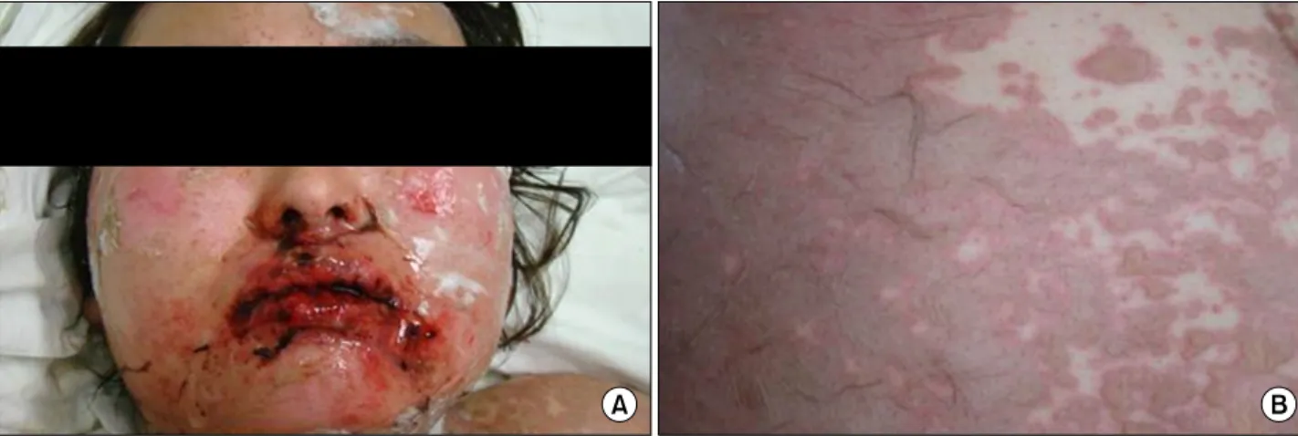
Figure 4. Day 8, Disrupted vesicles with crusting and spontaneous bleeding were evident during the re-epithelialization period.
Figure 5. Day 26, The skin lesions resolved with residual post-inflammatory hyperpigmentation.
(Figure 3). After eight days, confluent erythema and extensive blistering involving most of her back, abdomen, extremities, palms, and soles was noted, with a few disrupted vesicles and crusting and bleeding (Figure 4). The skin lesions eventually resolved over the course of approximately four weeks with re- sidual post-inflammatory hyperpigmentation (Figure 5). There was noticeable improvement in her general health. However, the patient had a visual defect due to severe corneal abrasion, and she underwent amniotic membrane transplantation.
Discussion
Leflunomide is a new immunomodulatory agent that is effec- tive in the treatment of rheumatoid arthritis. The mechanism of action has been proposed to be reversible inhibition of di- hydro-orotate dehydrogenase (DHODH). On an oral admin- istration, leflunomide is rapidly converted to its metabolite, teriflunomide, in the gut wall and the liver, and is excreted in both the urine and feces. Leflunomide has a long elimi- nation half-life of 15∼18 days, which is most likely due to enterohepatic circulation and biliary recycling. For this reason, oral colestyramine can be used to rapidly decrease plasma concentrations of the active metabolite and facilitate drug elimination (8).
Peripheral neuropathy and interstitial lung disease as side ef- fects of leflunomide have been reported in many countries in- cluding Korea. Cases of Stevens-Johnson syndrome or toxic epidermal necrolysis are rare. Among 5,163 patients treated with leflunomide in Japan, 13 experienced Stevens-Johnson syndrome (7). Only one case of drug hypersensitivity syn- drome induced by leflunomide has been reported in Korea.
However, there have been no cases of toxic epidermal necrol- ysis reported in either country (9). Only four cases of toxic epidermal necrolysis induced by leflunomide have been re- ported worldwide (4-7), and there are no reports of toxic epi- dermal necrolysis induced by leflunomide in Korea.
Stevens-Johnson syndrome and toxic epidermal necrolysis are acute and life-threatening mucocutaneous diseases that are always drug-related. Toxic epidermal necrolysis was first not- ed in 1956 by Alan Lyell, who described four patients with an eruption resembling scalding of the skin. Toxic epidermal necrolysis is associated with microbes (e.g., Staphylococcus) and certain drugs such as allopurinol, antibiotics, non-steroidal anti-inflammatory drugs, and anticonvulsants. Use of medi- cations is reported in over 95% of patients with toxic epi- dermal necrolysis. It is a rare disease with an annual incidence of 0.4∼1.2 per million.
As in our patient, the initial symptoms of toxic epidermal necrolysis can include fever, pain upon swallowing, lympha-
denopathy, hepatitis and cytopenias. Skin lesions appear as er- ythematous, macules of irregular size and shape in the early stage. Subsequent involvement of the epidermis progresses to- ward full-thickness necrosis. The necrotic epidermis detaches from the underlying dermis, giving rise to blisters and Nikolsky’s sign.
In this case, the patient had been diagnosed with seropositive rheumatoid arthritis and treated with prednisolone and hydroxychloroquine. Seventeen days before admission, le- flunomide had been added to her regimen due to poor prognosis. The patient visited the emergency room with a maculopapular rash, oral mucositis, and fever. Over the course of one week, the macules changed to bullae, and epidermal detachment and Nikolsky’s sign were evident. Based on the symptoms, history, and progress, the patient was diagnosed with toxic epidermal necrolysis. We suspected that the cause was the leflunomide that was started 17 days before her admission. The leflunomide was immediately discontinued, and the toxic epidermal necrolysis was treated with methyl- prednisolone, colestyramine and immunoglobulin. The skin le- sions eventually resolved over the course of approximately four weeks with residual post-inflammatory hyperpigmentation.
To our knowledge, this is the first reported case of toxic epi- dermal necrolysis induced by leflunomide in Korea. It is im- portant that clinicians be aware of toxic epidermal necrolysis induced by leflunomide. When clinicians treat patients with leflunomide, they should carefully monitor them for the pres- ence of skin lesions. If skin lesions are detected, leflunomide should be withdrawn immediately.
Summary
Here we describe a first case of toxic epidermal necrolysis induced by leflunomide in a patient with rheumatoid arthritis in Korea. Clinicians have to be aware of toxic epidermal nec- rolysis induced by leflunomide and should carefully monitor presence of skin lesion.
References
1. van Riel PL, Smolen JS, Emery P, Kalden JR, Dougados M, Strand CV, et al. Leflunomide: a manageable safety profile. J Rheumatol Suppl 2004;71:21-4.
2. Osiri M, Shea B, Robinson V, Suarez-Almazor M, Strand V, Tugwell P, et al. Leflunomide for the treat- ment of rheumatoid arthritis: a systematic review and metaanalysis. J Rheumatol 2003;30:1182-90.
3. Smolen JS, Emery P. Efficacy and safety of leflunomide in active rheumatoid arthritis. Rheumatology (Oxford) 2000;39 Suppl 1:48-56.
4. Schmutz JL, Barbaud A, Tréchot P. Leflunomide and Lyell syndrome. Ann Dermatol Venereol 2009;136:395.