대한소화기학회지 2007;50:131-135
접수: 2007년 2월 14일, 승인: 2007년 7월 13일 연락처: 이완식, 501-757, 광주광역시 동구 학동 8
전남대학교 의과대학 소화기내과학교실 Tel: (062) 220-6215, Fax: (062) 225-8578 E-mail: [email protected]
Correspondence to: Wan Sik Lee, M.D.
Department of Internal Medicine, Chonnam National University Hospital, 8, Hak-dong, Dong-gu, Gwangju 501-757, Korea Tel: +82-62-220-6215, Fax: +82-62-225-8578
E-mail: [email protected]
급성췌장염과 동반된 간문맥 내 가스 2예
전남대학교 의과대학 내과학교실
박형천ㆍ이완식ㆍ주소영ㆍ박선영ㆍ주영은ㆍ김현수ㆍ최성규ㆍ유종선
Hepatic Portal Venous Gas Associated with Acute Pancreatitis: Reports of Two Cases and Review of Literature
Hyeong Cheon Park, M.D., Wan Sik Lee, M.D., So Young Joo, M.D., Seon Young Park, M.D., Young Eun Joo, M.D., Hyun Soo Kim, M.D.,
Sung Kyu Choi, M.D., and Jong Sun Rew, M.D.,
Department of Internal Medicine, Chonnam National University Hospital, Gwangju, Korea
Hepatic portal venous gas (HPVG) is an uncommon disease entity that usually has grave prognosis. It is gen- erally associated with bowel necrosis, and has been reported in a wide variety of conditions such as ulcerative colitis, Crohn’s disease, diverticulitis, intestinal ischemia, or infarction. We experienced two cases of HPVG asso- ciated with acute pancreatitis. HPVG was found in patients with severe necrotizing pancreatitis and concurrent bowel ischemia. Despite aggressive resuscitation with fluids and broad spectrum antibiotics, each patient developed multiorgan failure, and died within few days. Acute pancreatitis is a potential cause of severe intraabdominal sys- temic catastrophe. Moreover, HPVG is associated with bowel ischemia in the setting of acute pancreatitis which could lead to rapid aggravation of symptom and complicated clinical course. Therefore, vigilant and aggressive management should be warranted in such condition. (Korean J Gastroenterol 2007;50:131-135)
Key Words: Hepatic portal venous gas; Pancreatitis; Intestinal ischemia
INTRODUCTION
Wolf and Evans first described HPVG in infants with necrotizing enterocolitis in 1955.1 Since then, HPVG has been reported with increasing frequency in the literature. HPVG has been found to be associated with a broad range of disease process, some of which are benign and insignificant, and resolve with only conservative treatment. However, in most cases, HPVG usually comes along with severe lethal conditions and requires emergency operation. Liebman et al. reviewed 64
cases of HPVG and reported that HPVG could be associated with necrotic bowel (72%), ulcerative colitis (8%), intra- abdominal abscess (6%), small bowel obstruction (3%), and gastric ulcer (3%).2 Most often, portal venous gas is associated with pneumatosis intestinalis although it may occur as an isolated finding.3
Acute pancreatitis has a possibility of incurring intraabdominal derangements and systemic catastrophes. In severe necrotizing pancreatitis, local and systemic complications can lead to renal failure, severe form of respiratory failure, and tissue hypoxemia.
There were few reports of HPVG associated with acute pan-
132 대한소화기학회지: 제50권 제2호, 2007
Fig. 1. Abdominal CT scan (case 1) shows gas within the supe- rior mesenteric vein and distal splenic vein (arrow) with pancre- atic head edema and peripancreatic fluid collections.
Fig. 2. Abdominal CT scan (case 1) shows diffuse dilated bowel loops with air-fluid levels and pneumatosis intestinalis.
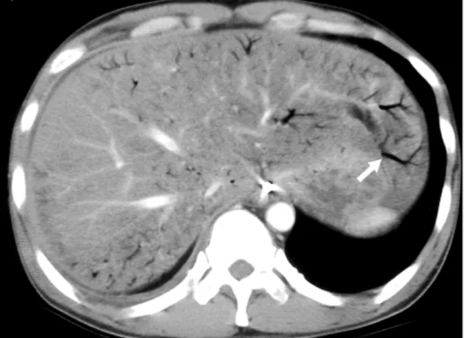
Fig. 3. Abdominal CT scan (case 1) demonstrates multiple air collection in the intrahepatic portal vein toward the hepatic pe- riphery (arrow).
creatitis.4-8 Some reported that conservative treatment was successful in certain situations. However, HPVG with bowel necrosis could contribute to poor prognosis that is associated with complicated clinical course and death.4
Herein, we report two cases of acute pancreatitis which showed gas in the hepatico-portal vein with pneumatosis in- testinalis.
CASE REPORT
1. Case 1
A 40-year-old man with a history of heavy alcohol
consumption visited to emergency ward complaining sudden onset of severe epigastric pain and vomiting. He had been on heavy alcohol binges for several days before admission.
Initially, the patient’s vital sign was insecure with his blood pressure (BP) 90/60 mmHg, pulse rate 128/min, and respiration rate 42/min. He was afebrile and physical examination revealed decreased bowel sounds, epigastric tenderness, and rigidity of abdominal wall. The plain radiographs showed diffuse bowel dilatations and multiple air-fluid levels in the abdomen. Labo- ratory data included a white blood cell count of 23,700/mm3, hemoglobin 14.0 g/dL, platelet count 105,000/mm3, AST > 1000 IU/L, ALT 368 IU/L, γ-GT 368 IU/L, glucose 529 mg/dL, total protein 4.6 g/dL, albumin 2.0 g/dL, total bilirubin 2.5 mg/dL, serum amylase >2,000 IU/L, serum lipase >2,000 IU/L, BUN 35 mg/dL, creatinine 2.1 mg/dL, and triglyceride 1,291 mg/dL. Arterial blood gas analysis showed pH 7.34, pCO2 19.9 mmHg, PO2 98.1 mmHg, and HCO3 10.9 mEq/L.
Computed tomography (CT) revealed a diffuse pancreatic edema and collections of peripancreatic fluids with air bubbles in the superior mesenteric vein and distal gastrosplenic vein (Fig. 1, 2). Multiple branched air density in peripheral portion of liver and portal vein confirms the presence of the HPVG (Fig. 3).
Emergency operation for necrotic bowel resection and pancreatic irrigation were urgently needed, but poor general conditions of the patient deterred any surgical treatment.
Despite aggressive supportive care using fluid resuscitations, broad-spectrum antibiotics, and respiratory support, his con- dition became worse rapidly. He died on the third hospital day.
박형천 외 7인. 급성췌장염과 동반된 간문맥 내 가스 2예 133
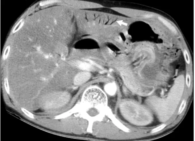
Fig. 4. Abdominal CT (case 2) demonstrates several linear air collection in the left hepatic portal veins of the periphery of liv- er (arrow).
Fig. 5. Abdominal CT (case 2) shows pancreatic necrosis (arrow), peripancreatic fluid collections, small bowel dilatations, and pneumatosis intestinalis.
2. Case 2
A 53-year-old man with heavy alcoholic binge history was admitted due to fever, rigour, and abdominal pain. His systolic BP was less than 60 mmHg, pulse rate 78/min, and respiratory rate 42/min. Laboratory data included white blood cell count of 16,300/mm3, hemoglobin 10.5 g/dL, platelet count 323,000/mm3, AST more than 1,000 IU/L, ALT 89 IU/L, serum amylase 1,207 IU/L, lipase 1,240 IU/L, glucose 486 mg/dL, and calcium 8.1 mg/dL. Arterial blood gas analysis showed pH 6.67 pCO2
18.7 mmHg, pO2 135 mmHg, and HCO3 2.1 mEq/L. After vigorous fluid replacements and infusion of inotropics and sodium bicarbonate, his blood pressure was temporarily stabilized to 120/70 mmHg. CT revealed diffuse pancreatic necrosis and peripancreatic fluid collections as well as bowel dilatations, air in the bowel walls, and HPVG (Fig. 4, 5). The patient died on the second hospital day due to multi-organ failure despite vigorous medical care.
DISCUSSION
HPVG is associated with a wide range of underlying pathologies, most commonly reported cases including mesen- teric ischemia, post-procedural complications, Crohn’s disease, intra- abdominal abscess, and bowel obstruction. Less common pathologies include colon cancer, gastric ulceration, acute pancreatitis, portal pyelophlebitis, infectious causes, and sigmoid diverticulitis.5
HPVG is also observed in few cases of acute pancreatitis.
First report was published in 1961.6 Thereafter, Faberman and Mayo-Smith reported three more cases and demonstrated that pancreatitis with HPVG is not fulminant as has been previously thought.7 In 2003, Iannitti et al. reported three cases and all of them had good clinical outcomes.5 Paran et al. reported one case of necrotizing pancreatitis with HPVG which had a benign course.8 More recently, Chan et al. reported two cases of HPVG occurring with pneumatosis intestinalis that showed fatal outcomes.4 This situation may suggest that the clinical outcome of acute pancreatitis with HPVG is variable. Fatal cases were invariably found to have a sign of intestinal ischemia, meaning that combining hypotensive episode, splanchnic vasoconstriction, and bowel ischemia were the determining factors.
The pathogenesis of HPVG is not fully understood. The primary factors that lead to portomesenteric gas are intestinal wall alterations, bowel dilatation, and sepsis.2 There are two theories - mechanical and bacterial theory for describing the pathogenesis of portomesenteric gas. The mechanical theory proposes that gas dissects into the bowel wall, damages mucosa and circulates into vein of bowel wall. Mucosal damage and bowel distension are important factors in this theory. The most common causes of intestinal wall alteration or mucosal damage are intestinal ischemia with bowel necrosis and inflammatory bowel disease (ulcerative colitis and Crohn’s disease).2 Bacterial theory suggests that gas-forming bacteria, such as Escherichia coli, Clostridial species, and other gas-forming bacteria, enter the submucosa through mucosal defect and produce gas within the intestinal wall and in the portal vein.2,9-12 Indeed, severe infectious abdominal processes, such as diverticulitis, abdominal
134 The Korean Journal of Gastroenterology: Vol. 50, No. 2, 2007
abscess and gangrene, cholecystitis, cholangitis, appendicitis, colitis, and abdominal tuberculosis can cause thrombophlebitis of mesenteric vein and produce portomesenteric gas.2,9-12 In acute pancreatitis, direct intestinal mucosal damage by pancreatic enzymes or secondary mucosal disruption due to transient ischemia and splanchnic hypoperfusion were offered as possible mechanisms of gas translocation from lumen through the bowel wall.8 Our cases would be clear examples of this phenomenon since both patients suffered from uncontrollable hypovolemia and shock. There was a possibility of gas forming bacteria as an etiology for HPVG in our cases as our patients could be inflicted with sepsis. However, conclusions regarding this postulation could not be drawn decisively due to poor evidence and lack of details of causing organisms.
The diagnosis of HPVG can be established mainly by radiological method. The presence of air in the portal vein can be detected by simple plain radiography, CT or ultrasono- graphy.5,13-15 CT is the most sensitive test for the detection of HPVG.15 The accumulation of gas in the hepatic portal vein produces tubular lucencies from the porta hepatis to the edge of the liver within 2 cm beneath the liver capsule. This is because the centrifugal flow of portal venous blood, which carries the portal venous gas to periphery. The differentiation of HPVG from pneumobilia is important. Gas in the biliary tree tends to collect in the large bile ducts at the hilum due to the centripetal flow of bile, and is usually found within the central portion of the liver more than 2 cm from the liver capsule.15 CT also discloses gas in bowel wall (pneumatosis intestinalis) and these findings may give a clue to the underlying intraabdominal pa- thology. Most cases of HPVG are associated with pneumatosis intestinalis, though this is not a constant finding.2 The ultras- onographic findings of HPVG are reported as 1) echogenic particles flowing within the portal vein or 2) poorly defined, highly echogenic patches within the hepatic parenchyma, especially in the non-dependent part.14 Kinoshita et al. reviewed 182 cases of HPVG in adults, and overall mortality rate was 39%, although the mortality is much higher to 79% in patient combined with bowel ischemia.15
In our cases, HPVG was found in patients with acute severe pancreatitis and pneumatosis intestinalis. The history of heavy alcohol consumption, and elevated level of serum amylase and lipase, and CT finding of pancreatic necrosis and peripancreatic fluid collections are sufficient for the diagnosis of acute pancreatitis. HPVG was diagnosed using CT with the typical findings that were described above.
HPVG should be treated according to their clinical status and underlying diseases. Urgent laparotomy is indicated in patients who undergo deteriorating clinical course or who develop other complications. Likewise, patients suspicious having ischemic mesenteric event should be explored.15 On the contrary, healthy patient who has a controllable risk factor for developing HPVG is not a surgical candidate. As for the HPVG associated with acute pancreatitis, we suggest that clinical characteristics in- cluding patient presentation, laboratory, and radiologic findings should be correlated with the severity of acute pancreatitis before deciding whether surgical intervention is warranted or not.
In conclusion, HPVG is not a specific disease but rather a diagnostic clue of acute intraabdominal pathologies. The treat- ment of patient with HPVG should be directed for underlying disease, and emergency operative treatment should be considered if bowel ischemia are suspected. Acute pancreatitis can be a possible cause of HPVG by developing intestinal ischemia and pneumatosis intestinalis.
요 약
간문맥 내 가스는 임상에서 흔하지 않으며 대부분 예후가 좋지 않다. 이는 대부분 장괴사와 연관되어 발생하는 것으 로 알려져 있으나, 이 외에도 궤양성 대장염, 크론병, 게실 염, 내시경 치료시술, 복부 외상 등의 다양한 원인으로 인해 발생할 수 있다. 현재까지 급성췌장염에서 동반된 간문맥 내 가스에 대한 보고는 드물다. 저자들은 급성췌장염 환자 에서 발생한 간문맥 내 가스 2예를 경험하였다. 다량의 음 주력을 가진 환자가 내원하였다. 임상 경과 및 생화학혈청 검사, 혈액가스검사, 전산화단층촬영에서 중증의 괴사 췌장 염 소견이었다. 간문맥 내 가스와 함께 장벽 내 가스를 보였 으며 보존 치료에도 불구하고 두 예 모두 다발 장기부전으 로 사망하였다. 급성췌장염은 복강 내 및 전신적인 합병증 을 유발할 수 있다. 간문맥 내 가스는 중증의 췌장염에서 동 반될 수 있으며, 특히 장허혈과 동반되었을 경우에는 주의 깊은 관찰과 수술과 같은 적극적인 치료가 필요하다.
색인단어: 급성췌장염, 간문맥 내 가스, 장허혈
REFERENCES
1. Wolfe JN, Evans WA. Gas in the portal veins of liver in in- fants; a roentgenographic demonstration with postmortem ana- tomical correlation. Am J Roentgenol Radium Ther Nucl Med
Park HC, et al. Hepatic Portal Venous Gas Associated with Acute Pancreatitis 135
1955;74:486-488.
2. Liebman PR, Patten MT, Manny J, Benfield JR, Hechtman HB. Hepatic portal venous gas in adults: Etiology, patho- physiology and clinical significance. Ann Surg 1978;187:
281-287.
3. Yamamuro M, Ponsky JL. Hepatic portal venous gas: report of a case. Surg Today 2000;30:647-650.
4. Chan SC, Wan YL, Cheung YC, Ng SH, Wong AM, Ng KK.
Computed tomography findings in fatal cases of enormous hepatic portal venous gas. World J Gastroenterol 2005;11:
2953-2955.
5. Iannitti DA, Gregg SC, Mayo-Smith WW, Tomolonis RJ, Cioffi WG, Pricolo VE. Portal venous gas detected by com- puted tomography: is surgery imperative? Dig Surg 2003;20:
306-315.
6. Wiot JF, Felson B. Gas in the portal venous system. Am J Roentgenol Radium Ther Nucl Med 1961;86:920-929.
7. Faberman RS, Mayo-Smith WW. Outcome of 17 patients with portal venous gas detected by CT. AJR Am J Roentgenol 1997;169:1535-1538.
8. Paran H, Epstein T, Gutman M, Shapiro Feinberg M, Zissin R. Mesenteric and portal vein gas: computerized tomography
findings and clinical significance. Dig Surg 2003;20:127-132.
9. Celoria G, Coe NP. Does the presence of hepatic portal ve- nous gas mandate an operation? A reassessment. South Med J 1990;83:592-594.
10. Chiu HH, Chen CM, Lu YY, Lin JC, Mo LR. Hepatic portal venous gas. Am J Surg 2005;189:501-503.
11. Wiesner W, Mortele KJ, Glickman JN, Ji H, Ros PR. Portal- venous gas unrelated to mesenteric ischemia. Eur Radiol 2002;12:1432-1437.
12. Haak HR, Kooymans-Coutinho MF, von Teeffelen ME, Adhin S, Falke TH. Portal venous gas in a patient with diver- ticulitis. Hepatogastroenterology 1990;37:528-529.
13. Lafortune M, Trinh BC, Burns PN, et al. Air in the portal vein: sonographic and Doppler manifestations. Radiology 1991;180:667-670.
14. Schulze CG, Blum U, Haag K. Hepatic portal venous gas.
Imaging modalities and clinical significance. Acta Radiol 1995;36:377-380.
15. Kinoshita H, Shinozaki M, Tanimura H, et al. Clinical fea- tures and management of hepatic portal venous gas: four case reports and cumulative review of the literature. Arch Surg 2001;136:1410-1414.