|Original Article|
Direct-Current Treatment as a Safe Sterilization Method for Electrospun
Biodegradable Polymer
Hye-Lee Kim1,2, Jeong Hyun Lee1,2, Mi Hee Lee1, Hak Hee Kim1, Jungsung Kim1, Inho Han1, Bong Joo Park3, Jeong Koo Kim3, Dong Wook Han4, Soo Hyun Kim5, Seung Jin Lee6, and Jong-Chul Park1,2*
1Department of Medical Engineering, 2Brain Korea 21 Project for Medical Science, Yonsei University College of Medicine, Shinchon-dong, Seodaemun-gu, Seoul, 120-752 Korea
3Department of Biomedical Engineering College of Biomedical Science and Engineering, Inje University, Obang-dong, Gimhae, Gyungnam, 621-749 Korea
4Department of Nanomedical Engineering, College of Nanoscience & Nanotechnology, Pusan National University, Geumjeong-gy, Busan, 609-735 Korea
5Biomaterials Research Center, Korea Institute of Science and Technology, P.O. Box 131, Cheongryang, Seoul, 130-650 Korea 6College of Pharmacy, Ewha Womans University, Daehyun-dong, Seodaemun-gu, Seoul, 120-750 Korea
(Received: April 5th, 2011; Accepted: May 11th, 2011)
Abstract : Sterilization is an essential process for biodegradable polymers to be used as biomaterials or tissue engi-neered-scaffolds. The characteristics of biodegradable scaffolds can change due to decomposition of constituent polymers due to high temperature, pressure, or moisture during sterilization. This study investigated direct–current (DC) treatment as a safe method that can prevent structural change and deformation. Treatment of electrospun poly (lactic-co-glycolic acid) (PLGA) with DC showed a bactericidal effect within 40 sec at 4 V. When DC was applied at 6 V to the electrospun PLGA, the bactericidal effect emerged within 30 sec. The morphology of fibers and molec-ular weight of PLGA polymer was maintained after DC treatment. In contrast, electrospun PLGA exposed to ethyl-ene oxide showed fiber degradation, and gamma or e-beam irradiation resulted in decreased molecular weight. The demonstrated improvement in chemical and physical stability of biodegradable polymers after DC sterilization may help extend their application.
Key words: direct-current, biodegradable polymer, poly(lactic-co-glycolic acid), sterilization, electrospinning
1. Introduction
In tissue engineering, scaffolds usually have a three-dimensional (3D) structure to replace damaged tissue and are porous to support cell attachment and growth. The scaffolds can attain 3D and porous structure through several methods, such as electrospinning,1-4 solvent casting,5 and gas forming/salt-leaching.6 Prior to application in tissue engineering, porous scaffolds need to be sterilized.7 However, porous scaffolds made of polymers can be deformed after sterilization due to instability of the composing polymer at high temperature, pressure, or moisture. Therefore, ethylene oxide (EO) is widely used due to low processing temperature and humidity.8 EO is the most commonly used sterilization technique due to its
bactericidal effectiveness at low temperature, high penetration, and compatibility with a wide range of materials. However, owing to their large surface area, porous scaffolds can attain harmful quantities of EO residues.8-11 Another conventional method that can be applied to scaffolds is irradiation, which does not use chemical reagents, heat, or moisture. However, irradiation also has the problem of causing changes in chemical bonds and solubility, and resultant changes in degradation rates.12-13 For these reasons, further investigation into safe sterilization methods for biodegradable polymers is required.
In previous studies, microorganism membranes were broken down by electrical treatment. Electrical treatment involves direct (DC) or alternating (AC) current flow through the electrolyte containing microorganisms without chemical reagents or high temperature. This method ensures sterilization in a very short time even with low current. In the case of DC treatment, Vibrio parahaemolyticus (V. parahaemolyticus) in *Tel: +82-2-2228-1917; Fax: +82-2-363-9923
treatment to sterilize bacteria in electrospun poly(lactic-co-glycolic acid) (PLGA), one of the most widely used biodegradable polymers. Characterization was completed to determine the effect of the electrical treatment sterilization on the morphology and chemical properties.
2. Materials and Methods
2.1 Preparation of Electrospun PLGA
PLGA polymer (lactide/glycolic = 75/25) was purchased from Lakeshore Biomaterials (Birmingham, AL, USA). The number and weight average of molecular weight (Mn and Mw)
of the PLGA were 80 kDa and 120 kDa, respectively. PLGA solution for electrospinning was prepared by dissolving a measured amount of PLGA in 1,1,1,3,3,3-hexafluoro-2-propanol at room temperature. The concentration of PLGA in solution was maintained at 20% (w/v). The solution was delivered at a flow rate of 7 ml/h via a 23-gauge needle connected to 18 kV. The average thickness of the electrospun PLGA was approximately 0.38 mm, and was controlled by injected solution volume. Each experimental sample was prepared as 1 cm×1 cm squares shorn from the electrospun PLGA.
2.2 Treatment Method with DC and Traditional Sterilization Methods
The electrolysis vessel used for treatment with DC is shown in Fig 1. The electrolysis vessel was made of two platinum electrodes (25 mm×25 mm), which were 10 mm apart. Each platinum electrode was connected to a computer-based timing control through a parallel port interface, which was used to control the power transistor. The electrolysis vessel was filled with 3 ml of saline solution, and the inoculated sample was soaked in the solution. The inoculated sample in the electrolysis vessel was treated with DC at 4 V or 6 V for 10 to 60 sec.
2.3 Traditional Sterilization Methods
The traditional sterilization methods used for comparison to DC treatment methods were EO gas, gamma ray, and electron beam. For the group of samples sterilized by EO, the fibrous PLGA membranes were exposed to EO at 37oC for 4 h in a 3M Steri-Vac 5XL sterilizer (3M Health Care Ltd., St. Paul, USA), and vented over 12 h at room temperature in the same space. The other groups of samples were irradiated with gamma or e-beam. Gamma irradiation was carried out in air with a 60Co source at a dose rate of 1 kGy/h for a final dose of 25 kGy at room temperature (Greenpia Tech. Inc., Yujoo-Kun, Korea). For e-beam irradiation, fibrous PLGA membranes on a tray were exposed to a surface dose of 15-25 kGy at room temperature in air, using electron acceleration (EB Tech Co., Ltd., Daejeon, Korea). The electron energy was maintained at 1 eV and with a tray speed of 1 cm/min.
2.4 Sterility Tst
Escherichia coli (E. coli, ATCC 8739) and Staphylococcus aureus (S. aureus, ATCC 6358p) were used for the sterilization test because these bacteria cause significant public health problems worldwide.17-20 The bacteria were prepared for inoculation into samples by suspension in saline solution (0.9% NaCl in distilled water). The density of microorganism suspensions was 1×10⁶ colony forming units/ml. Each electrospun PLGA was inoculated by immersion in each microorganism suspension and shaking for 10 min. The inoculated samples were then treated with DC or traditional sterilization methods. After treatment, the samples were incubated in standard agar media (Becton Dickinson, NJ, USA) at 36oC for 14 days. The sterilization efficiency of DC treatment was determined by the number of colonies and the methods are shown in Fig 2. The samples inoculated with E. coli and S.
aureus are shown in Fig 2 after incubation in standard agar at 36oC for 5 days. Non-sterilized samples inoculated with E. coli and S. aureus were used as controls (Fig 2A and D). Appearance of colonies on the agar media after 14 days indicated contamination and inefficient sterilization (Fig 2B and E); while a clear, uncontaminated media indicated efficient sterilization (Fig 2C and F), producing a sterile product. Five days after putting the inoculated samples on the agar media pictures were taken using a digital camera (DSC-H5, Sony Inc., Tokyo, Japan). All the treatment conditions were independently repeated 10 times.
2.5 Cytotoxicity
To determine the cytotoxicity of the sterilized samples, cell viability was determined using a direct contact method.21 L-929 cells were maintained in Dulbecco’s modified Eagle’s medium (WellGENE Inc., Daegu, Korea) supplemented with 10% fetal bovine serum (WellGENE Inc.) and 1% antibiotic antimycotic solution (WellGENE Inc.) at 37oC and 5% CO2 in a humid environment. The treated samples (n=3) were placed at the center of the confluent L-929 cells monolayer and incubated for 24 h in a CO2 incubator. Latex and an untreated sample were used as positive and negative controls, respectively. After incubation, the samples were removed from the cultures. The viability of the cells in contact with samples was determined by
the MTT [3-(4,5-dimethylthiazol-2-yl)-2,5-diphenyltetrazolium bromide] assay. The cells were incubated with MTT reagent for 4 h at 37oC in the dark. The media was decanted and washed twice with phosphate buffered saline. The produced formazan crystals were dissolved in dimethylsulfoxide, and the absorbance was measured by an enzyme-linked immunosorbent assay reader at 570 nm. The cytotoxicity test was independently repeated three times.
2.6 Characterization of Fibrous PLGA Membranes The shapes of samples were photographed by a digital camera (DSC-H5, Sony Inc.). Scanning electron microscopy (SEM, Hitachi S-4700, Tokyo, Japan) was used to observe surface morphology of the electrospun PLGA and bacteria. The inoculated samples were fixed using 2.5% glutaraldehyde solution. After fixation, samples were dehydrated using graded ethanol, and dried. The dried samples were coated with gold before observing by SEM. The number and weight average of molecular weight (Mn and Mw) of each sample (n=5) were determined by gel permeation chromatography (GPCmax VE2001, Viscotek Europe, Irigny, France) with chloroform.
3. Results and Discussion
Different DC treatment times and voltages were investigated
Figure 2. Digital photograph of the electrospun PLGA inoculated with Escherichia coli (A-C) and Staphylococcus aureus (D-F) after incubation on standard agar at 36oC for 7 days. (A, D), untreated electrospun PLGA; (C, E), electrospun PLGA treated with 6 V for 10 s; and (D, F), electrospun PLGA treated with 6 V for 50 sec (n=10).
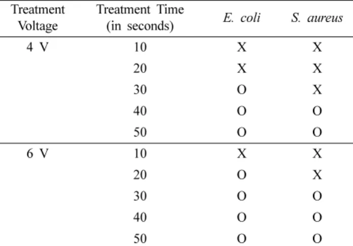
as summarized in Table 1. Complete sterilization was achieved over 30 sec for E. coli and 40 sec for S. aureus at 4 V. At 6 V, E. coli was sterilized in 20 sec and S. aureus was sterilized in 30 sec. At these treatment conditions, no electrolysis was observed, and thus the detected sterilization effects were due to DC. The treatment time for complete sterilization should be inversely related to the treatment voltages. Thus, the treatment times for sterilization could be reduced by increasing the treatment voltage. However, both bacteria were completely sterilized within 40 sec independent of treatment voltage. DC treatment achieved complete sterilization in a brief period compared to traditional sterilization methods. Traditional sterilization requires long periods for processing. After sterilization using EO gas, several days are required to remove the residual EO gas. Irradiation methods also require long exposure periods, depending on the size and quantity of the medical devices.8,10 Therefore, the treatment time with DC was an advantage over traditional sterilization methods using high-temperature steam, EO gas, or gamma irradiation. Based on these results, the treatment conditions are 6 V for 50 sec.
The morphology of E. coli and S. aureus in membranes are shown in Fig 3. Bacteria had been infiltrated into membranes and evenly distributed. Before treatment with DC, the inoculated bacteria showed regular morphology in membranes (Fig 3A and C). After DC treatment, all inoculated bacteria in membranes were reduced in size and exhibited transformed morphology (Fig 3B and D). These results suggested that DC treatment is an effective sterilization method. In previous studies, treatment with electric current caused breaks and ruptures in bacterial membranes14-16, resulting in release of cellular contents onto surrounding surfaces and shrinkage of the microorganisms
following release of their contents. This phenomenon is likely the main mechanism of sterilization of microorganisms by DC treatment. Therefore, DC treatment has a potential for application as a sterilization method for contaminated devices. The cytotoxicity results (Fig 4) confirmed the sterilized membranes to be innocuous to cells. The specialized nanofibrous feature of electrospinning which employs massive surface area did not influence the toxic effects of residues remaining on sterilized electrospun PLGA. DC treatment and traditional sterilization methods did not leave any cytotoxic residues on the sterilized membranes. Sterilization using EO gas is reported to produce toxic residues and can react with polymeric functional groups rendering an innocuous polymer toxic. Dimitrievskaet et al. described that EO sterilization induced
40 O O
50 O O
X, inefficient sterilization after treatment with DC; O, efficient sterilization after treatment with DC; n=10.
Figure 3. Scanning electron micrograph of Escherichia coli (A, B) and Staphylococcus aureus (C, D) in the electrospun PLGA. (A, C), untreated; (B, D), treated with 6 V for 60 sec.
Figure 4. Cytotoxicity of electrospun PLGA as evaluated by MTT assay (n=3).
alkylation of the PET polymer and significantly increased TNF-α release and macrophage activation.11-12 This result was due to the toxic effects of EO. Thus, biodegradable polymers sterilized with EO gas should be regarded as potentially cytotoxic. In contrast, DC treatment does not have the potential for cytotoxicity because this method does not use to any toxic reagent. Consequently, DC treatment is a biocompatible method suitable for sterilization of porous biodegradable polymers.
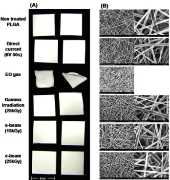
Digital photography is shown in Fig 5A and morphology by SEM is shown in Fig 5B. After EC treatment, the shape and the fibers of electrospun PLGA was preserved. However, electrospun PLGA sterilized using EO gas stuck to each other as in Fig 5B. In the report of Olde Damink et al., the deformation of the microparticles was affected by the conditions of the sterilization process with EO gas, such as temperature, pressure and exposure time.11 The processing temperature was near the glass transition temperature (40–60oC) for PLGA (Friess et al., 2006). Thus, the report concluded that the sterilization conditions using EO gas were incompatible for biodegradable polymers and resulted their deformation. After sterilization using EO, the membranes were transforming during imaging at magnification higher than 1,000 using SEM. It was also reported that EO treatment reduced the physical properties of the membranes, including the yield stress and break stress.8 The physical properties of biopolymers should be preserved because they are
connected to long-term stability. Therefore, to preserve shape, DC treatment is suitable for sterilization of biodegradable polymers.
Fig 6 presents the changes of the Mn and Mw of the electrospun PLGA. The Mn and the Mw of membranes after
treatment with DC were preserved. These molecular weights were reduced after irradiation with gamma or e-beam and the decrease was proportional to the radiation doses of e-beam. Many researchers have reported that gamma and e-beam irradiation caused polymer degradation through chain scission. In particular, backbone-chain scission results from exposure to gamma and e-beam radiation, and is the reason for the rapid decrease in the molecular weights. The decrease in the molecular weights of polymers is also related to significantly less crystalline structure. It has been demonstrated that the crystal structure can delay degradation because the crystalline regions in the polymer have resistant chains that are more oriented and closely packed compared to amorphous regions.23-23 Thus, the decrease in molecular weight should be avoided for the long-term stability because of accelerated biodegradation rates. DC treatment has the benefit of preserving the molecular weight of biodegradable polymers after sterilization.
Of the four methods compared, DC treatment is the most suitable sterilization method for biodegradable polymers with extensive surface areas. The electrospun PLGA membranes treated with DC were the only ones that maintained morphology and molecular weight.
4. Conclusion
The results of this study confirmed that DC treatment has the
Figure 5. Shape and surface morphology of electrospun PLGA. (A) Digital photograph, and (B) scanning electron micrograph.
Figure 6. The number and weight average of molecular weight (Mw and Mn) of the electrospun PLGA (n=5).
Ministry of Education, Science and Technology (grant no. 2010-0002089), and a grant from the Korea Health 21 R&D Project, Ministry of Health & Welfare, Republic of Korea (grant no. A050082).
References
1. S Sant, CM Hwang, S-H Lee, et al., Hybrid PGS–PCL microfibrous scaffolds with improved mechanical and biological properties, J Tissue Eng Regen Med, 5, 283 (2010). 2. J Stitzel, J Liu, SJ Lee, et al., Controlled fabrication of a
biological vascular substitute, Biomaterials, 27, 1088 (2006). 3. W He, Z Ma, T Yong, et al., Fabrication of collagen-coated
biodegradable polymer nanofiber mesh and its potential for endothelial cells growth, Biomaterials, 26, 7606 (2005). 4. RA Miller, JM Brady, DE Cutright, Degradation rates of oral
resorbable implants (polylactates and polyglycolates): rate modification with changes in PLA/PGA copolymer ratios, J Biomed Mater Res, 11, 711 (1977).
5. DC Miller, A Thapa, KM Haberstroh, et al., Endothelial and vascular smooth muscle cell function on poly(lactic-co-glycolic acid) with nano structured surface features, Biomaterials, 25, 53 (2004).
6. JJ Yoon, TG Park, Degradation behaviors of biodegradable m acroporous scaffolds prepared by gas foaming of effervescent salts, J Biomed Mater Res B: Appl Biomat, 55, 401 (2001).
7. H Lu, G Pei, P Zhao, et al., Cyclosporine-impregnated allograft bone sterilized with low-temperature plasma, J Tissue Eng Regen Med, 4, 638 (2010).
13. AG Hausberger, RA Kenley, PP Deluca, Gamma irradiation effects on molecular weight and in vitro degradation of poly (D,L-Lactide-co-glycolide) microparticles, Pharm Res, 12, 851 (1995).
14. JC Park, MS Lee, DW Han, et al., Inactivation of bacteria in seawater by low-amperage electric current, Appl Environ Microbiol, 70, 2405 (2003).
15. MH Lee, D-W Han, YI Woo, et al., Inactivation of Listeria monocytogenes in brine and saline by alternating high-voltage pulsed current, J Microbiol Biotechnol, 18, 1274 (2008). 16. SC Jin, H Yoo, YI Woo, et al., Selective sterilization of Vibro
parahaemolyticus from a bacterial mixture by low-amperage electric current, J Microbiol Biotechnol, 19, 537 (2009). 17. MT Madigan, JM Martinko, J Parker, Biology of microorganisms,
Prentice-Hall Inc., UK, USA (1997).
18. SC Clarke, RD Haigh, PPE Freestone, et al., Virulence of enteropathogenic Escherichia coli, a global pathogen, Clin Microbiol Rev, 16, 365 (2003).
19. D Metry, R Katta, New and emerging pediatric infections, Dermatol Clin, 21, 269 (2003).
20. KL Hiramatsu, CM Kuroda, T Ito, The emergence and evolution of methicillin-resistant Staphylococcus aureus, Trends Microbiol, 9, 486 (2001).
21. JV Luis, E Francese, A Elaine, et al., Cellular adhesion, proliferation and viability on conducting polymer substrates, Macromolecular Bioscience, 8, 1144 (2008).
22. S Dimitrievska, A Petit, CJ Doillon, et al., Effect of sterilization on non-woven polyethylene terephthalate fiber structures for vascular grafts, Macromol Biosci, 11, 13 (2010).
23. JSC Loo, CP Ooi, MLT Tan, et al., Isothermal annealing of poly(lactide-co-glycolide) (PLGA) and its effect on radiation degradation, Polym Int, 54, 636 (2005).