GSK3β의 선택적 저해제인 Kenpaullone의 B16 멜라노마 및 인간 멜라노사이트에서의 영향
김 해 종⋅이 유 리⋅호앙구엔⋅이 향 복⋅김 은 기†
인하대학교 바이오피부신소재실험실
(2011년 6월 20일 접수, 2011년 9월 16일 수정, 2011년 9월 19일 채택)
Effect of Kenpaullone, a Specific Inhibitor of GSK3β, on Melanin Synthesis in B16 Melanoma and Human Melanocytes
Hae Jong Kim, You Ree Lee, Dung Hoang Nguyen, Hyang Bok Lee, and Eun-Ki Kim†
Department of Biological Engineering, National Research Lab of Skin Bioactive Material, Inha University, 253 Younghyun-dong, Nam-Gu, Incheon 402-751, Korea
(Received June 20, 2011; Revised September 16, 2011; Accepted September 19, 2011)
요 약: Glycogen synthase kinase 3 beta (GSK3β)의 선택적 저해제인 Kenpaullone가 B16 멜라노마 및 사람의 멜라노 사이트에 미치는 멜라닌 합성능을 조사하였다. Kepaullone은 B16 멜라노마 및 사람의 멜라노사이트에 대하여 세포증식 에는 영향이 없는 범위 내에서 농도 의존적으로 멜라닌 합성을 촉진시켰다. B16 멜라노마 세포에 Kenpaullone을 첨가 48 h 후 tyrosinase 활성이 증가하였으며, 농도별 처리에 대하여 tyrosinase 단백질의 발현 및 tyrosinase mRNA양이 증가함을 관찰하였다. 결론적으로 Kenpaullone는 B16 멜라노마 세포에서 tyrosinase 효소의 발현을 증가시켜 멜라닌 합성을 촉진하는 것으로 판단되어진다. 따라서 GSK3β 저해제가 멜라닌 합성을 촉진시키는 결과는 백반증과 같은 저 색소관련 질병의 치료제 개발의 가능성을 갖고 있는 소재로서 응용가능하리라고 판단되어진다.
Abstract: Effects of Kenpaullone, a specific inhibitor of GSK3β, on melanin synthesis in B16 melanoma cells and human melanocytes were investigated. Kenpaullone showed a melanogenesis stimulation activity in a concentration- dependent manner in murine B16 melanoma cells and human melanocytes without any significant effects on cell proliferation. Tyrosinase activity was increased 48 h after treatment of B16 cells with Kenpaullone. The protein ex- pression level of tyrosinase was dose-dependently enhanced after the treatment with Kenpaullone. At the same time, the expression level of tyrosinase mRNA was also increased after addition of Kenpaullone. The stimulatory effect of Kenpaullone mainly resulted from increased expression of tyrosinase. These findings suggest that the application of GSK3β inhibitors may be a potential therapeutic agent for the treatment of hypopigmentation disorder.
Keywords: GSK3β, melanogenesis, tyrosinase, vitiligo, kenpaullone
1. Introduction
1)
Skin pigmentation results from melanin synthesized by melanocytes and is caused by exposure to ultraviolet (UV) radiation. Melanin plays an important role in the
† 주 저자 (e-mail: [email protected])
prevention of sun-induced skin injury and is a major determinant of skin color[1,2]. Tyrosinase is a key en- zyme in melanin synthesis that catalyzes three differ- ent chemical reactions; the hydroxylation of tyrosine to 3,4-dihydroxyphenylalanine (DOPA), the oxidation of DOPA to DOPAquinone, and the oxidation of 5,6-dihy- droxyindole (DHI) to indole-quinone[3]. In the absence
of thiols, DOPAquinone changes to DOPAchrome and then to DHI or indole 5,6-quinone 2-carboxylic acid (DHICA). There are two other factors in this melano- genic pathway, one is tyrosinase-related protein-2 (TRP-2; DOPAchrome tautomerase), which catalyzes the conversion of DOPAchrome to DHICA, and the other is TRP-1 (DHICA oxidase) that catalyzes the oxidation of DHICA[4-6].
The loss of skin pigmentation can also result in com- promised cutaneous immunity, resulting in conditions such as vitiligo. Vitiligo is an acquired condition charac- terized by depigmented, cutaneous lesions that result from the death of pigment-producing cells, melano- cytes, in delimited areas of the skin[7].
It affects about 1 % of the world’s population and has significant impact on both the physical and mental health of patients[8,9]. Following melanocyte loss, the skin is deprived of pigment protection, leaving it more susceptible to solar damage, and occasionally, compro- mised immunity may result[10].
Melanin synthesis is stimulated by various effectors, including α-melanocyte-stimulating hormone (α-MSH), theophylline, cyclic AMP (cAMP)-elevating agents (forskolin, isobutyl methylxathine, glycyrrhizin), pla- cental total lipid fraction (PTLF) and ultraviolet light[11-13]. α-MSH binds to its specific receptor (MC1R), resulting in activation of stimulatory GTP- binding protein (Gs), which in turn stimulates ad- enylate cyclase to produce cAMP[14].
cAMP undergoes melanogenesis mainly via activa- tion of microphthalmia-associated transcription factor (MITF), a melanocyte-specific transcription factor, thereby leading to induction of melanogenic enzymes expression[14].
Kenpaullone is one of the inhibitors of GSK3β, which is a useful target for the drug development for several diseases including cancer. Previous reports showed Kenpaullone and its derivatives were able to prevent the development of cancer of the breast and lung. These biological effects of Kenpaullone de- rivatives are the results of their anti-cancer ability, by inducing apoptosis and cell cycle arrest[15]. Although many reports on the anti-cancer effect of Kepaullone
and its derivatives have been published, its effect on melanogenesis remains unknown.
In this study, we demonstrated that Kenpaullone, a GSK3β inhibitor, showed a potent melanogenesis stim- ulation activity in cultured murine B16 melanoma cells and normal human melanocytes without any significant effects on cell proliferation, suggesting the involvement of GSK3β in melanogenesis.
2. Materials and Methods
2.1. Materials
Kenpaullone, dimethyl sulfoxide (DMSO), Arbutin, Phenylthiourea, 3-(4, 5-dimethylthiazol-2-yl)-2, 5- di- phenyltetrazolium bromide (MTT) was purchased from Sigma Chemical Co. (MO, U.S.A). DMEM, Medium 254, human melanocyte growth supplement, fetal bo- vine serum (FBS), trypsin EDTA, phosphate buffered saline (PBS), penicillin/streptomycin were purchased from Invitrogen Corp. (CA, U.S.A). The antibody to tyrosinase and anti-rabbit and anti-goat antibodies conjugated with horseradish peroxidase were from Santa Cruz Biotechnology (SantaCruz, CA). Antibody against MITF was obtained from NeoMarkers (Beverly, MA). Enhanced chemilluminescence (ECL) kit (Supex) was purchased from Takara (Japan).
2.2. Cell Cultures
B16F10 melanoma cells were cultured in DMEM with 10 % fetal bovine serum and penicillin/strepto- mycin (100 IU/50 ug/mL) in a humidified atmosphere containing 5 % CO2 in air at 37 ℃. Primary cultures of normal human epidermal melanocytes were isolated from neonatal foreskins and maintained in Medium254 (M-254-500; Cascade Biologics) supplemented with human melanocyte growth supplement (HMGS; S-002-5;
Cascade Biologics). HMGS contains bovine pituitary extract, fibroblast growth factor, hydrocortisone, hep- arin and phorbol 12-myristate13-acetate.
2.3. Tyrosinase Activity Assay
For measurement of tyrosinase activity according to the previous method, the cells were washed with ice-
cold PBS and then lysed by incubating at 4 ℃ for 30 min in RIPA buffer (10 mM Tris-HCl, pH 7.5, 1 % NP-40, 0.1 % sodium deoxycholate, 0.1 % SDS, 150 mM NaCl, and 1 mM EDTA) containing protease in- hibitors (Complete TM protease inhibitor mixture). The lysates were centrifuged at 15,000 g for 30 min to ob- tain a supernatant as source of tyrosinase. The reaction mixture in which contained 50 mM phosphate buffer, pH 6.8, 0.05 % L-dopa and the supernatant (tyrosi- nase) was incubated at 37 ℃ for 20 min. After in- cubation, dopachrome formation was assayed by meas- uring absorbance at 475 nm. Tyrosinase activity was shown in percentage values. Each percentage value in the treated cells was calculated with respect to that in the pretreated cells.
2.4. Melanin Content Assay
Cells were seeded into a 6-well plate at an appro- priate density. After 24 h of cultivation, the medium was replaced with fresh medium containing various concentrations of Kenpaullone. The harvested cells were washed twice with PBS. 1 N NaOH containing 10
% DMSO was added and heated at 80 ℃ for 1 h. The absorbance of extracted melanin was read at 405 nm using an ELISA microplate reader to determine the contents of melanin. Phenylthiourea (PTU) was used as a positive control.
2.5. MTT Assay
The general viability of the cultured cells was de- termined by measuring the reduction of 3-(4,5-dime- thylthiazol-2yl)-2,5-diphenyltetrazolium bromide (MTT) to formazan[16]. After incubation with Kenpaullone for 24 h, the cells were washed twice with PBS. MTT (100 ug in 0.1 mL PBS) was then added to each well.
Cells were incubated at 37 ℃ for 1 h before dimethyl sulfoxide (100 mL) was added to dissolve the formazan crystals. The absorbance was measured with a spec- trophotometer at 570 nm.
2.6. Reverse Transcription-Polymerase Chain Reaction (RT-PCR)
Total cellular RNA was prepared using RNeasy mini
kit (Qiagen, Valencia, CA, U.S.A.) according to the manufacturer’s instructions. After preparing cDNA from the extracted RNA using oligo d(T)16 as an RT primer, PCR amplification was performed usinga Gene Amp Kit according to the manufacturer’s instructions.
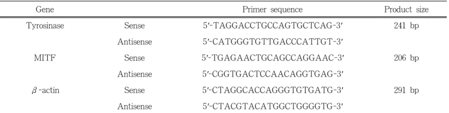
The oliogonucleotide primers used for PCR were as de- scribed in Table 1. Amplification conditions were 94 ℃ (30 s), 55 ℃ (30 s), 72 ℃ (30 s) for 30 cycles. The PCR products were electrophoresed on 2 % agarose gel containing ethidium bromide.
2.7. Western Blot Analysis
The B16 cells (5 × 10⁴ cells) were plated on 6-well plate and incubated in the presence or absence of 100 nM α-MSH. The cells were then incubated for 48 h with various concentrations of Kenapullone and washed twice in PBS at 4 ℃. Total cell lysates were lysed in lysis buffer [40 mM Tris (pH 8.0), 120 mM NaCl, 0.5
% Nonidet P-40 (NP-40), 0.1 mM sodium orthovana- date, 2 mg/mL aprotinin, 2 mg/ml leupeptin, and 100 mg/ml phenylmethylsulfonyl fluoride (PMSF)]. The supernatant was collected and protein concentrations were then determined with protein assay reagents (Pierce, Rockford, IL, U.S.A.). For the Western blot- ting, equal amount of proteins were boiled for 2 min and chilled on ice, subjected to 10 % sodium dodecyl sulfate polyacrylamide gelelectrophoresis (SDS-PAGE), and electro phoretically transferred to a PVDF mem- brane. The proteins were visualized with the enhanced chemiluminescence (ECL) detection system (Takara, Japan).
3. Results
3.1. Effect of Kenpaullone on Melanin Amount and Cell Proliferation in B16 Cells and in Normal Human Melanocytes
To investigate the effects of Kenpaullone, a specific inhibitor of GSK3β on melanin synthesis, B16F10 mel- anoma cells were incubated with various concentrations of Kenpaullone for 48 h. Kenpaullone increased melanin content in dose-dependent manner (Figure 1A). These findings suggest that Kenpaullone is essential for en-
(A) (B)
Figure 1. Effects of kenpaullone on melanogenesis in B16 melanoma cells. (A) The cells were treated with various concen- trations of Kenpaullone for 48 h. (B) The cells were treated with 2.5 µM kenpaullone, 500 nM α-MSH or 50 µM PTU for 48 h. Data represents the mean ± S.D. of two different experiments, each carried out in triplicate.
Figure 2. Effects of kenpaullone on melanogenesis in nor- mal human melanocytes. The cells were treated with 0, 1.25, 2.5 µM Kenpaullone, 500 nM α-MSH for 48 h.
hancing melanogenesis. Its pigmentation was similar to the effect of α-MSH, used as a positive control for the stimulation of melanogenesis (Figure 1B). The viability itself as assessed by MTT was unaffected by the pres- ence of Kenpaullone at this concentration (Figure 1A and 1B).
To confirm Kenpaullone's melanogenesis-stimulating activity, primary melanocytes, separated from foreskin, were incubated with Kenpaullone and melanin assay was performed. As shown in Figure 2, Kenpaullone stimulates melanogenesis in normal human melanocytes in dose-dependent manner. α-MSH was used as the positive control. α-MSH induced 3.5 fold increased in melanin synthesis in human melanocytes, comparing with control group.
3.2. Effect of Kenpaullone on Tyrosinase Expression in B16 Melanoma Cells
To elucidate the mechanism of melanogenesis stimu- lated by Kenpaullone, B16 cells were treated with Kenpaullone, arbutin, or α-MSH. Since melanin is de- rived from the precursor dopaquinone that is formed by tyrosinase oxidation of L-tyrosine, tyrosinase plays an important role in melanin synthesis. Thus, we exam- ined the effect of Kenpaullone on tyrosinase activity.
Each percentage value of tyrosinase activity in the
treated cells was calculated with respect to that the untreated cells. After the treatment of B16 cells with Kenpaullone, tyrosinase activity was enhanced 48 h af- ter the addition of Kenpaullone (Figure 3).
To clarify the further mechanism of melanogenesis regulation by Kenpaullone, the levels of tyrosinase ex- pression in B16 cells were examined by Western blot analysis. As shown in Figure 4, the tyrosinase ex- pression was increased after the treatment of Ken- paullone. Increased expression in tyrosinase was en-
Figure 3. Effect of kenpaullone on tyrosinase activity in B16 melanoma cells. The cells were treated with 0, 2.5, 5 µM kenpaullone, 500 nM α-MSH or 200 µg/mL arbutin for 48 h. Tyrosinase activity was determined by measur- ing the formation of dopachrome. Tyrosinase activity was expressed as percentage values. Each percentage value of tyrosinase activity in the treated cells was calculated with respect to that in the untreated cells.
Figure 4. Effect of kenpaullone on expression of melano- genic proteins in B16 melanoma cells. The cells were treated with 0, 2.5, 5 µM kenpaullone in the presence or absence of α-MSH for 48 h. The expression levels of mela- nogenic proteins were examined by Western blot analysis using specific antibodies.
Gene Primer sequence Product size
Tyrosinase Sense 5'-TAGGACCTGCCAGTGCTCAG-3' 241 bp
Antisense 5'-CATGGGTGTTGACCCATTGT-3'
MITF Sense 5'-TGAGAACTGCAGCCAGGAAC-3' 206 bp
Antisense 5'-CGGTGACTCCAACAGGTGAG-3'
β-actin Sense 5'-CTAGGCACCAGGGTGTGATG-3' 291 bp
Antisense 5'-CTACGTACATGGCTGGGGTG-3'
MITF: Microphthalmia-associated transcription factor
Table 1. Primer Sequences Used for Reverse Transcription Polymerase Chain Reaction hanced when the cells were cotreated with Kenpaullone
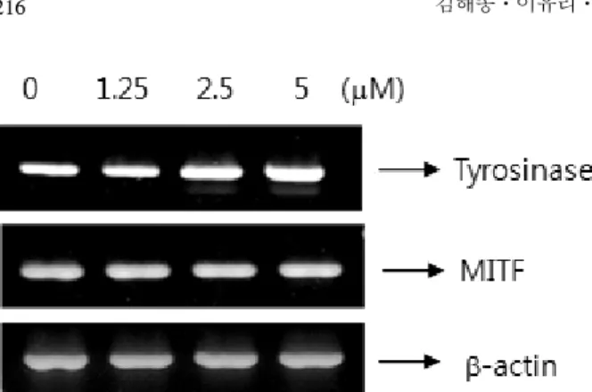
and α-MSH a known melanogenesis stimulator (Figure 4). Next, we also examined the effects of Kenpaullone treatment on mRNA levels. The cells were treated with Kenpaullone for 48 h and the total cellular RNA was extracted. Specific mRNA was amplified af- ter reverse transcription with PCR using specific pri- mers (Table 1) for tyrosinase and MITF. As shown in Figure 5, mRNA of tyrosinase increased after treat- ment with Kenpaullone. These results suggest that Kenpaullone-induced stimulation of melanogenesis oc-
curs at the transcriptional level.
3.3. Discussion
In this study, we report the pigmenting effect of Kenpaullone on melanoma cells. Kenpaullone stimulated melanin production in B16 melanoma cells and human normal melanocytes.
Melanin is a unique pigmented biopolymer synthe- sized by specialized cells known as melanocytes, the dendritic cells in the skin. Melanin has a number of im- portant functions including determination of phenotypic appearance, protective coloration[17]. Melanogenesis it- self is a complex process. In fact, the number of genes
Figure 5. Effect of kenpaullone on mRNA expression of tyrosinase. B16 cells incubated with various concentrations of kenpaullone for 24 h. The resulting cDNA was sub- jected to 30 cycles of polymerase chain reaction using spe- cific primers that gave amplified products of 241 bp for tyrosinase, 206 bp for MITF, 291 bp for actin. PCR prod- ucts were electrophoresed on a 2 % agarose gel and stained with ethidium bromide.
involved in regulating mammalian pigmentation is quite large, and at least 125 genetic loci have been identified for the regulation of melanogenesis either directly or in- directly[18]. Mutations of these genes have been shown to be associated with a number of different pig- mentary diseases. For example, substitution of the MITF Ser298 has been associated with Waardenburg syndrome type 2, a condition characterized by pig- mentary disorders[19,20].
GSK3β is implicated in many biological events, in- cluding embryonic development, cell differentiation, apoptosis, and the insulin response[21]. GSK3β also plays a key role in the Wnt signaling and MITF is one of its targets. GSK3β could activate the function of MITF through phosphorylation at Ser298[22]. Howev- er, the relationship between GSK3β activity and mela- nogenesis is controversial. Khaled and co-workers dem- onstrated that GSK3β activation induced by cAMP elevation leads to melanogenesis[17], while Bellei and coworkers demonstrated GSK3β inhibition promotes melanogenesis[21]. In our system, GSK3β inhibition by Kenpaullone induced melanogenesis, accompanied by the increase of activity, mRNA and protein ex- pression of tyrosinase, consistent with Bellei et al.[21].
Currently many research efforts have focused on the specific mechanisms involved in melanogenesis in order
to develop new therapeutic agents for skin pigmenta- tion abnormalities. In this regard, the agents that stim- ulate tyrosinase activity and melanin production can al- so be used as skin tanning agents. Dihydroxyacetone (DHA)-containing preparations have been used for more than 50 years and are still very popular for tem- porary pigmentation resembling a UV-induced tan[23, 24]. The tan produced by UVB and/or UVA radiation is photoprotective against subsequent UV exposure.
DHA also produce cosmetically acceptable pigmenta- tion of vitiliginous skin[25]. Application of the stim- ulatory effects of Kenpaullone on melanogenesis might be useful as an adjunctive therapy for treatment of hy- popigmentation-related disorders, as well as for tanning.
4. Conclusions
In summary, we have demonstrated that Kenpaullone stimulates melanogenesis and tyrosinase activity. In ad- dition, we have also found that Kenpaullone-induced stimulation of melanogenesis occurs through increased expression of tyrosinase.
Acknowledgments
This study was supported by a grant of the Korea Healthcare technology R&D Project, Ministry of Health
& Welfare, Republic of Korea (Grant No. : A103017).
References
1. A. Korner and J. Pawelek, Mammalian tyrosinase catalyzes three reactions in the biosynthesis of mel- anin, Science, 217(4565), 1163 (1982).
2. S. C. Taylor, Skin of color: biology, structure, func- tion, and implications for dermatologic disease, J.
Am. Acad. Dermatol., 46, S41 (2002).
3. V. J. Hearing and K. Tsukamoto, Enzymatic control of pigmentation in mammals. FASEBJ, 5(14), 2902 (1991).
4. I. J. Jackson, D. M. Chambers, K. Tsukamoto, N. G.
Copeland, D. J. Gilbert, N. A. Jenkins, and V. J.
Hearing, A second tyrosinase-related protein, TRP-2,
maps to and is mutated at the mouse slaty locus, EMBO J., 11(2), 527 (1992).
5. K. Kameyama, T. Takemura, Y. Hamada, C. Sakai, S. Kondoh, S. Nishiyama, K. Urabe, and V. J.
Hearing, Pigment production in murine melanoma cells is regulated by tyrosinase, tyrosinase-related protein 1 (TRP1), DOPAchrome tautomerase (TRP2), and a melanogenic inhibitor, J. Invest. Dermatol., 100(2), 126 (1993).
6. P. Aroca, F. Solano, C. Salinas, J. C. Garcia-Borron, and J. A. Lozano, Regulation of the final phase of mammalian melanogenesis: The role of dopachrome tautomerase and the ratio between 5,6-dihydrox- yindole-2-carboxylic acid and 5,6-dihydroxyindole, Eur. J. Biochem., 208(1), 155 (1992).
7. A. Arrunátegui, C. Arroyo, L. Garcia, C. Covelli, C.
Escobar, E. Carrascal, and R. Falabella, Melanocyte reservoir in vitiligo, Int. J. Dermatol., 33(7), 484 (1992).
8. P. Manga, D. Sheyn, F. Yang, R. Sarangarajan, and R. E. Boissy. A role for tyrosinase-related protein1 in 4-tert-butylphenol-induced toxicity in melano- cytes: Implications for vitiligo, Am. J. Pathol., 169(5), 1652 (2006).
9. A. B. Lerner, On the etiology of vitiligo and gray hair, Am. J. Med., 51(2), 141 (1971).
10. J. J. Nordlund, The pigmentary system and in- flammation, Pigment. Cell. Res., 5(5), 362 (1992).
11. W. Englaro, R. Rezzonico, M. Durand-Clement, D.
Lallemand, J. P. Ortonne, and R. Ballotti, Mitogen- activated protein kinase pathway and AP-1 are ac- tivated during cAMP-induced melanogenesis in B16 melanomacells, J. Biol. Chem., 270(41), 24315 (1995).
12. J. Lee, E. Jung, J. Park, K. Jung, E. Park, J. Kim, S. Hong, J. Park, S. Park, S. Lee, and D. Park, Glycyrrhizin induces melanogenesis by elevating a cAMP level in B16 melanoma cells, J. Invest.
Dermatol., 124(2), 405 (2005).
13. S. K. Singh, C. Sarkar, S. Mallick, B. Saha, R. Bera, and R. Bhadra, Human placental lipid induces mel- anogenesis through p38 MAPK in B16F10 mouse melanoma, Pigment.Cell. Res., 18(2), 113 (2005).
14. R. Busca and R. Ballotti, Cyclic AMP a key mes- senger in the regulation of skin pigmentation, Pigment. Cell. Res., 13(2), 60 (2000).
15. L. Brault, E. Migianu, A. Nelquesque, E. Battaaglia, D. Bagrel, and G. Kirsch, New thiophene analogues of kenpaullone: synthesis and biological evaluation in breast cancer cells, Eur. J. Med. Chem., 40(8), 757 (2005).
16. V. J. Hearing, Mammalian monophenol mono- oxygenase (tyrosinase): purification, properties, and reactions catalyzed, Method. Enzymol., 142, 154 (1987).
17. M. Khaled, L. Larribere, K. Bille, E. Aberdam, J. P.
Ortonne, R. Ballotti, and C. Bertolotto, Glycogen synthase kinase 3beta is activated by cAMP and plays an active role in the regulation of melano- genesis, J. Biol. Chem., 277(37), 33690 (2002).
18. H. J. Park, Y. J. Kim, K. Leem, S. J. Park, J. C.
Seo, H. K. Kim, and J. H. Chung, Coptis japonica root extract induces apoptosis through caspase3 ac- tivation in SNU-668 human gastric cancer cells, Phytother. Res., 19(3), 189 (2005).
19. A. E. Hughes, V. E. Newton, X. Z. Liu, and A. P.
Read, A gene for Waardenburg syndrome type 2 maps close to the human homologue of the micro- phthalmia gene at chromosome 3p12-p14.1, Nat.
Genet., 7(4), 509 (1994).
20. M. Tassagehji, V. E. Newton, and A. P. Read, Waardenburg syndrome type 2 caused by muta- tions in the human microphthalmia (MITF) gene, Nat. Genet., 8(3), 251 (1994).
21. B. Bellei, E. Flori, E. Izzo, V. Maresca, and M.
Picardo, GSK3beta inhibition promotes melano- genesis in mouse B16 melanoma cells and normal human melanocytes, Cell. Signal., 20(10), 1750 (2008).
22. K. Takeda, C. Takemoto, I. Kobayashi, A. Wata- nabe, Y. Nobukuni, D. E. Fisher, and M. Tachiba- na, Ser298 of MITF, a mutation site in Waarden- burg syndrome type 2, is a phosphorylation site with functional significance, Hum. Mol. Genet.
9(1), 125 (2000).
23. V. J. Hearing, Biochemical control of melanogenesis and melanosomal organization, J. Investig. Dermatol.
Symp. Proc., 4(1), 24 (1999).
24. V. Del Marmol and F. Beermann, Tyrosinase and related proteins in mammalian pigmentation, FEBS Lett., 381(3), 165 (1996).
25. S. B. Levy, Dihydroxyacetone-containing sunless or self-tanning lotions, J. Am. Acad. Dermatol., 27(6), 989 (1992).