서 론
담수환경은 육상에서 일어나는 다양한 오염원의 방출 로 인해 내분비계장애물질(endocrine disrupting chemi- cals, EDCs)를 비롯한 다양한 독성물질에 의한 오염이 증가하고 있다. 내분비계장애물질은 먹이연쇄를 통해 생 체 내에 축적될 뿐 아니라 세포 내에서 정상적으로 진행 되는 ligand-receptor 상호작용, 단백질-단백질 상호작용
등을 변화시켜 그 결과 생물체의 생식, 면역, 신경을 포괄 하는 다양한 생리적 기능을 변화시킬 수 있다(DeRosa et al., 1998). 특히 담수에서 EDCs의 오염은 먹는 물자원을 직접적으로 위협하므로 간과할 수 없는 환경 현안으로 대두되었으며 따라서 담수환경에서 EDCs에 대한 위해 성 평가 및 관리의 중요성이 증대되고 있다. 이에 따라 담수환경의 관리 및 수생 생물자원의 보호를 위해 내분 비계 장애물질에 대한 생태적, 분자생물학적 표식자 기준 이 필요하게 되었다. 다양한 내분비계 장애물질 가운데
─
─ 122 ──
수환경 내 Estrogen 에스트로젠 활성 검출을 위한 누치 난황전구단백질 유전자 발현의 RT-PCR 시험법
계 명 찬
(한양대학교 자연과학대학 생명과학과)
Analysis of Vitellogenin Gene Expression by RT-PCR in Hemibarbus labeo (Cyprinidae) for the Analysis of Estrogenic Activity in Aquatic Environment. Gye, Myung Chan (Department of Life Science, College of Natural Sciences, Hanyang University, Seoul 133-791, Korea)
In an effort to develop the biomarker for monitoring the contamination of xenoe- strogen in the freshwater environment of Korea, reverse transcription-polymerase chain reaction (RT-PCR) analysis of vitellogenin (VTG) gene expression was optimized in Hemibarbus labeo. Based on the homology of the VTG cDNA sequences between the common carp and zebra fish, a set of PCR primers for VTG mRNA amplification for H. labeo was designed. VTG mRNA level in livers from female and male fishes was analyzed by RT-PCR following single injection of 17 beta estradiol (E210 mg kg-1B.W.). As an internal control, beta actin mRNA was amplified. One ug of total liver RNA was subjected to RT-PCR. In female the amount of PCR product of VTG gradually increased in the range from 16 to 34 cycles of amplification. On the contrary, in control male, PCR product first detected at 32 cycles of amplification and linearly increased up to 40 cycles of amplification. In E2injected male liver, the VTG mRNA level was similar to that in the female. Taken together, this result sug- gests that liver of male H. labeo expresses minute amount of VTG mRNA which are 2-16equivalent of female and that induction of VTG mRNA occurs in male liver after estrogen treatment. In conclusion, the optimized protocol for RT-PCR analysis of VTG mRNA expression in liver of male H. labeo will provide the environmental monitoring method for the xenoestrogen contamination in the rivers in Korea.
Key words : vitellogenin, RT-PCR, estrogen, Hemibarbus labeo
* Corresponding author: Tel: 02) 2290-0958, Fax: 02) 2298-9646, E-mail: [email protected]
여성호르몬과 유사한 생리활성을 갖는 합성에스트로젠 (xenoestrogen)들을 검색하기 위한 다양한 생물학적 지 표가 이용되고 있는데 난생 척추동물 수컷의 간 또는 혈 액에서 발견되는 난황전구단백질(vitellogenin)은 xenoe- strogen의 노출을 검색에 중요한 분자생물학적 지표로 이용되고 있다(계와 한, 2000). 어류 개체 수준에서 간조 직 또는 간세포 배양체 수준에서 이들biomarker 유전자 의 발현을 RNA 및 단백질 수준에서 검출함으로서 새로 운 xenoestrogen의 검색 수단을 개발하기 위한 연구 및 내분비계 장애효과를 유발하는 최소한의 농도 등 환경 내EDCs의 적절한 안전 및 허용기준을 제시하기 위한 연 구들이 진행되었다(Sumpter and Jobling, 1995; Heppell et al., 1995; Lee et al., 2002; Hennies et al., 2003;
Rotchell and Ostrander, 2003). 이들 대부분의 연구는 송 어 등의 실험 어종을 대상으로 하였지만 최근에는 자국 의 수환경 내에 서식하는 야생 어종에 대한 내분비계 장 애물질 위해성을 평가하기 위한 연구가 강화되고 있다 (Harrison et al., 1997; 환경부, 2001; Jobling and Tyler, 2003; Robinson et al., 2003). 그러나 한국의 야생의 담수 어류를 대상으로 EDCs 가운데xenoestrogen의 위해성을 biomarker 유전자 수준에서 추적하는 기술은 일부 어류 이외에는 확보되어 있지 않았다(최와 계, 2003).
누치(Hemibarbus labeo)는 잉어목, 잉어과의 어류로 한반도의 서남해로 유입되는 하천, 만주, 중국동남부, 일 본 남서부에 분포한다(김, 1997) (Figs. 1, 2). 누치가 분포 하는 한반도 뿐 아니라 누치가 분포하는 중국 내륙은 지 난 수십년간 급속히 인구 및 공업활동이 증가하고 있는 지역으로 이 지역 담수환경은 필연적으로 다양한 내분비 계 장애물질에 의해 오염되었을 것으로 추측할 수 있다. 누치가 주로 분포하는 이 지역의 대형 하천들은EDCs를 포함한 오염 부하량이 비교적 큰 담수역이다. 따라서 누 치는 EDCs에 의한 생체영향을 평가하기에 좋은 생물학 적 지표종으로서의 특징을 갖는다. 한국에서는 최근 같은 잉어과 어류인 붕어가 정부(환경부) 내분비계 장애물질 위해성 평가사업의 어류 모델 어종으로 지정되어 수년간 단백질 수준에서VTG의 발현과 환경매체 내 특정EDCs
농도와의 인과관계에 대한 연구들이 진행되고 있다(환경 부, 2001). 붕어의 경우 떡붕어(Carassius cuvieri) 등 유 전적으로 근계의 어류가 국내의 하천에 붕어와 혼서하며 일부 하천과 저수지에서 붕어보다 더 우세하게 출현하고 있어 종의 식별을 기본으로 하는EDCs 위해성 모니터링 연구의 과학적 신뢰성에 문제가 제기된다. 또한 VTG mRNA의 발현을 추적함으로서VTG 항체를 이용하여 추 적하는 기법 보다 좀더 신속하고도 정교하게xenoestro- gen에 의한 VTG 유도현상을 추적 할 수 있는 연구기법 은 시도되고 있지 않고 있다. 본 연구는 한국의 담수환경 내에서xenoestrogen 오염을 정밀하게 추적하기 위한 기 술 개발의 일환으로 누치의VTG cDNA 염기서열에 근거 한 reverse transcription and polymerase chain reaction (RT-PCR) 시험법을 최적화하였다.
재료 및 방법
1. 채집, 사육 및 전처리
누치 암수 성어는 2003년 하계 팔당호에서 정치망을 이용하여 채집한 개체를 사용하였다. 채집된 개체들은 실 Fig. 1. Hemibarbus labeo.
Fig. 2. Distibution of H. labeo.
험실로 운반한 후 18±1�C의 수조에서 14시간 조명 하 에1주간 사육하여 적응시켰다. VTG mRNA 발현의 분석 에는 전장 300 mm 이상의 개체를 사용하였다. 수컷에서 VTG의 유도를 위해 corn oil에 희석한 17 beta estradiol 을 10 mg kg-1체중 농도로 복강 주사하였다. 주사 2일 후RNA 분석에 공시하였다.
2. RNA 분리 및 역전사
누치의 두부를 타격하여 도살한 후 복강을 해부하여 암컷 및 수컷의 간 조직을 절취하였다. 분리된 간 조직은 20배 부피의 TRIzol 용액(Life Technologies, USA)에 옮 긴 후 4�C에서 전동마쇄기(Polytron, USA)를 이용하여 균질화 하였다. 제조사의 방법에 따라 total RNA를 분리 한 후0.1% DEPC로 처리된 증류수에 3 mg mL-1농도로 용해하였다. 역전사 반응에는 murine leukemia virus [MuLV] reverse transcriptase (Applied Bioscience, USA) 을 이용하였고 제조사의 용법에 따라 RNA 1µg을 이용 하여 20µL 부피의 역전사 반응을 수행하여 cDNA를 합 성하였다.
3. PCR의 최적화
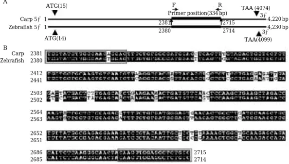
누치 VTG의 RT-PCR에 이용할 수 있는 염기서열을 확보하고자 Blast 2 program을 이용하여 잉어(Cyprinus
carpio)와 제브라피시(Danio rerio)의 VTG cDNA 염기서 열 상동성을 비교한 후 PCR primer design program (Oligo software)를 이용하여 VTG primer는 5′-TGGTA- TCTTGCCAACAGCAG-3′(forward), 5′-GGCAGAGCC- TCCA CCTTGTA-3′(reverse)를 제작하였다(Fig. 3). 역 전사 제조한 간조직의 cDNA 0.5µL을 Ex Taq (Takara, Japan) PCR kit를 표준 PCR 조건(20 mM Tris, pH 8.4, 50 mM KCl, 1.25 u i-Taq, 0.2 mM dNTP, 1.5 mM MgCl2, 0.5 pM each primer, 1/20 RT reaction)에서 증폭하였다. 각 PCR 단계는 denaturation: 95�C, 30 sec; annealing:
55�C 60 sec; extension: 72�C, 30 sec로 하였고 i-Cycler (BioRad, USA)를 이용하였다. 증폭산물의 염기서열을 결 정을 위해 RT-PCR 반응산물은 2% agarose gel에서 전 기영동한 후 ethydium bromide 염색하여 해당 밴드를 오려내어 QIAquick Gel Extraction Kit (Qiagen, USA)로 DNA를 추출하였다. 정제한 DNA 절편은 T vector에 클 로닝하여competent cell (E. coli DH5α)에 형질도입 하였 다. Amphicilin 저항성 positive clone을 선발하여 plas- mid를 분리하고 제한효소 처리를 통해insert의 삽입 유 무를 전기영동을 통해 확인한 다음 확인된clone의 경우 플라스미드를 자동염기서열 분석기를 이용 Dye Termi- nator Cycle Sequencing Ready Reaction Kit with Ampli- Taq DNA polymerase법으로 염기서열을 분석하였다.
각 시료에 존재하는 cDNA의 상대적인 정량을 위해
Fig. 3. Primer design for RT-PCR of vitellogenin of H. labeo. (A) Primer positions. F, forward primer; R, reverse primer.
(B) Primers for RT-PCR of VTG mRNA of H. labeo was designated by multiple sequence alignment of the carp (Cyprinus carpio) and zebrafish (Danio rerio) partial vitellogenin sequences.
A
B
ATG(15)
ATG(14)
Primer position(334 bp)
F R
Carp 5′ 1 Zebrafish 5′ 1
Carp 2381 Zebrafish 2380 2412 2441 2503 2502 2564 2563 2652 2651 2686 2685
2715 2714
2381 2715
2380 2714
4,220 bp 4,230 bp TAA (4074)
TAA(4099) 3′
3′
beta actin을 증폭하여 비교하였다. beta actin primer는 5′-ATGGATGATGAAATTGCCGCACTTG-3′(forward),
5′-CTGGGTCATCTTCTCCCTGTTGGC-3′(reverse)를
사용하였다(Lee et al., 2000). 간 cDNA로 부터 VTG 및 beta actin mRNA 발현 정량을 위한 최적 annealing 온 도를 확인하기 위해 암컷 간을 이용하여 제작한 cDNA 를 이용하여 55.0~64.8�C의 온도 구배 조건에서 40회 PCR을 수행하였다. 최적의 PCR 반응 회수를 결정하기 위해 annealing 온도 55�C에서 12~40 cycle의 PCR을 진행하였다. 고안한primer에 의한genomic DNA의 증폭 을 확인하기 위해 TRIzol 용액을 사용하여 분리한 geno- mic DNA 0.1µg을 표준조건에서 40회 증폭하였다. VTG mRNA의 상대적 정량을 위한RT-PCR 실험에서는PCR 반응산물의 양을 Gel documentation system (Vilber Lourmat, France)을 이용하여 밴드의 강도를 측정한 후 PCR 반응 회수-반응량 곡선을 얻었다. 차후 정량적 분석 을 위해 PCR cycle 수의 증가에 따라 PCR 반응산물의 증가가 뚜렷한 구간을 설정하였다(Gye and Ohsako, 2003). 확인된 누치 VTG cDNA의 부분적 염기서열을 Clustal W program (GenomeNet, Kyoto, Japan) 를 이용
하여 잉어, 제브라피시와 염기서열 상동성을 분석하였다.
결 과
RT-PCR 결과로 나타난 334 bp amplicon의 염기서열 분석을 통해 확인된 누치 VTG cDNA의 부분적 염기서 열에 대한 염기서열 상동성 분석결과 잉어와 94%, 제브 라피시와89%의 염기서열 상동성을 보였다.
최적 annealing 온도를 확인하기 위해 암컷 간을 이용 하여 제작한cDNA를 이용하여55.0~64.8�C의 온도 구 배 조건에서 40회 PCR을 수행한 결과 VTG과 beta actin 모두55�C에서 최적화되었다(Fig. 4A).
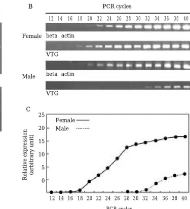
암컷 간의 경우 16~32 cycle 범위의 증폭 시 반응산물 이 일정하게 증가하였으며 이후PCR 반응이 포화되었다. Estrogen을 처리하지 않은 수컷 간에서는 32~40 cycle 사이에서 반응산물이 일정하게 증가하였다. RT-PCR 증 폭산물이 관찰되는 최저 반응 횟수는 암컷 16, 수컷 32 cycle 이므로 수컷에서 미량 발현되는 VTG mRNA의 상 대적 발현량은 암컷의 2-16배에 상당하였다. 함께 증폭한
Fig. 4. Optimization of RT-PCR of vitellogenin and beta actin mRNA in H. labeo. (A) Optimization of annealing temperature for RT-PCR products of VTG mRNA and beta actin. (B) Increase in RT-PCR products of VTG mRNA and beta actin according to amplification cycle in both sexes. (C) Amplification curve of RT-PCR-products of VTG and beta actin mRNA in both sexes.
A B
C
Annealing temperature (�C) PCR cycles
SM 55.0 56.4 57.8 59.2 60.6 62.0 63.4 64.8 12 14 16 18 20 22 24 26 28 30 32 34 36 38 40
12 14 16 18 20 22 24 26 28 30 32 34 36 38 40 PCR cycles
Relative expression (arbitrary unit)
beta-actin Female
Male
Female Male 25 20 15 10 5 0
beta-actin VTG
VTG
beta actin
VTG
beta actin mRNA의 경우 20~34 cycle에서 일량하게 증 가하였다(Fig. 4B, C).
암수 간조직의genomic DNA를 분리하여 같은 primer
로 수행한40회의PCR 반응 결과 약500 bp의 증폭산물 을 확인하였다(Fig. 5).
30회 PCR 반응을 수행한 결과 암컷과 17 beta estra- diol (10 mg kg-1B.W.)을 일회 주사한 후2일 된 수컷에 서VTG mRNA의RT-PCR 산물이 확인되었다(Fig. 6).
고 찰
최근 수환경 내의 EDCs 오염을 추적을 위해 어류에서 RT-PCR혹은 ELISA법 등으로 VTG을 비롯한 다양한 biomarker 유전자의 발현을 분석함으로서 이들 어류의 서식 환경의xenoestrogen 오염 여부를 확인하고 있으며 VTG를 이용한 검사법은 다양한 biomarker 들 가운데 OECD 표준으로 채택되었다(OECD, 1992). 누치와 같은 잉어과 어류를 이용한 연구로 carp (Cyprinus carpio), major carp (Cirrhinus mrigala), goldfish, fathead min- nows (Pimephales promelas) 등에서 estrogen, androgen 및xenoestrogen과VTG 발현과의 인과관계에 대한 연구 가 활발하지만(Hori et al., 1979; Nath et al., 1992; Nath and Maitra, 2001; Villeneuve et al., 2002; Jobling et al., 2003; Giesy et al., 2003; Pickford et al., 2003; Sole et al., 2002, 2003) 대부분VTG 항체를 이용하여 혈중 VTG 농 도를 조사한 것으로 본 연구에서처럼 미량의 mRNA 발 현 까지도 정밀하게 검출할 수 있는 연구는 male carp (Cyprinus carpio), sheepshead minnows (Cyprinodon variegatus) 등 일부 실험 어류에 불과하다(Denslow et al., 2001; Lattier et al., 2001). 이처럼 담수에서 현지조사 를 위한 연구모델로 잉어과 어류들이 주목받는 이유는 이들이 비교적 오염물질의 부하량이 큰 곳에 서식할 뿐 아니라 하상 또는 하상의 침전물 속 생물을 주요 먹이원 으로 하는 취이 특징을 갖기 때문이다. 한국의 경우 아직 까지 육수환경인 호소 및 강에 서식하는 야생 경골어류 를 대상으로xenoestrogen 등의 내분비계 장애물질의 위 해성을 유전자 발현 수준에서 평가할 수 있는biomarker 기술은 개발되지 않았다. 본 연구는 이와 관련된 최초의 연구로 의미를 갖는다.
다양한 경골어류들에서 에스트로젠 처리에 따른 VTG 발현의 유도는 어종에 따라 수온과 번식계절의 영향을 받는 것으로 알려졌다(Atlantic salmon, Korsgaard et al., 1986; carp, Hernandez et al., 1992; trout, Mackay and Lazier, 1993). VTG 발현을 위해서는 간조직에서 estro- gen 수용체의 발현이 선행되어야 하며 비번식기 어류에 서는 간조직 내의 estrogen 수용체의 발현이 매우 낮아 Fig. 5. Genomic PCR using primer set for RT-PCR of
vitellogenin mRNA in H. labeo. Genomic DNA from liver of H. labeo was subjected to 40 cycles of PCR for verification of primer. Arrow indicates PCR product of VTG gene. SM, size marker (100 bp ladder); M, male; F, female.
Fig. 6. Induction of vitellogenin mRNA by estrogen in male fish in H. labeo. RT-PCR of VTG mRNA in female, control male, and 17 beta estradiol-inject- ed (1 mg kg-1B.W.) male livers (30 cycle). Arrow indicates RT-PCR product of VTG mRNA. SM, size marker (100 bp ladder); M, male; F, female.
ME2, 17 beta estradiol-injected (1 mg kg-1 B.W.) male; F-RT, Female liver RNA without RT reac- tion.
SM M F
SM M F ME2 F-RT
estrogen 처리 시에도 VTG는 유도되지 않는다. 따라서 계절적으로 변동하는 뇌-생식소 축 상에 작용하는 내분 비호르몬 변동이 난황형성기에 estrogen의 감수성을 담 보하는 것으로 사료된다. 누치의 경우 암컷의 난소의 발 달은 동계에 서서히 발달하여 춘계에 최고조에 달하며5 월에 산란한다. 따라서 춘계와 하계를 통해 번식이 이뤄지 는 누치의 경우 이 시기에 채집한 개체를 대상으로 VTG
mRNA 분석을 할 경우 결과의 신뢰도를 높일 수 있을
것이다.
본 연구에서 이용한 누치는 실험실 조건 14시간 조명, 18�C 조건에서 순치한 개체로 암컷의 해부과정에서 난자 를 포함하고 있는 난소를 육안으로 확인할 수 있어 난황 형성기임을 알 수 있었다. 따라서 estrogen에 대한 감수 성 즉, estrogen 수용체의 발현이 일어나는 상태로 추정 된다. 누치 수컷의 생식내분비학적 특징이 암컷과 동기화 되었다고 가정할 때 수컷에서 에스트로젠 또는 xenoes- trogen에 의해 VTG mRNA 유도를 확인하고자 할 때는 암컷의 난황형성기로 수컷에서도 estrogen 감수성이 유 지되는 늦은 동계 또는 춘계에 채집한 개체를 이용하여 VTG 발현을 검사하는 것이 적합할 것이다.
PCR 반응 사이클 수에 따른VTG 증폭산물의 증가 양 상은 암컷에서는 최저 16 cycle에서 확인된 반면 수컷에 서는 32 cycle에서 확인된다. 따라서 산술적으로 누치 수 컷에서VTG mRNA의 발현량은 암컷의2-16에 상당한다. PCR 반응의 횟수를 32 cycle 이상으로 증가할 경우 40 cycle까지 반응산물이 일량하게 증가한다. 따라서 수컷에 서도 미량의 VTG mRNA가 발현됨을 알 수 있다. 한편 E2(10 mg kg-1B.W.)주사2일 후 누치 수컷 간에서 암컷 과 같은 정도의 VTG mRNA 발현이 확인되어 estrogen 에 의한 VTG mRNA 유도의 감수성이 있음을 알 수 있 다. 이와 유사하게 실험어종인 sheephead minnow 수컷 의 간에서도 미량(0.04 ng µg-1total RNA)의 VTG RNA 가 발현되며 estrogen (5 mg kg-1B.W.) 주사 후 35배 증 가한다(Bowman et al., 2000; Hemmer et al., 2002). 이처 럼 수컷에서 VTG가 발현되는 이유는 수컷 체내에 존재 하는 estrogen 때문으로 추정된다. 비록 수컷이라도 혈중 에는 미량의 estrogen이 존재하며 이들은 spermatogo- nial stem cell의 증식을 촉진하는 등의 정상적인 정자형 성에 중요한 역할이 있다(Hess et al., 1997; Miura et al., 1999). 또한 척추동물 수컷의 정소 뿐 아니라 뇌조직에도 testosterone을 estrogen으로 전환하는 aromatase의 발 현이 확인된다(Schlinger et al., 1992; Nitta et al., 1993).
따라서 간 조직에서 estrogen 수용체를 발현할 경우 미 량의 estrogen에 의해 VTG 전사가 가능하며RT-PCR과
같이 민감한 검출방법을 사용하게 되면 미량의 VTG mRNA의 존재를 확인할 수 있을 것이다.
어류 수컷을 외인성 estrogen 또는 xenoestrogen에 노 출한 경우VTG 유전자의 전사 뿐 아니라 전사체의 단백 질로 전환 및 정소조직 내에 축적이 일어난다(Folmar et al., 2002). 그러나 미량의 VTG mRNA가 검출되었다고 해서 본 연구에 사용된 수컷 개체가xenoestrogen에 의 해 오염되었다고 단정하기는 어렵다. 본 연구에서 estro- gen을 처리하지 않은 수컷에서 확인되는 VTG의 발현량 은 결코 무시할 수 있는 양이 아니다. 따라서 독성학적으 로 xenoestrogen에 노출된 개체를 판정하기 위한 VTG mRNA 발현량의 기본 값 설정에 신중할 필요가 있다. Xenoestrogen에 의한 오염에 대한 명확한 결론은 수컷 체내에 존재하는 생리적 농도의 estrogen에 의한 VTG mRNA의 발현정도 및 생성된 VTG 단백질의 반감기 등 체내 동태에 대한 정보에 기초하여야 할 것이다. 본 연구 에서 시행한 것과 같은 방법으로 누치 수컷의 간에서 발 현되는VTG mRNA의 RT-PCR 반응을 수행할 경우 32
~40 사이클에서 VTG 증폭산물이 일량하게 증가하므로 이 범위 내에서RT-PCR반응을 수행할 경우 estrogenic 활성을 갖는 다양한 xenoestrogen의 검색 뿐 아니라 특 정 담수역에서 VTG mRNA의 RT-PCR법에 의해 xenoestrogen에 의한 오염의 추적이 가능할 것이다. 특히 genomic PCR 결과 RT-PCR 반응 산물과는 다른 크기 (500 bp)의 amplicon이 검출되므로 genomic PCR 산물과 구분되었다. 따라서 고안된 primer set 사이에 적어도 1 개 이상의 intron이 위치하고 있은 것으로 추정되며 본 연구에서 이용한 primer를 사용할 경우 누치 VTG mRNA의RT-PCR 검출결과의 신뢰성을 담보할 수 있을 것이다. 향후 bisphenol, nonylphenol 등 이미 다양한 어 류에서 VTG 발현을 유도하는 것으로 알려진 다양한 외 인성 estrogen에 의한 영향을 확인하기 위해서는 물질 별로 투여 농도-발현량 관계에 근거한 구체적인 기준이 설정되어야 할 것이며 누치 이외에도 다양한 담수 어류 들을 대상으로 다양한 biomarker gene의 발굴과 xenoe- strogen을 포함한 내분비계 장애물질 검색을 위한 표준 화 연구가 필요할 것이다.
결 론
어류 vitellogenin (VTG) cDNA의 염기서열 상동성에 근거하여 고안한 RT-PCR primer를 이용하여 누치 VTG mRNA의 발현 검출에 필요한 RT-PCR 시험법을
최적화하였다. 누치 암수 성어를 이용하여 조사한 결과 수컷은 암컷의 2-16배에 상당하는 미량의 VTG mRNA를 발현하였다. 17 beta estradiol (10 mg kg-1B.W.)을 1회 주사한 수컷에서 암컷의 발현량에 해당하는 VTG mRNA 가 발현되었다. 본 시험법을 이용하여 estrogenic활성을 갖는 어류 내분비계장애물질의 민감하고도 특이적인 검 색 및 국내 대형 하천의 특정 수역에서의 xenoestrogen 노출 여부의 판정이 가능할 것이다.
사 사
본 연구는 해양수산부 수산특정연구비(Proj. No.
20010060) 및 2003년 한양대학교 특성화사업단 연구비 의 일부지원에 의해 수행되었음.
인 용 문 헌
계명찬, 한명수. 2000. 척추동물의 난황형성과 환경에스트로젠. 한국환경생물학회지18: 291-298.
김익수. 1997. 제37권 동물편(담수어류), pp.221-223. 한국동 식물도감. 교육부.
최동주, 계명찬. 2003. Nonylphenol에 의한 누치(Hemibar-
bus labeo) 수컷의 간조직 내난황전구단백질 유전자 발현
의 유도. Kor. J. Biol. Sci. 7: 138.
환경부. 2001. 내분비계 장애물질에 의한 생태영향조사. Bowman, C.J., K.J. Kroll, M.J. Hemmer, L.C. Folmar and
N.D. Denslow. 2000. Estrogen-induced vitellogenin mRNA and protein in sheepshead minnow (Cyprinodon variegatus). Gen. Comp. Endocrinol. 120: 300-313.
Denslow, N.D., C.J. Bowman, R.J. Ferguson, H.S. Lee, M.J. Hemmer and L.C. Folmar. 2001. Induction of gene expression in sheepshead minnows (Cyprinodon varie- gatus) treated with 17 beta-estradiol, diethylstilbe- strol, or ethinylestradiol: the use of mRNA fingerprints as an indicator of gene regulation. Gen. Comp. Endo- crinol. 121: 250-260.
DeRosa, C., P. Richter, H. Pohl and D.E. Jones. 1998. Envi- ronmental exposures that affect the endocrine system:
public health implications. J. Toxicol. Environ. Health B Crit. Rev. 1: 3-26.
Folmar, L.C., M.J. Hemmer, N.D. Denslow, K. Kroll, J.
Chen, A. Cheek, H. Richman, H. Meredith and E.G.
Grau. 2002. A comparison of the estrogenic potencies of estradiol, ethynylestradiol, diethylstilbestrol, nonylphe- nol and methoxychlor in vivo and in vitro. Aquat. Toxi-
col. 60: 101-110.
Giesy, J.P., E.M. Snyder, K.M. Nichols, S.A. Snyder, S.A.
Villalobos, P.D. Jones and S.D. Fitzgerald. 2003. Exa- mination of reproductive endpoints in goldfish (Caras- sius auratus) exposed in situ to municipal sewage trea- tment plant effluent discharges in Michigan, USA.
Environ Toxicol Chem. 22: 2416-2431.
Gye, M.C. and S. Ohsako. 2003. Effects of flutamide in the rat testis on the expression of occludin, an integral member of the tight junctions. Toxicol. Lett. 143: 217- 222.
Harrison, P.T., P. Holmes and C.D. Humfrey. 1997. Repro- ductive health in humans and wildlife: are adverse trends associated with environmental chemical expo- sure? Sci. Total Environ. 205: 97-106.
Hemmer, M.J., C.J. Bowman, B.L. Hemmer, S.D. Friedman, D. Marcovich, K.J. Kroll and N.D. Denslow. 2002.
Vitellogenin mRNA regulation and plasma clearance in male sheepshead minnows (Cyprinodon variegatus) after cessation of exposure to 17 beta-estradiol and p-nonyl- phenol. Aquat. Toxicol. 58: 99-112.
Hennies, M., M. Wiesmann, B. Allner and H. Sauerwein.
2003. Vitellogenin in carp (Cylrinus carpio) and perch (Perca fluviatilis): purification, characterization and development of an ELISA for the detection of estro- genic effects. Sci. Total Environ. 309: 93-103.
Hernandez, I., A. Poblete, R. Amthauer, R. Pessot and M.
Krauskopf. 1992. Effect of seasonal acclimatization on estrogen-induced vitellogenesis and on the hepatic estrogen receptors in the male carp. Biochem. Int. 28:
559-567.
Heppell, S.A., N.D. Denslow, L.C. Folmar and C.V. Sullivan.
1995. Universal assay of vitellogenin as a biomarker for environmental estrogens. Environ. Health Perspect. 103 Suppl. 7: 9-15.
Hess, R.A., D. Bunick, K.H. Lee, J. Bahr, J.A. Taylor, K.S.
Korach and D.B. Lubahn. 1997. A role for oestrogens in the male reproductive system. Nature 390: 509-512.
Hori, S.H., T. Kodama and K. Tanahashi. 1979. Induction of vitellogenin synthesis in goldfish by massive doses of androgens. Gen. Comp. Endocrinol. 37: 306-320.
Jobling, S., D. Casey, T. Rodgers-Gray, J. Oehlmann, U.
Schulte-Oehlmann, S. Pawlowski, T. Baunbeck, A.P.
Turner and C.R. Tyler. 2003. Comparative responses of molluscs and fish to environmental estrogens and an estrogenic effluent. Aquat. Toxicol. 65: 205-220.
Jobling, S. and C.R. Tyler. 2003. Endocrine disruption, parasites and pollutants in wild freshwater fish. Para- sitology. 126 Suppl. S103-108.
Korsgaard, B., T.P. Mommsen and R.L. Saunders. 1986.
The effect of temperature on the vitellogenic response in Atlantic salmon post-smolts (Salmo salar). Gen.
Comp. Endocrinol. 62: 193-201.
Lattier, D.L., D.A. Gordon, D.J. Burks and G.P. Toth. 2001.
Vitellogenin gene transcription: a relative quantitative exposure indicator of environmental estrogens. Envi- ron. Toxicol. Chem. 20: 1979-1985.
Lee, C., J.G. Na, K.C. Lee and K. Park. 2002. Choriogenin mRNA induction in male medaka, Oryzias latipes as a biomarker of endocrine disruption. Aquat. Toxicol. 61:
233-241.
Lee, J.S., S.H. Lee and M.C. Gye. 2000. The beta-actin gene of two species of southern top mouth minnow (Pseudorasbora parva) and the common fat minnow (Rhynchocypris oxycephalus) from the family Cyprini- dae. DNA Seq. 11: 301-307.
Mackay, M.E. and C.B. Lazier. 1993. Estrogen responsi- veness of vitellogenin gene expression in rainbow trout (Oncorhynchus mykiss) kept at different temperatures.
Gen. Comp. Endocrinol. 89: 255-266.
Miura, T., C. Miura, T. Ohta, M.R. Nader, T. Todo and K.
Yamauchi. 1999. Estradiol-17β stimulates the renewal of spermatogonial stem cells in males. Biochem. Bio- phys. Res. Commun. 264: 230-234.
Nath, P., M. Bhakta and K. Mitra. 1992. Demonstration of two forms of vitellogenin in serum of estradiol-17 beta -treated Indian major carp, Labeo rohita. Indian J.
Exp. Biol. 30: 464-469.
Nath, P. and S. Maitra. 2001. Role of two plasma vitello- genins from Indian major carp (Cirrhinus mrigala) in catfish (Clarias batrachus) vitellogenesis. Gen. Comp.
Endocrinol. 124: 30-44.
Nitta, H., D. Bunick, R.A. Hess, L. Janulis, S.C. Newton, C.F. Millette, Y. Osawa, Y. Shizuta, K. Toda and J.M.
Bahr. 1993. Germ cells of the mouse testis express p450 aromatase. Endocrinology. 132: 1396-1401.
OECD. 1992. Guideline 204: OECD Guidelines for Testing of Chemicals, Organization for Economic Cooperation
and Development. Paris.
Pickford, K.A., R.E. Thomas-Jones, B. Wheals, C.R. Tyler and J.P. Sumpter. 2003. Route of exposure affects the oestrogenic response of fish to 4-tert-nonylphenol.
Aquat. Toxicol. 65: 267-279.
Robinson, C.D., E. Brown, J.A. Craft, I.M. Davies, C.F.
Moffat, D. Pirie, F. Robertson, R.M. Stagg and S.
Struthers. 2003. Effects of sewage effluent and ethynyl oestradiol upon molecular markers of oestrogenic expo- sure, maturation and reproductive success in the sand goby (Pomatoschistus minutus, Pallas). Aquat. Toxicol.
62: 119-134.
Rotchell, J.M. and G.K. Ostrander. 2003. Molecular mar- kers of endocrine disruption in aquatic organisms. J.
Toxicol. Environ. Health B Crit. Rev. 6: 453-496.
Schlinger, B.A. and A.P. Arnold. 1992. Circulating estrogen in a male songbird originate in the brain. Proc. Natl.
Acad. Sci. USA. 89: 7650-7653.
Sole, M., D. Barcelo and C. Porte. 2002. Seasonal variation of plasmatic and hepatic vitellogenin and EROD acti- vity in carp, Cyprinus carpio, in relation to sewage treatment plants. Aquat Toxicol. 60: 233-248.
Sole, M., D. Raldua, D. Barcelo and C. Porte. 2003. Long- term exposure effects in vitellogenin, sex hormones, and biotransformation enzymes in female carp in rela- tion to a sewage treatment works. Ecotoxicol. Environ.
Saf. 56: 373-380.
Sumpter, J.P. and S. Jobling. 1995. Vitellogenesis as a bio- marker for estrogenic contamination of the aquatic en- vironment. Environ. Health Perspect. 103 Suppl. 7:
173-178.
Villeneuve, D.L., S.A. Villalobos, T.L. Keith, E.M. Snyder, S.D. Fitzgerald and J.P. Giesy. 2002. Effects of water- borne exposure to 4-nonylphenol on plasma sex steroid and vitellogenin concentrations in sexually mature male carp (Cyprinus carpio). Chemosphere. 47: 15-28.
(Manuscript received 17 January 2004, Revision accepted 28 February 2004)