α-Asarone Modulates Activity of Matrix Metalloproteinase as well as Antioxidant Activity
Hye-Jung Park and Moon-Moo Kim*
Department of Chemistry, Dong-Eui University, Busan 614-714, Korea
Received June 24, 2015 /Revised August 6, 2015 /Accepted August 13, 2015
α-Asarone is the main component of Acorus gramineus, which is a widely used oriental traditional medicine. A. gramineus is known to have a variety of medicinal effects, such as anti-gastric ulcer, anti- allergy and antioxidant activity. It is also known to inhibit the release of histamine. However, the mechanism of its action remains unclear in humans. In this study, the effects of α-asarone on matrix metalloproteinase (MMP) and its antioxidant effect in a cell-free system were examined in HT1080 cells. In an MTT assay, the effect of α-asarone on cell viability showed no cytotoxicity below 16 μM.
In an antioxidant assay, α-asarone increased reducing power in a dose-dependent manner but not the scavenging activity of 1,1-diphenyl-2-picrylhydrazyl (DPPH) radicals. In addition, α-asarone exhibited the protective effect against DNA oxidation induced by hydroxyl radicals produced by the Fenton reaction. Furthermore, in a gelatin disk assay, α-asarone enhanced collagenase activity. It also in- creased the activities of MMP-2 and MMP-9 stimulated by phorbol 12-myristate 13-acetate (PMA) in a gelatin zymography. On the other hand, the activity of MMP-9 stimulated by phenazine metho- sulfate (PMS) but not that of MMP-2 was increased in the presence of α-asarone. These findings sug- gest that α-asarone could be a candidate for the prevention and treatment of pathological diseases re- lated to oxidative stress and MMPs.
Key words : α-Asarone, Acorus gramineus, antioxidant, collagenase, matrix metalloproteinase
*Corresponding author
*Tel : +82-51-890-1511, Fax : +82-51-890-2620
*E-mail : [email protected]
This is an Open-Access article distributed under the terms of the Creative Commons Attribution Non-Commercial License (http://creativecommons.org/licenses/by-nc/3.0) which permits unrestricted non-commercial use, distribution, and reproduction in any medium, provided the original work is properly cited.
Journal of Life Science 2015 Vol. 25. No. 9. 1000~1006 DOI : http://dx.doi.org/10.5352/JLS.2015.25.9.1000
서 론
최근에는 천연에서 유래한 flavonoid와 polyphenol과 같은 phytochemical을 이용하여 암전이, 치매 및 고혈압 등의 다양 한 질병 치료에 관한 연구가 진행되고 있다. 그 중에서도 α- asarone (4,5-trimethoxy-1-pro- penylbenzene)은 뇌기능과 치 매에 효과 있다고 알려진 동양의 전통 약재인 석창포(Acorus
gramineus)의 정유성분이다[3]. α-Asarone은 뇌의 여러 부위에서 항산화, 산화질소 생성 억제효과를 통한 신경 보호와 해마 에서 항산화 효과로 superoxide dismutase, catalase, and glu- tathione peroxidase 수준을 감소시킨다고 보고되었다[11, 12, 17]. 최근에는 α-asarone이 항염증 효과로 염증을 유발시킨 쥐 에서 기억력 개선 효과가 있는 것으로 나타났다[22]. 특히 α- asarone은 고콜레스테롤증과 치매에 관련해 많은 연구가 진행 되고 있고, 기질금속단백질분해효소의 발현을 촉진시켜 신혈 관 형성을 촉진시킨다는 보고가 있다[11, 19]. 뿐만 아니라,
asarone이 함유되어 있는 Acorus calamus의 에탄올추출물이 상처가 치료되었다는 보고가 있다[9]. 하지만 α-asarone의 항 산화 효과와 상처 치유와 관련된 연구는 부족한 실정이다.
체내에 축적되는 유해 활성산소종(Reactive oxygen spe-
cies)은 다양한 원인에 의해 생성되며 superoxide dismutase
(SOD), catalase, peroxidase 그리고 glutathione reductase와
같은 체내 항산화 효소에 의해 제거된다[14]. 하지만 유해 활성
산소 종이 제거되지 못하고 체내에 축적되면서 효소의 활성을
상실 시키거나, DNA, RNA, 효소 및 세포막을 손상시키고,
결국 세포사를 일으켜 성인병이나 암 등의 여러 가지 질병을
야기시킨다[1, 24]. 따라서 많은 건강기능성 식품 및 의약품들
은 체내에 축적되는 활성산소종 제거를 목표로 한 연구 개발
에 집중되고 있다. 적정량의 유해 활성산소종은 체내 살균 효
과 뿐만 아니라 세포의 재생과 상처치료에 있어 필수적인
Matrix metalloproteinases (MMPs) 효소 생성에 있어 중요한
역할을 한다고 알려져 있다[9]. 최근의 연구결과는 phenazine
methosulfate (PMS)에 의한 지속된 H
2O
2의 생성이 HT1080
세포에서 MT1-MMP 발현 유도를 통하여 pro-MMP-2의 활성
을 유도한다고 보고하였다[9]. MMPs는 상처 치료와 재생, 신
혈관 형성 그리고 암전이에 있어 핵심 요소가 되는 효소로써
세포외 기질(extracellular matrix, ECM)과 기저막(besement
membrane,BM)의 중요한 단백질 구성 성분들을 분해한다[2,
13]. 이러한 MMPs는 피부 조직의 재생에 있어 매우 중요하지
만 반면 암세포의 전이를 위해서도 필수적인 효소이기 때문에
적절한 수준을 유지하는 것이 매우 중요하다. 체내에서는 단 백질분해 억제제인 tissue inhibitors of matrix metal- loproteinases (TIMPs)와 복합체를 형성하여 활성이 조절된다 [4, 25]. 선행연구에서 α-asarone은 MMP-2 및 MMP-9의 발현 을 촉진하고 SOD-1과 glutathione reductase의 발현을 증가시 켜 신혈관 형성을 촉진시킨다고 보고하였다[18]. 그러므로 활 성산소와 MMP발현사이에는 밀접한 관계가 있는 것으로 추 정되나 현재까지도 상세한 기전은 밝혀진 바가 없고 α-asar- one이 MMP활성을 촉진시키는 효과에 대하여는 아직까지 보 고된 바가 없다. 또 다른 연구에서는 활성산소가 전사인자인 NF-kB를 활성화시켜 MMPs의 발현을 촉진시킨다고 보고하 였다[10].
따라서 α-asarone의 유해 활성산소종 제거효과와 피부재생 과정에 관련되어 있는 MMPs에 대한 효과를 조사하기 위해 PMS와 PMA (phorbol-12-myristate-13-acetate)으로 자극한 사람의 섬유아육종세포인 HT1080 세포주를 이용하여 본 연구 를 수행하여, MMPs와 활성산소 관련 기전을 밝히는데 있어 α-asarone이 MMPs활성에는 어떠한 영향을 주는지 조사하고 자 하였다.
재료 및 방법
재료 및 시료의 제조
세포배양을 위한 Dulbecco’s Modified Eagle’s Medium (DMEM), Trypsin-EDTA, penicillin / streptomycin / am- photericin (각각 10,000 U/ml, 10,000 μg/ml 및 2,500 μg/ml), fetal bovine serum (FBS) 시약은 Gibco BRL, Life Technolo- gies (Paisley, Scotland)로 부터 구입하였다. HT1080 세포는 ATCC (American Type Culture Collection, USA)으로부터 제 공받았다. α-Asarone (Sigma 231282), MTT reagent, gelatin, agarose와 기타시약은 Sigma Chemical Co. (St. Louis, MO, USA)로 부터 구입하였다.
DPPH radical assay
Brand-Williams [6] 실험방법을 변형하여 1,1-diphenyl-2- picrylhydrazyl (DPPH) radical에 대한 α-Asarone의 소거능력 을 측정하였다. 시험농도의 α-Asarone을 DPPH 용액을 가하 여 10 sec 동안 잘 혼합한 다음, 실온에서 20 min 동안 반응시 킨 후 525 nm에서 흡광도를 측정하였다. DPPH radical 함량 은 시료 첨가군과 대조군의 흡광도 비를 % 값으로 환산하여 나타내었다.
환원력 assay
Oyaizu [16]의 방법에 따라 측정하였다. 시료 1 ml에 pH 6.6의 200 mM 인산 완충액 및 1%의 potassium ferricyanide를 각 1 ml씩 차례로 가하여 교반한 후 50℃의 수욕상에서 20
min 동안 반응시켰다. 여기에 10% TCA (trichloroacetic acid) 용액을 1 ml가하여 13,500× g에서 15 min 동안 원심분리하여 상등액 1 ml에 증류수 및 ferric chloride 각 1 ml을 혼합한 후 700 nm에서 흡광도를 측정하였다. 시료의 환원력은 시료 첨가군과 대조군의 흡광도 비를 % 값으로 환산하였다.
세포배양
Human fibrosarcoma cells (HT1080) 세포는 5% CO
2및 37
℃에서 95% 이상의 습도를 유지한 배양기에서 10% fetal bo- vine serum, 2 mM glutamine and 100 μg/ml penicillin -streptomycin을 포함하는 DMEM 배지에서 배양하였다.
MTT assay
Hansen [5]의 방법에 따라 HT1080 세포에 대한 α-Asarone 의 세포독성을 MTT (3-(4,5-dimethyl-2-yl)-2,5-diphenylte- trazolium bromide)를 이용하여 측정하였다.
Hydroxyl radical에 의한 DNA 손상을 전기영동법으로 분석
Genomic DNA는 약간 변형된 표준 과정에 따라 HT1080 세포로부터 추출 하였다[20]. Fenton반응에 의하여 발생된 hy- droxyl radical에 노출된 DNA 산화 는 기존에 실험된 방법에 따라 수행되었다[14]. 먼저 100 μl의 DNA 용액에 시험농도의 α-Asarone, 200 μM FeSO
4, 1 mM H
2O
2및 50 μg/ml genomic DNA를 첨가하였다. 반응 혼합물을 30 min 동안 상온에서 반 응시킨 후 10 mM EDTA를 첨가하여 반응을 종결시킨다. 1 μg의 DNA를 포함하는 20 μl의 반응혼합물을 1% agarose gel 에서 100 V로 30 min 동안 전기영동 하였다. Gel은 1 mg/
ml ethidium bromide로 염색하여 물로 세척하여 UV로 LAS3000® image analyzer (Fujifilm Life Science, Tokyo, Japan)를 이용하여 관찰하였다.
Collagenase activity 조절 측정
Kim [8]의 방법에 따라 0.15% gelatin, collagenase buffer (50 mM Tris-HCl, 10 mM CaCl2, 0.15 M NaCl, 0.02% sodium azide)을 함유하는 1% agarose gel에 paper disk를 일정 간격 으로 올린다. Paper disk 위에 0.1 mg/ml collagenase와 시험 농도의 α-asarone를 처리하고 상온에 1시간 동안 방치 후 34℃
에서 24시간 반응시켰다. Gelatin이 분해된 부분은 청색배경 에 투명한 band로 나타난다. Band의 진하기는 collagenase 활 성에 비례하여 나타난다.
Gelatin zymography
Kim [8]의 방법에 따라 MMP-2와 MMP-9의 효소 활성은
FBS를 첨가하지 않은 DMEM 배지에서 배양한 HT1080 세포
에 α-asarone를 처리한 후에 PMA 1 ng/ml와 PMS 2 μM 농도
Fig. 1. Effect of α-asarone on viability of HT1080 cells. HT1080 cells were treated with α-asarone at the indicated con- centration and cell viability was determined by MTT as- say after 24 hr. Data are given as means of values ± SD from three independent experiments. Level of sig- nificance was identified statistically (p<0.05) compared with blank group using Student’s t test.
0 20 40 60 80 100 120
Blank Vit.C 1 2 4 8 16
DPPH radical (%)
α-Asarone (μM)
A
0 50 100 150 200 250 300 350 400
Blank Vit.C 1 2 4 8 16
Reducing power (%)
α-Asarone (μM)
*** ***
***
** ** **
B A
B
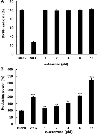
Fig. 2. Scavenging activities of α-asarone on DPPH radical and reducing power. DPPH radical were evaluated in the presence of α-asarone. After α-asarone at the indicated concentration was reacted with each radical, the optical density of each reaction mixture was measured at a spe- cific wavelength with spectrophotometer. Scavenging ac- tivities of DPPH radical and reducing power were eval- uated in the presence of α-asarone. Data are given as means of values ± SD from three independent experiments.
Level of significance was identified statistically (**, p<0.01; ***, p<0.001 ) compared with blank group using Student’s t test.
로 자극한 뒤 50 μg의 총단백질을 함유하는 세포배양액을 1.5 mg/ml gelatin을 포함하는 비환원조건의 10% poly- acrylamide gels를 이용하여 전기영동하였다. Gelatin이 분해 된 bands는 청색배경에 투명한 bands로 나타난다. Bands의 진하기는 MMPs의 활성에 비례하여 나타나는데 LAS3000®
image analyzer (Fujifilm Life Science, Tokyo, Japan)를 관찰 한 후 촬영하였다.
통계처리
각 실험은 3회 이상 반복실험을 통하여 그 결과를 얻어 각각 의 시료농도에 대해 평균±표준편차로 나타내었다. 각 시료 농 도군에 대한 유의차 검정은 대조군과 비교하여 Student’s test 한 후 p<0.05 값을 통계적으로 유의성 있는 결과로 간주하였다.
결 과
α-Asarone의 세포 독성 조사
본 실험에서는 α-asarone이 세포성장에 미치는 영향을 관 찰하기 위하여 세포 독성 유무를 조사하였다. MTT assay를 이용하여 α-asarone의 세포독성 효과를 조사하였으며, Fig. 1 에서 보여지는 바와 같이 HT1080 세포에서 16 μM 이하의 농 도에서 blank군과 비교하여 어떠한 독성 효과도 없는 것으로 판 정 되었다. 따라서, 16 μM 이하의 농도에서 α-asarone은 본 연구 에서 사용된 HT1080세포에 대하여 안전하다는 것을 나타낸다.
Cell free systems에서 α-asarone의 DPPH radical 소거능 및 환원력
생체에서 산화적 스트레스와 관련되어 있는 DPPH radi-
cal에 대한 α-asarone의 소거능력 및 환원력에 대하여 조사하 였다. DPPH radical 소거법은 DPPH radical이 항산화 물질로 인해 환원되면, 자색이 탈색되는 정도를 지표로 하여 지질과 산화의 초기 억제 정도를 예측 할 수 있다. α-asarone의 항산화 효과를 알아보기 위해 DPPH를 이용하여 항산화 작용을 측정 한 결과 Fig. 2A에서 보는 바와 같이, DPPH에 대하여 α-asar- one은 blank군과 비교하여 소거효과가 없는 것으로 나타났다.
양성 대조군은 vitamin C는 100 μg/ml를 사용하였는데 73%
의 소거효과를 나타내었다. 그 다음 α-asarone의 환원력 측정
에는 potassium ferrycyanide reduction법을 사용하여 α-
asarone의 금속이온을 환원시키는 환원력을 조사하기 위하여
흡광도를 측정한 결과, vitamin C는 0.01 μg/ml의 농도에서
대조군에 비교하여 197%의 환원력을 나타냈으며, α-asarone
0 50 100 150 200 250 300 350
Blank Control 1 2 4 8
Inhibition of DNA oxidation (%)
α-Asarone (µM)
·OH radical - + + + + + α-Asarone (µM) 1 2 4 8
***
*** ***
***
Fig. 3. Protective effect of α-asarone on DNA oxidative damage induced by hydroxyl radical. Genomic DNA purified HT1080 cells were pre-treated with α-asarone for 1 hr exposed to ․OH using Fenton reaction. After 30 min, reaction mixture containing about 1 μg of DNA was elec- trophoresed on a 1% agarose gel for 30 min at 100 V and visualized by UV light after stained with 1 mg/ml ethidium bromide. Data are given as means of values
± SD from three independent experiments. Level of sig- nificance was identified statistically (***, p<0.001 ) com- pared with control group using Student’s t test.
0 50 100 150 200 250 300 350
Blank Control 1 2 4 8 16
Collagenase activity (%)
α-Asarone (μM)
***
- + + + + + + α-Asarone (µM) 1 2 4 8 16 Collagenase
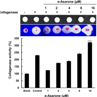
Fig. 4. Inhibitory effect of α-asarone on activity of collagenase.
Inhibitory effect of α-asarone on bacterial collagenase assessed by gelatin digestion assay. Bacterial collage- nase-1 was reacted with 0.1% DMSO (A) as control, with doxycycline as positive control, and with α-asarone at 1, 2, 4, 8 and 16 μM; and then 10 μl of reaction products were loaded onto paper disks placed on agarose gel con- taining gelatin and incubated for 18 hr. Enzyme activity of remaining bacterial collagenase-1 was calculated by densitometric determination of the gelatin digested clear zone visualized by Coomassie Blue staining. Data are given as means of values ± SD from three independent experi`ments. Level of significance was identified statisti- cally (***, p<0.001 ) compared with control group using Student’s t test.
은 1 μM 이상의 농도에서 환원력이 blank군과 비교하여 농도 의존적으로 증가하였다(Fig. 2B). α-Asarone은 1, 2, 4, 8, 16 μM의 농도로 처리한 경우 각각의 환원력은 115%, 130%, 153%, 208%, 324%로 나타나 그 효과가 우수한 것으로 나타났 다.
α-Asarone의 Hydroxyl radical에 의한 DNA 손상에 대 한 항산화 효과
Fenton reaction에 의해 생성되는 Hydroxyl radical에 ge- nomic DNA가 노출되면 분해가 일어난다. α-asarone의 DNA 손상에 대한 항산화 효과를 조사하기 위하여 HT1080 세포로 부터 genomic DNA를 분리하여 Fenton reaction에 의하여 생 성된 Hydroxyl radical에 노출시켰다. Fig. 3에서 보는 바와 같이, control군과 비교하여 α-asarone 1 μM 의 이상의 농도에 서 DNA 분해가 억제되어, α-asarone이 이 농도 이상에서 hy- droxyl radical에 의한 DNA의 손상을 유의성 있게 감소시킴 을 확인할 수 있었다.
α-Asarone의 Collagenase 활성 조절 효과
피부의 주요 구성성분인 Collagen을 분해하는 collagenase 의 활성을 측정하였다. Collagenase와 α-asarone을 paper disk 에 도포하고 34℃에서 24시간 동안 반응시켰다. Fig. 4에서 보
는 바와 같이 blank군과 비교하여 α-asarone의 농도가 16 μM 에서는 collagenase의 활성이 증가하여 collagen 분해가 활발 히 이루어 진 것을 확인하였다.
PMA/PMS로 자극된 HT1080세포에서 α-asarone의 MMP-2, -9 활성 조절 효과
Gelatin과 fibronectin 같은 세포외 기질을 분해한다고 알려
져 있는 MMP-2, -9은 형태형성 및 상처치유와 같은 생리적인
과정에 필수적인 요소임으로, α-asarone의 MMP-2, -9 활성 조
절에 미치는 영향을 조사하기 위하여 gelatin zymography를
수행하였다. Fig. 5A에서는 HT1080세포에 α-asarone을 처리
한 후 PMA로 자극하여 72시간 동안 배양한 상등액을 이용하
였고, Fig. 4B에서는 PMS로 자극하였다. Fig. 5A에서 보는
바와 같이, blank군과 비교하여 PMA군에서는 비활성효소인
proMMP-2가 활성화되어 활성형태의 MMP-2로 전환된 것을
명확히 확인 할 수 있었다. 그리고 1 μM이상의 농도에서 α-
asarone은 농도의존적으로 MMP-2 및 MMP-9의 활성을 증가
AA BB
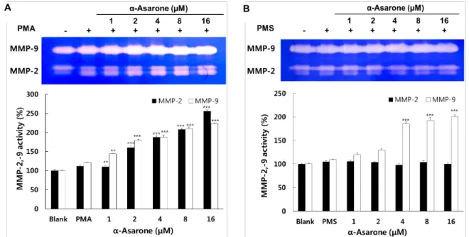
Fig. 5. Effect of α-asarone on activities of MMP-2 and MMP-9 in HT1080 cells treated with PMA/PMS. The effect of α-asarone on activities of MMP-2, -9 without PMA (A) with PMS (B) was evaluated in HT1080. The cells stimulated with PMA/PMS to induce MMPs expression were treated with α-asarone at 1, 2, 4, 8 and 16 μM under serum-free condition for 72 hr.
MMP-2 and MMP-9 activities in conditioned media were determined by gelatin zymography. Data are given as means of values ± SD from three independent experiments. Level of significance was identified statistically (**, p<0.01; ***, p<0.001) compared with PMA group (A) or with PMS group (B) using Student’s t test using Student’s t test.
시키는 것으로 나타났다. 하지만 Fig. 4B에서는 blank군과 PMS군의 비교시 MMP-2 및 MMP-9 차이가 없었으나 α-asar- one처리군에서는 PMS군과 비교시 MMP-2의 활성에서는 차 이가 없었으나 4 μM이상의 농도에서 α-asarone은 MMP-9의 활성을 증가시키는 것으로 나타났다.
고 찰
체내에 제거되지 못하고 축적되는 유해 활성산소종은 세포 막, DNA 및 RNA, 단백질 등에 손상을 주어 다양한 질병을 야기시킨다[1]. 뿐만 아니라 피부 조직의 재생, 상처 치유, 암 전이 등에 필수적인 요소로 작용하는 단백질 분해효소인 MMPs의 활성도 유해 활성산소종에 의해 조절된다는 연구결 과가 보고된 바, 그 기전은 세포내의 H
2O
2의 생성이 Ras 단백 질의 활성화를 유발시키고 그 다음 mitogen activated protein (MAP) kinases를 활성화 시키고, 몇 가지 유전자의 활성화에 의하여 MMPs의 발현을 촉진한다고 보고하였다[9]. 따라서 이 러한 유해 활성산소종의 유의성 있는 제거는 다양한 분야에서 주목하고 있다. 본 연구에서는 예로부터 항알러지, 고콜레스 테롤증 그리고 치매 등에 효과가 있다고 알려진 동양의 전통 한약재인 석창포의 유효 활성성분인 α-asarone의 항산화 효과 와 MMPs 효소에 대한 효과를 조사하였다. 연구에 사용된 HT1080 세포주는 사람의 섬유아육종세포로서 α-asarone 16 μM 농도에서 아무런 독성을 나타내지 않아 16 μM 이하의 농
도에서 계속적인 실험을 수행하였다. α-asarone의 항산화 효
과는 DPPH radical 소거능, 환원력 그리고 DNA 산화 억제능
을 통해 평가되었다. 이 중에서도 특히 환원력이 매우 우수하
게 나타났으며 H
2O
2와 FeSO
4를 이용한 fenton reation에 의한
DNA 손상으로부터 보호효과를 나타냈다. 뿐만 아니라 α
-asarone의 항산화 효과는 이전에 발표된 α-asarone이 쥐의
뇌에서 glutathione reductase 조절을 통해 antioxidant로 작용
한다는 연구결과와도 일치한다[18]. 이는 PMS 자극에 의해
H
2O
2의 생성이 지속되어 HT1080 세포에 가해지면, 외부 스트
레스로부터 방어하기 위하여 α-asarone에 의해 항산화 효과의
발현이 유도 된다고 사료된다. 항산화 효소에 의하여 체내 방
어기전에 의해 활성산소종이 적정량으로 유지된다면 살균효
과와 더불어 피부재생 과정에 중요한 역할을 하는 MMPs의
활성 또한 증가된다. 일반적으로 MMPs와 활성산소의 관련
기전으로 자외선이나 염증 및 물리적 손상에 의하여 피부조직
내에서 생성된 활성산소는 핵속의 전사인자인 NF-kB 혹은
AP-1과 같은 전사인자를 활성화시켜 MMPs의 발현을 촉진시
킨다고 받아들여지고 있다. HT1080 세포에 PMS로 자극하여
H
2O
2생성을 지속적으로 가하면, MT1-MMP 발현 유도를 통
하여 pro-MMP-2의 활성이 유도된다[9]. α-asarone을 전 처리
하여 MMP-2 및 MMP-9의 활성에 대한 효과를 조사한 결과,
α-asarone이 PMA 자극 하에서 MMP-2 및 -9의 활성 수준을
증가시켰다. 그러나 α-asarone은 MMPs 자극제인 PMS 자극
하에서는 MMP-9의 활성 수준을 증가시키고 MMP-2의 활성
에는 영향을 주지 않았다. 위의 결과로 부터 α-asarone은 PMS 자극군과 PMA 자극군에서 활성 양상이 다르게 나타난다는 것을 알 수 있었다. 이러한 결과는 Fenton reaction에 의해 유 도된 산화를 억제를 나타내는 α-asarone의 항산화 효과와도 관련성이 있는 결과로서 α-asarone이 H
2O
2에 유의적으로 반 응하는 것으로 사료된다. 이러한 α-asarone의 H
2O
2억제 효과 는 H
2O
2유도된 cell death로부터 α-asarone이 세포를 보호한 다는 연구 결과와도 일치한다[3]. 뿐 만 아니라 PMS 자극군에 서 MMP-2, -9의 활성 증가는 α-asarone의 항산화 효과와 연관 되어 직접적으로 조절 된 것으로 사료되지만 PMA 자극군에 서 MMP-2의 활성 증가는 간접적인 신호경로를 통하여 이루 어 진 것으로 판단된다. α-Asarone이 나타내는 MMP-2 및 MMP-9 활성 증가 효과는 피부 재생에 있어 매우 중요한 역할 할 것으로 사료된다. 특히 MMP-2 및 MMP-9는 상처 치료의 초기단계에서 피부세포로 부터 분비되어져 상처 치료에 있어 매우 중요한 역할을 한다고 알려져 있다[20]. MMP-2 및 MMP-9은 세포외기질(ECM), 기저막(BM)의 중요한 구성요소 인 콜라겐과 같은 결합조직이 특이적으로 분해하여 새로운 세포가 자랄 수 있도록 해준다. 이러한 피부재생과 관련하여 α-asarone의 효과는 collagenase 활성 측정실험에서도 일치하 게 나타났다. α-asarone이 collagen을 분해하는 collagenase의 활성을 높임으로써 세포의 이동과 재생을 돕는 것으로 기대된 다. 따라서 α-asarone은 H
2O
2와 유의적으로 반응함으로써, 유 해 활성산소종의 축적을 억제하고 세포 보호효과를 나타내며, 피부재생 과정에서 결합조직의 기질을 분해하는데 필수적인 효소인 MMP-2 및 MMP-9의 활성을 증가시키는 것으로 나타 났다. 결론적으로 MMPs와 활성산소와의 관련성에 대한 기전 을 설명하자면, α-asarone은 항산화 효과에 의하여 세포내에 서 생성되는 과량의 활성산소를 억제하여 세포내에 그 수준을 적절하게 유지하면서 MMPs의 활성을 촉진시키는 것으로 추 정된다. 따라서 α-asarone은 산화적 스트레스 및 MMPs와 관 련된 병리학적 질환의 예방 및 치료에 효과가 있을 것으로 기대된다.
감사의 글
이 논문은 2015년도 정부(미래창조과학부)의 재원으로 한 국연구재단의 기초연구사업 지원을 받아 수행된 것(No.
2013R1A1A1A05005160)과, 2015학년도 동의대학교 교내연구 비에 의해 연구되었음(2015AA021).
References
1. Apel, K. and Hirt, H. 2004. Reactive oxygen species: metabo- lism, oxidative stress, and signal transduction. Annu. Rev.
Plant Biol. 55, 373-399.
2. Bergers, G., Brekken, R., McMahon, G., Vu, T. H., Itoh, T.,
Tamaki, K., Tanzawa, K., Thorpe, P., Itohara, S., Werb, Z.
and Hanahan, D. 2000. Matrix metalloproteinase-9 triggers the angiogenic switch during carcinogenesis. Nat. Cell Biol.
2, 737-744.
3. Cho, J., Kim, Y. H., Kong, J. Y., Yang, C. H. and Park, C.
G. 2002. Protection of cultured rat cortical neurons from ex- citotoxicity by asarone, a major essential oil component in the rhizomes of Acorus gramineus. Life Sci. 71, 591-599.
4. Coussens, L. M., Fingleton, B. and Matrisian, L. M. 2002.
Matrix metalloproteinase inhibitors and cancer-trials and tribulations. Science 295, 2387-2392.
5. Hansen, M. B., Nielsen, S. E. and Berg, K. 1989. Re-examina- tion and further development of a precise and rapid dye method for measuring cell growth/cell kill. J. Immunol.
Methods 119, 203-210.
6. Imai, J., Ide, N., Nagae, S., Moriguchi, T., Matsuura, H. and Itakura, Y. 1994. Antioxidant and radical scavenging effects of aged garlic extract and its constituents. Planta Medica 60, 417-420.
7. Jain, N., Jain, R., Jain, A., Jain, D. K. and Chandel, H. S.
2010. Evaluation of wound-healing activity of Acorus cala- mus Linn. Nat. Prod. Res. 24, 534-541.
8. Kim, M. M., Ta, Q. V., Mendis, E., Rajapakse, N., Jung, W.
K., Byun, H. G., Jeon, Y. J. and Kim, S. K. 2006.
Phlorotannins in Ecklonia cava extract inhibit matrix metal- loproteinase activity. Life Sci. 79, 1436-1443.
9. Lee, K. W. and Lee, H. J. 2006. Biphasic effects of dietary antioxidants on oxidative stress-mediated carcinogenesis.
Mechsm Age. Dev. 127, 424-431.
10. Lee, S. J., Seo, K. W., Yun, M. R., Bae, S. S., Lee, W. S., Hong, K. W. and Kim, C. D. 2008. 4-Hydroxynonenal enhan- ces MMP-2 production in vascular smooth muscle cells via mitochondrial ROS-mediated activation of the Akt/NF- kappaB signaling pathways. Free Radic. Biol. Med. 45, 1487- 1492.
11. Limón, I. D., Mendieta, L., Díaz, A., Chamorro, G., Espinosa, B., Zenteno, E. and Guevara, J. 2009. Neuroprotective effect of alpha-asarone on spatial memory and nitric oxide levels in rats injected with amyloid-β (25-35). Neurosci. Lett. 453, 98-103.
12. Manikandan, S. and Devi, R. S. 2005. Antioxidant property of alphaasarone against noise-stress-induced changes in dif- ferent regions of rat brain. Pharmacol. Res. 52, 467-474.
13. Martin, P. 1997. Wound healing--aiming for perfect skin regeneration. Science 276, 75-81.
14. Mittler, R. 2002. Oxidative stress, antioxidants and stress tolerance. Trends Plant Sci. 7, 405-410.
15. Neufeld, G., Cohen, T., Gengrinovitch, S. and Poltorak, Z.
1999. Vascular endothelial growth factor (VEGF) and its receptors. FASEB J. 13, 9-22.
16. Oyaizu, M. 1986. Studies on product of browning reaction prepared from glucose amine. Jpn. J. Nutr. 44, 307-315.
17. Pages, N., Maurois, P., Delplanque, B., Bac, P., Stables, J.
P., Tamariz, J., Chamorro, G. and Vamecq, J. 2010. Activities of α-asarone in various animal seizure models and in bio- chemical assays might be essentially accounted for by anti-
초록:α-Asarone이 항산화 활성 및 기질금속단백질 분해효소 활성 조절에 미치는 영향
박혜정․김문무*
(동의대학교 화학과)
α-Asarone은 동양의 전통적인 약재로 잘 알려진 석창포(Acorus gramineus)의 주된 성분이다. 석창포는 항위궤 양, 항알러지, 히스타민 방출 억제 그리고 항산화 효과와 같이 다양한 효과를 나타내는 것으로 잘 알려져 있다.
그러나 석창포 역할에 대한 기전연구는 아직 부족한 실정이다. 본 연구에서는, HT1080 세포주에서 α-asarone의 항산화 효과뿐만 아니라 matrix metalloproteinase에 대한 효과를 조사하였다. 가장 먼저 α-asarone의 세포 생존에 대한 효과를 조사하기 위해 MTT assay를 이용하여 16 μM이하에서 세포독성이 없음을 나타내었다. α-asarone이 환원력과 fenton reaction에 의해 유도된 DNA 산화로부터 보호효과를 나타내는 것을 확인하였다. 더욱이, α- asarone은 collagenase 활성을 증가시키고 phorbol 12-myristate 13-acetate (PMA)로 자극된 MMP-2 및 MMP-9의 활성을 증가시켰다. 한편 phenazine methosulfate (PMS) 로 자극된 경우 MMP-9의 활성은 α-asarone의 존재하에 서 증가되었으나 MMP-2 활성에는 변화가 없었다. 그러므로 우리의 연구결과는 α-asarone이 산화적 스트레스 및 MMPs와 관련된 병리학적 질환의 예방 및 치료제로 개발이 기대된다고 제안한다.
oxidant properties. Neurosci. Res. 68, 337-344.
18. Park, H. J., Lee, S. J. and Kim, M. M. 2015. Effect of α -asarone on angiogenesis and matrix metalloproteinase.
Environ. Toxico.l Pharmacol. 39, 1107-1114.
19. Rodríguez-Páez, L., Juárez-Sanchez, M., Antúnez-Solís, J., Baeza, I. and Wong, C. 2003. α-Asarone inhibits HMG-CoA reductase, lowers serum LDL-cholesterol levels and reduces biliary CSI in hypercholesterolemic rats. Phytomedicine 10, 397-404.
20. Salo, T., Mäkelä, M., Kylmäniemi, M., Autio-Harmainen, H.
and Larjava, H. 1994. Expression of matrix metal- loproteinase-2 and-9 during early human wound healing.
Lab. Invest. 70, 176-182.
21. Sambrook, J., Fritsch, E. F. and Maniatis, T. 1989. Molecular cloning: a laboratory manual: Cold Spring Harbor Laboratory
Press, Cold Spring Harbor, NY, 1977.
22. Shin, J. W., Cheong, Y. J., Koo, Y. M., Kim, S., Noh, C. K., Son, Y. H., Kang, C. and Sohn, N. W. 2014. Alpha-asarone Ameliorates memory deficit in lipopolysaccharide-treated mice via suppression of pro-inflammatory cytokines and microglial activation. Biomol. Ther. 22, 17-26.
23. Simon, H. U., Haj-Yehia, A. and Levi-Schaffer, F. 2000. Role of reactive oxygen species (ROS) in apoptosis induction.
Apoptosis 5, 415-418.
24. Taniyama, Y. and Griendling, K. K. 2003. Reactive oxygen species in the vasculature molecular and cellular mecha- nisms. Hypertension 42, 1075-1081.
25. Vihinen, P. and Kähäri, V. M. 2002. Matrix metal- loproteinases in cancer: prognostic markers and therapeutic targets. Int. J. Cancer 99, 157-166.