Use of the Synthetic Gene Encoding the Truncated Human Rotavirus VP8* Protein in Escherichia coli for Production of Vaccine Candidates or Development of Diagnostic Antibodies
Sang-Rae Kim and Bheong-Uk Lee*
Major in Biomedical Sciences, Division of Biological Sciences and Chemistry, Kosin University, 194 Wachi-ro, Yeongdo-gu, Busan 49104, Korea Received December 2, 2017 /Revised December 29, 2017 /Accepted December 31, 2017
Human rotavirus is a causative agent of acute diarrhea among children. The artificial gene encoding the truncated VP8* protein of human rotavirus A (serotype 1 strain WA) was synthesized according to the Escherichia coli codon preference. The synthetic VP8* gene also possessed the NdeI and HindIII restriction sites for the convenient in-frame cloning for translation and a 6-histidine tag at C-terminus for Ni+ affinity purification. Molecular weight of the truncated VP8* protein deduced from the nucleo- tide sequences of the artificial gene was a 19.7-kDa. This synthetic VP8* DNA fragment was inserted into the pT7-7 expression vector and transformed into E. coli BL21 (DE3). Transformants harboring the synthetic gene encoding the VP8* protein was induced by supplement of a final concentration of 0.05 mM ITPG at 20℃. Protein crude extract from the E. coli transformants was subjected to Western blot- ting with the mouse anti-rotavirus capsid antibody, showing ~20-kDa VP8* protein band. The trun- cated Vp8* protein band was also observed by Western blotting using the rabbit polyclonal antibody serum made against the truncated VP8* protein. This study suggested that the synthetic gene could be used as an easy way to produce the antigenic vaccine candidate for control of virus-associated diseases or to develop antibodies for diagnostic purpose.
Key words : Codon preference, Escherichia coli, rotavirus VP8*, synthetic gene
*Corresponding author
*Tel : +82-51-990-2321, Fax : +82-51-911-2520
*E-mail : [email protected]
This is an Open-Access article distributed under the terms of the Creative Commons Attribution Non-Commercial License (http://creativecommons.org/licenses/by-nc/3.0) which permits unrestricted non-commercial use, distribution, and reproduction in any medium, provided the original work is properly cited.
Journal of Life Science 2018 Vol. 28. No. 4. 478~482 DOI : https://doi.org/10.5352/JLS.2018.28.4.478
서 론
인간 rotavirus는 어린이 특히 영아에서 설사를 일으키는 바이러스이다[14]. Rotavirus는 이중나선 RNA 바이러스로서 A, B, C, D, E, F, G 및 H형의 serotype이 존재하며 인간에게는 대부분 A형이 질병을 일으킨다[8]. 감염이 되면 소장 표면 세 포에 손상을 일으켜서 장염의 원인이 된다. 영아 사망을 일으 키는 바이러스였지만 최근에 백신의 개발로 사망률이 크게 감소하였다[2, 6]. 6종의 구조적인 바이러스 단백질(VP)을 발 현하는데 각각 VP1, VP2, VP3, VP4, VP6 및 VP7으로 명명되 어있다[8, 13]. 이중에서 크기가 87 kDa인 VP4는 비리온 표면 의 돌기(spike)를 형성하며 숙주 세포의 수용체에 결합하여 세포 내로 침입하는데 필수적이다[16]. VP4는 단백질 분해효 소인 트립신에 의해서 VP5* 및 VP8*로 절단이 되며 이중에서 VP8* 단백질이 숙주 세포의 표면 단백질과의 부착에 필수적인 것으로 알려져 있다. 따라서 VP8*의 부착 기능을 억제하면 바
이러스는 소장 세포 내로 침입을 할 수가 없으며 질병이 예방 될 것으로 판단된다. 기존에 인간 rotavirus를 예방하기 위한 백신의 생산을 위해서 다양한 생물 종(species)들을 이용한 연 구가 있었다[7, 10, 15, 17]. 하지만 Escherichia coli 균주를 이용 한 발현 연구에서는 VP8* 단백질 전체를 사용하여 발현이 용 이하지 않거나 정제를 위해서 융합된 단백질이 거대하여 제거 에 어려움이 있었다[11].
본 연구에서는 E. coli의 코돈 선호도에 일치하도록 VP8*
단백질의 일부만을 생산하도록 크기를 축소시킨 인공 DNA를 합성하여 pT7-7 발현 벡터에 삽입하고 발현을 유도하였다.
Rotavirus capsid에 대한 단일 항체를 이용하거나 토끼로부터 생산된 다가항체 혈장을 사용한 Western blotting을 수행한 결과를 보면, 발현된 VP8* 부분 단백질이 항원성을 보유하고 있으며 또한 진단 시약으로 사용될 수 있는 항체의 신속한 개발에도 응용될 수 있다는 것을 확인할 수 있다.
재료 및 방법
인간 rotavirus VP8* 부분 서열 유전자의 합성
인간 rotavirus A (serotype 1 Wa)의 VP8* 단백질 서열(Gen Bank accession: VPXRWA)을 분석을 통하여 164개의 아미노 산 서열을 인위적으로 선택하고 E. coli 코돈 선호도를 기반으 로 인공 유전자를 합성하였으며, 5‘-말단에 제한효소 NdeI 절 단 서열 CATATG 및 3’- 말단에 HindIII 서열 AAGCTT를 추 - Note -
Fig. 1. The sequences of the truncated 164-amino acid VP8* protein. The original VP8* protein of the rotavirus A (serotype1 Wa) strain is composed of 280 amino acids. (GenBank accession: VPXRWA). The amino acid sequence of the truncated VP8*
protein are capitalized.
Fig. 2. The nucleotide sequence of the synthetic gene encoding the truncated VP8* protein. Total DNA length is 533 bp. The truncated VP8* gene is 516-base pair long and encodes a putative 19.7 kDa protein composed of 171 amino acids including 6-histidine tag for convenient affinity purification with Ni+ column. The nucleotide sequence for 6-histidine tag is underlined. The artificial DNA fragment also has NdeI and HindIII restriction sites at 5’- and 3’- ends respectively. Two restriction sites are dou- ble-underlined. The start and stop codons are capitalized.
가하였다. VP8* 단백질의 C-말단에 6개의 히스티딘을 암호화 하는 서열이 배치되도록 합성하여 필요 시에는 Ni+ 친화 칼럼 을 사용하여 정제가 가능하도록 하였다(Fig. 2).
합성 VP8* 유전자의 클로닝 및 발현
합성된 VP8* 유전자를 NdeI 및 HindIII 제한효소로 절단하 여 동일한 제한효소들로 처리된 pT7-7 발현 벡터에 도입하였 다[3]. 조립된 발현 플라스미드를 TSS 방법[4]으로 E. coli BL21 (DE3) 균주에 형질전환한 후에 ampicillin 100 μg/ml이 포함 된 LB 고체 배지에서 선별하였다. 선별된 균주들을 37℃의 LB 액체배지에서 OD600=0.6까지 배양한 후에, IPTG를 최종 농도 가 0.05 mM가 되도록 첨가하여 20℃로 배양 온도를 낮추어서 15시간 더 배양하고 원심분리를 이용하여 수거하였다. 수거된 배양 세포들을 초음파로 파쇄한 후에 원심분리로 상등액을 수집하여 –20℃에 보관하였다.
발현된 VP8* 단백질의 Western blotting
합성 VP8* 유전자로부터 단백질의 항원성을 확인하기 위하 여 mouse anti-rotavirus capsid monoclonal antibody (Santa Cruz Biotechnology)를 1차 항체로 사용하고 goat anti-mouse IgG–peroxidase (Sigma Immunochemicals)를 2차 항체로 사 용하여 Western blotting을 수행하였다. 항체 면역 반응을 유 도하는지 조사하기 위해서 VP8* 단백질을 이용하여 토끼로부
터의 VP8* 단백질에 대한 polyclonal antibody serum 생산을 조사하였다. 이 실험을 위해서 SDS-PAGE로 발현이 확인된 VP8* 단백질을 acrylamide gel과 함께 절단하고 절편을 미세 하게 파쇄한 후에 뉴질랜드 화이트 토끼의 피하에 4주 간격으 로 3회 주입하였고 채혈 후에 혈장을 분리하였다[12]. 2차 항체 로 goat anti-rabbit IgG-HRP conjugate와 결합시킨 후에, lu- minol/enhancer solution을 사용하여 형광을 만들고 x-ray 필 름(Kodak Co.)에 노출 시켜서 인화하였다.
기타 실험 방법
기타 세균의 배양, DNA 조립 및 단백질 실험 등에 관련된 방법들은 Short Protocols in Molecular Biology 및 Short Protocols in Protein Science (Wiley Co.)를 따라서 수행하였다 [1, 5].
결과 및 고찰
인간 rotavirus VP8* 부분 서열 유전자의 합성
인간 rotavirus A (serotype1 Wa)의 VP8* 단백질은 총 280 개의 아미노산으로 구성되어 있는데 이중에서 164개의 아미 노산 서열을 인위적으로 선택하고(Fig. 1), E. coli 코돈 선호도 를 기반으로 인공 유전자를 합성하였다(Fig. 2; Fig. 3B). 합성 된 DNA 절편의 총 길이는 533 bp이며 pT7-7 발현 벡터로의
A
B C
Fig. 3. Expression of the truncated VP8* protein. (A) Nucleotide sequence shows the junction region of the pT7-7 pro- moter and the synthetic DNA fragment. Slash (/) in- dicates junction. SD stands for the underlined Shine- Dalgarno sequence. NdeI restriction site is double-under- lined. The start codon is written in bold letters and a part of NdeI restriction site. (B) Picture of the agarose gel elec- trophoresis with the synthesized 533 bp-long DNA frag- ment containing the truncated human rotavirus VP8*
gene. Arrow at lane 2 indicates the synthetic DNA. Lane 1 shows HindIII-digested λ bacteriophage DNA (C) SDS- PAGE showing the proteins from uninduced (lane 2) and induced (lane 3) E. coli BL21 (DE3) cells, respectively.
Protein size markers are at lane 1. Arrow indicates the
~20-kDa truncated Vp8* protein.
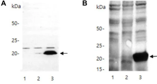
A B
Fig. 4. Antigenicity of the truncated VP8* proteins and induction of its antibody. (A) Picture of Western blotting using the mouse anti-rotavirus capsid antibody as the first anti- body and goat anti-mouse IgG–peroxidase as the secon- dary antibody (B) Western blotting using the rabbit poly- clonal antibody serum induced against the VP8* protein produced from E. coli cells. Arrows indicate ~20-kDa VP8* protein bands. Lane 1, cells without expression vec- tor; Lane 2 cells with vector, but not induced; Lane 3, cells with vector and induced.
삽입 시에 편이성을 제공하고, 또한 필요가 없는 아미노산이 추가로 번역되는 것을 방지하기 위하여 개시 코돈, ATG를 보 유한 제한효소 NdeI 절단 서열 CATATG를 5‘-말단에 위치하 도록 설계하였다(Fig. 3A). VP8* 단백질의 C-말단에 6개의 히 스티딘을 암호화하는 CATCATCACCATCACCAC 서열을 배 치하여 단백질의 빠르고 쉬운 정제가 필요한 경우에 Ni+ 친화 칼럼을 사용할 수 있도록 하였다(Fig. 2). E. coli 코돈 선호도에 최적화되도록 조립된 VP8* 염기 서열은 아미노산의 번역 서열 은 동일하지만 GenBank에 저장된 원래의 인간 rotavirus의 염기 서열과는 72%의 일치를 보였다(data not shown).
합성 VP8* 유전자의 단백질 발현 및 항원성 조사 NdeI 및 HindIII 제한효소로 절단된 합성 VP8* 유전자는 동일한 제한효소들로 처리된 pT7-7 발현 벡터에 도입하였다.
이 경우에 NdeI 제한효소의 인식 서열인 CATATG의 ATG를 개시 코돈으로 사용하기 때문에 생산된 단백질의 N- 말단에 잉여로 존재하는 아미노산이 없게 된다(Fig. 3A). IPTG를 최종 농도가 0.05 mM이 되도록 첨가하여 37℃에서 형질전환된 E.
coli BL21 (DE3) 균주를 단백질을 발현 유도한 경우에는 세포 잔존물(debris)을 제거한 crude extract 상등액에서는 합성 VP8* 단백질의 농도가 매우 낮았다(data not shown). 이런 경 우에 대부분의 단백질이 3차 구조를 가지 못한 inclusion body 의 형태로 존재할 것으로 추측하였다. 합성 VP8* 단백질의 solubility를 증가시키기 위하여 E. coli 형질전환 세포의 배양
온도를 20℃로 내리고 동일한 농도의 IPTG를 첨가하여 발현 을 유도하였다. 낮은 온도에서 배양된 E. coli에서 추출한 단백 질들의 SDS-PAGE 결과를 보면 성공적으로 생산된 ~20-kDa 크기의 단백질을 확인할 수 있다(Fig. 3C).
합성 VP8* 유전자로부터 단백질의 항원성을 확인하기 위하 여 인간 rotavirus의 capsid를 인식하는 mouse monoclonal antibody를 1차 항체로 사용하고 2차 항체로는 mouse anti- body를 인식하는 goat antibody-peroxidase를 사용하여 Wes- tern blot을 수행하였다(Fig. 4A). 결과를 보면 인공적으로 합 성된 유전자의 발현이 유도된 경우에 뚜렷한 밴드를 확인할 수 있었는데, 이는 164개의 아미노산으로 구성된 VP8* 단백질 이 rotavirus capsid를 인식하도록 생산된 monoclonal anti- body에 의해서 인식이 된다는 의미이다. 합성된 유전자로부 터 발현된 VP8* 단백질이 항체 생성을 유도하는지 조사하기 위하여 단백질이 피하에 4주 간격으로 3회 주입된 토끼에서 채혈된 혈액에서 분리된 혈장을 사용하여 Western blot을 수 행하였다(Fig. 4B). SDS-PAGE 상에 전개된 VP8* 단백질 밴드 부분을 절단해 낸 후에, 피하주사가 가능할 정도로 미세하게 파쇄를 하여 adjuvant의 첨가가 없이 피하에 주입하였다. 토끼 혈장 내의 polyclonal antibody들을 1차 항체로 사용하고 rab- bit antibody를 인식하는 goat antibody-peroxidase를 사용한 결과를 보면 SDS-PAGE 상에 전개된 단백질들에서 VP8*를 인식하는 것을 관찰할 수가 있었다(Fig. 4B). 이는 인공 VP8*
단백질이 적절한 adjuvant의 첨가가 없이도 토끼로부터 항체 형성을 유도할 있다는 의미이다.
본 연구는 병원성 바이러스의 spike를 형성하는 야생형 단 백질에 비하여 크게 축소된 인공 단백질이 항원성을 보유할 수 있다는 것과 이를 이용하여 생쥐 및 토끼 등에서 항체 생성
실험들을 수행할 수 있다는 것을 보여주었다. 이는 비교적 합 성이 용이하고 시간의 소비가 적은 방식으로 생산된 인공 단 백질들이 질병을 일으키는 다른 세균 혹은 바이러스를 예방하 는 백신들과 혼합되거나 혹은 융합 단백질(fusion protein) 형 태의 백신을 생산하기 위한 후보 물질로 고려되는 것도 가능 하다는 의미이다[9, 18]. 또한 비교적 빠르게 합성할 수 있는 유전자들로부터 생산된 부분적인 capsid 단백질들을 사용하 여 항체를 생산하고, 이 항체들을 병원체 진단을 위한 용도에 적용할 수도 있을 것이다.
감사의 글
본 연구는 2017년도 고신대학교 교내연구비로 수행되었습 니다.
References
1. Ausubel, F. M., Brent, R., Kingston, R. E., Moore, D. D., Scidman, J. G., Smith, R. A. and Struhl, K. 2002. Short Protocols in Molecular Biology. (John Wiley & Sons, New York, NY, USA).
2. Babji, S. and Kang, G. 2012. Rotavirus vaccination in devel- oping countries. Curr. Opin. Virol. 2, 443-448.
3. Brown, W. C. and Campbell, J. L. A., 1993. New cloning vector and expression strategy for genes encoding proteins toxic to Escherichia coli. Gene 127, 99-103.
4. Chung, C. T., Niemela, S. L. and Miller, R. H. 1989. One-step preparation of competent Escherichia coli: transformation and storage of bacterial cells in the same solution. Proc. Natl.
Acad. Sci. USA. 86, 2172-2175.
5. Coligan, J. E., Dunn, B. M., Speicher, D. W. and Wingfield, P. T. 2003. Short Protocols in Protein Science. (John Wiley &
Sons, New York, NY, USA).
6. Dóró, R., László, B., Martella, V., Leshem, E., Gentsch, J., Parashar, U. and Bányai, K. 2014. Review of global rotavirus strain prevalence data from six years post vaccine licensure surveillance: is there evidence of strain selection from vac- cine pressure? Infect. Genet. Evol. 28, 446-461.
7. Favacho, A. R., Kurtenbach, E., Sardi, S. I. and Gouvea, V.
S. 2006. Cloning, expression, and purification of recombi- nant bovine rotavirus hemagglutinin, VP8*, in Escherichia
coli. Protein Expr. Purif. 46, 196-203
8. Hoshino, Y. and Kapikian, A. Z. 2000. Rotavirus serotypes:
classification and importance in epidemiology, immunity, and vaccine development. J. Health Popul. Nutr. 18, 5-14.
9. Kang, D. K., Kim, P. H., Ko, E. J., Seo, J. Y., Seong, S. Y., Kim, Y. H., Kwon, I. C., Jeong, S. Y. and Yang, J. M. 1999.
Peroral immunization of microencapsulated human VP8* in combination with cholera toxin induces strong systemic and intestinal antibody responses. Mol. Cells 9, 609-616 10. Kim, H. S., Lee, B., Han, S. Y. and Jung, Y. T. 2017. Expres-
sion of bovine rotavirus VP8 and preparation of IgY anti- bodies against recombinant VP8. Acta Virol. 61, 143-149.
11. Kovacs-Nolan, J., Sasaki, E., Yoo, D. and Mine, Y. 2001.
Cloning and expression of human rotavirus spike protein, VP8*, in Escherichia coli. Biochem. Biophys. Res. Commun. 282, 1183-1188.
12. Leenaars, P. P., Hendriksen, C. F., de Leeuw, W. A., Carat, F., Delahaut, P., Fischer, R., Halder, M., Hanly, W. C., Hartinger, J., Hau, J., Lindblad, E. B., Nicklas, W., Out- schoorn, I. M. and Stewart-Tull, D. E. 1999. The Production of polyclonal antibodies in laboratory animals. The report and recommendations of ECVAM workshop 35. Altern. Lab.
Anim. 27, 79-102.
13. Lepault, J. 2001. Structural polymorphism of the major cap- sid protein of rotavirus. EMBO J. 20, 1498-1507.
14. Parashar, U. D., Hummelman, E. G., Bresee, J. S., Miller, M. A. and Glass, R. I. 2003. Global illness and deaths caused by rotavirus disease in children. Emerg. Infect. Dis. 9, 565- 572.
15. Pêra, F. F., Mutepfa, D. L., Khan, A. M., Els, J. H., Mbewana, S., van Dijk, A. A., Rybicki, E. P. and Hitzeroth, II. 2015.
Engineering and expression of a human rotavirus candidate vaccine in Nicotiana benthamiana. Virol. J. 12, 205-215.
16. Ruiz, M. C., Leon, T., Diaz, Y. and Michelangeli, F. 2009 Molecular biology of rotavirus entry and replication. Sci.
World J. 9, 1476-1497.
17. Wen, X., Cao, D., Jones, R. W., Li, J., Szu, S. and Hoshino, Y. 2012. Construction and characterization of human rotavi- rus recombinant VP8* subunit parenteral vaccine candidates.
Vaccine 30, 6121-6126.
18. Xue, M., Yu L., Jia, L., Li, Y., Zeng, Y., Li, T., Ge, S. and Xia, N. 2016. Immunogenicity and protective efficacy of ro- tavirus VP8* fused to cholera toxin B subunit in a mouse model. Hum. Vaccin. Immunother. 12, 2959-2968.
초록:합성 유전자를 이용하여 Escherichia coli에서 백신 후보의 생산 혹은 진단용 항체의 개발을 위 한 인간 rotavirus VP8* 부분 단백질의 발현
김상래․이병욱*
(고신대학교 생명과학.화학부 의생명과학 전공)
인간 rotavirus는 영아에게 급성 설사를 일으키는 병원체의 하나이다. 본 연구에서는 Escherichia coli의 코돈 선 호도를 따라서 인간 rotavirus A (serotype 1 strain WA)의 VP8* 단백질을 일부분 암호화하도록 인공적인 유전자 를 합성하였다. 합성된 VP8* 유전자는 코돈을 번역틀에 일치시키고 클로닝이 용이하도록 하기 위한 NdeI 및 HindIII 제한효소 절단 부위와 친화적 정제를 위한 6-히스티딘 암호화 서열을 C-말단에 보유하고 있다. 합성된 VP8* DNA 절편을 pT7-7 발현 벡터에 삽입하여 E. coli BL21 (DE3)로 형질전환한 후에 최종 농도 0.05 mM IPTG 로 생산을 유도한 결과 예상했던 대로 19.7-kDa 크기의 VP8* 단백질이 고농도로 발현되었다. SDS-PAGE에 전개 된 단백질들을 대상으로 mouse anti-rotavirus capsid antibody를 사용한 Western blotting의 결과 ~20-kDa VP8*
단백질 밴드가 관찰되었다. 인공 Vp8* 단백질이 피하 주사된 토끼의 polyclonal antibody 혈장을 이용한 조사에서 도 동일한 크기의 단백질 밴드를 확인할 수 있었다. 이는 합성된 유전자가 바이러스성 질환을 통제할 항원성 백신 후보의 생산 혹은 진단용 항체를 개발하기 위한 쉽고 빠른 방법을 제공할 수 있다는 의미이다.