LPS로 유도된 대식세포 활성에서 발효 산약 추출물의 억제 효과
임성원·이선희·허정무*·이영미**·김대기#
전북대학교 의과대학 및 의과학연구소, *종근당 건강(주), **원광대학교 약학대학 한약학과 및 원광한약연구소 (Received September 23, 2011; Revised September 23, 2011; Accepted September 30, 2011)
The Inhibitory Effect of Fermented Dioscoreae batatas Extract on Lipopolysaccharide-induced Macrophage Activation
Sung-Won Lim, Sun-Hee Lee, Jung-Mu Hur*, Young-Mi Lee** andDae-Ki Kim#
Department of Immunology and Institute of Medical Science, Chonbuk National University Medical School, Jeonju 561-756, Korea
*Chong Kun Dang Healthcare Corp. Research Center, Dangjin 343-827, Korea
**Department of Oriental Pharmacy, College of Pharmacy and Wonkwang-Oriental Medicines Research Institute, Wonkwang University, Iksan 570-744, Korea
Abstract — This study was to elucidate the anti-inflammatory activities of a methanol extract derived from the fermented bark of Dioscoreae batatas on LPS-induced activation in macrophages. It was fermented with Lactobacillus fermentum and L. plantarum and then analyzed to identify the contents of methanol extract and diosgenin. The fermented product showed 3-fold increase in the extraction yield by methanol, and 1.8-fold increase in diosgenin contents, compared to that from the dried bark of D. batatas. Although the methanol extract from the unfermented D. batatas inhibited lipopolysaccharide (LPS)- induced production of nitric oxide (NO) and TNF-α in J774 A.1 cells, the methanol extract from the fermented product revealed significantly the enhanced the inhibitory activities on LPS-induced production of NO and TNF-α. Taken together, our results indicate that fermentation of bark of D. batatas elevates the functional activity inhibiting macrophage activation through the increase of the content of anti-inflammatory compounds. Thus, its methanol extract may be useful as a func- tional material for the therapy of inflammatory diseases.
Keywords □ Dioscoreae batatas, Fermentation, Lactobacillus, Macrophages, TNF-α, Nitric oxide
백합목 마과(Dioscoreacea)에 속하는 마(Yam)는 우리나라 전 국 산야에 자생하는 덩굴성 다년생 식물로서 국내에는 1속 9종 이 있으며 주로 재배되는 마는 Dioscorea batatas Decne.(마) 및 D. japonica Thunb.(참마) 2종이다. 한방에서는 지하부를 산약 (山藥)이라 하며 약용으로 뿌리줄기의 주피를 벗겨 그대로 또는 쪄서 말린 것을 설사, 구리, 식욕부진, 해수, 소갈, 유정, 대하, 빈 뇨 등의 치료 목적으로 오랜 세월 동안 사용되어 왔다.1-3)산약 의 구성성분은 대부분이 식이섬유이며, 탄수화물(10~15%), 아 미노산, 점액성 당단백질, diosgenin, mucilages, allantoin, dioscorins, batatasins, phenanthrene glycoside, soyacerebroside, beta-sitosterol, palmitic acid, choline, sitosterol계 화합물들이 보고되었다.4,5)최근의 보고에 의하면 산약에서 분리 추출한 저
장성 단백질 dioscorin, 점액성 다당류 및 polyphenol성 화합 물 등이 중금속(Co, Cr, Cu) 흡착 제거능과 항산화 활성이 뛰 어나다고 보고되었다.6,7) 또한 위장관 기능 강화작용,8,9) 혈당 강하작용,10)혈중 콜레스테롤 저하작용,11)항고혈압작용,12)인 지능력 및 치매개선효과,13)항비만작용14)등의 다양한 효능이 보고되었으며, 이에 근거하여 기능성식품 소재로 관심이 높아 지고 있다.
대식세포는 골수로부터 생산되는 주요한 면역세포이며 자연면 역 뿐 만 아니라 획득면역에도 관여하면서 인체 내에 침입하는 세균, 바이러스, 노화세포 등의 이물질을 포식하고 제거하는 식균작용을 하며, 병원균과 같은 외부 자극원에 의해 야기되는 초기 염증반응에서 활성아민류, arachidonic acid 대사산물 (prostaglandins, leukotrienes), 사이토카인류(TNF-α, IL-1β) 등 다양한 염증매개물질들을 분비하여 면역반응을 활성화하고 생체 내 방어기능과 항상성 유지 기능을 조절하는 중심적인 역할을 한 다고 알려져 있다.15-17)최근에 산약추출물이 대식세포활성을 억
#본 논문에 관한 문의는 저자에게로 (전화) 063-270-3080 (팩스) 063-855-6807 (E-mail) [email protected]
종설
제하며, 특히 산약의 지표성분 diosgenin에 의해 대식세포로 부 터 염증매개물질 분비를 억제한다고 보고되었다.18,19) 우리가 섭 취하는 식품 중에는 수많은 발효식품이 존재한다. 일반적으로 식 품이 발효과정을 거치면 유용성분의 함량증가, 새로운 생리활성 증강, 흡수율 증가, 잔류농약의 감소 또는 제거, 장내 유용미생 물의 증가 등 다양한 생리적 작용을 강화시킬 수 있다. 초기 발 효식품은 젖산균을 이용한 젖산 생성을 통하여 식품의 pH를 낮 추고 식품의 저장성을 높이는데 중요한 역할을 하였으나, 최근 건강기능성 식품을 선호하는 추세에 따라 발효식품에 대한 관심 도가 높아지면서 Lactobacillus, Bifidobacterium, Lactococcus 등 과 같은 젖산균들을 활용하여 건강 기능성을 강화시키는 식품들 이 새롭게 대두되고 있다.20-25)산약의 경우 특유의 점액성성분 으로 생산성 및 기호성을 나쁜 단점 때문에 생물적 변환을 통한 개선이 요구됨에도 불구하고,26)현재 산약을 활용한 발효식품에 대한 연구는 전무한 상태이다.
본 연구에서는 젖산균을 활용하여 산약을 발효시키고, 그 발 효물의 methanol 추출물에서 대식세포 활성에 미치는 영향을 연 구하므로써 기능성식품으로의 가능성을 검토하고자 하였다.
실험방법
실험 재료
본 실험에 사용한 시료는 국내에서 재배한 마(Dioscorea batatas)를 익산 금마농협으로부터 구입하여, 물로 수세 후 표면 의 물기를 제거, 열풍건조기를 이용하여 건조(60oC) 시킨 뒤 100 mesh 이내로 분쇄하여 분말화 하였다. 발효를 위해 사용한 젖산 균주는 MRS 배지에 Lactobacillus fermentum(KCCM40026) 와 L. plantarum(KCCM11322)를 한국미생물보존센터(Seoul, Korea)에서 분양받았다. 마우스 대식세포주 J774 A.1세포는 한 국세포주은행(Seoul, Korea)으로 부터 분양받았으며, 세포배양을 위한 Dulbecco's modified Eagle's media(DMEM) 및 fetal bovine serum(FBS) 는 Hyclone(Logan, UT)에서 구입하였고, Penicillin(100 units/ml)-streptomycin(100 µg/ml)은 WelGENE (Daegu, Korea)에서, 세포증식 분석시약 Cell Counting Kit-8 (CCK-8)은 Dojindo사(Kumamoto, Japan)에서, Lipopolysaccharide (LPS)는 Sigma사(St. Louis, MO)에서 구입하였다. TNF-α 및 IL-1β 분석용 ELISA Kit는 eBioscience(San Diego, CA)에서 구 입하였다. 그밖의 시약과 용매들은 Sigma사(St. Louis, MO)에서 구입하여 사용하였다.
산약 발효물 및 메탄올 추출물 제조
산약 발효물은 (주)한풍제약연구소에서 다음과 같이 제조하 여 제공받았다. 발효균주 L. fermentum(KCCM40026)와 L.
plantarum(KCCM11322)를 각각 MRS plate에서 2회 계대배양
후 MRS 액상 배지에 1 loop 접종하여 37oC, 48시간 배양하였 다. 배양된 각 균들을 1 : 1 비율로 혼합하였다. 산약건조분말을 정제수 10배 용량에 균질하게 현탁하고, 균주 배양액을 산약 발 효물 현탁액의 4%가 되도록 혼합하여 37oC에서 교반하면서 3 일간 배양하였다. 발효상태는 pH의 변화로 확인하였으며, 발효 과정에서 합성된 유기산의 산도를 측정하였다. 최종 발효물은 60oC에서 수분함량 6% 이하가 되도록 열풍 건조하였으며, 이 를 분쇄하여 산약발효분말 시료를 제조하였다. 최종발효물의 pH 와 유기산의 산도를 측정하고자, 시료 10 g에 끓여 식힌 증류수 100 ml를 가하고 pH를 측정하였으며, 이에 페놀프탈레인시약 0.5 ml를 가하여 0.1 N NaOH로 30초간 홍색이 지속될 때까지 적정하여, 다음 식에 따라 산도를 산출하였다.
유기산 산도(%)(젖산으로서)=S×(f×0.009/시료채취량(g))×100 S: 0.1 N 수산화나트륨액의 소비량(ml)
f: 0.1 N 수산화나트륨액의 역가
산약 건조분말(NFD) 및 발효물 건조분말(FD) 50 g에 methanol 500 ml을 가하여 상온에서 24시간 교반하여 추출한 후 Whatman 여과지로 걸러 불용물을 제거하였다. 여과액을 rotary evaporator (EYELA, Tokyo, Japan)로 농축한 후 동결 건조하여 메탄올추출 물을 얻었으며 이를 본 연구에 사용하였다.
세포독성측정
본 실험에서 사용한 시료들에 의한 세포독성을 측정하기 위해 CCK-8을 이용하여 세포생존율을 측정하였다. 대식세포주 J774 A.1을 96 well plate에 1×10⁴cells/well로 분주하고 24시간 동안 세포를 안정시킨 후 시료를 농도별로 희석한 배지를 처리하여 5% CO2, 37oC 조건의 incubator에서 48시간을 배양하였다. 배 양 후 CCK-8시액 10 µl를 각 well에 첨가하여 다시 1시간 배양 하고 그 배양액을 450 nm에서의 흡광도를 측정하였다.
Nitric Oxide(NO) 측정
세포를 24 well plate 에 1×10⁵cells/well로 분주하고, phenol red가 포함되지 않은 DMEM 배지에서 배양하였다. 24시간 배양 하여 세포를 안정시킨 후 시료를 세포에 30분간 전처리하고, 각 well에 LPS(0.1 µg/ml)를 넣고 24시간 배양하였다. 배양액을 Tube 에 수집하여 12000 rpm, 4oC, 10분간 원심분리하여 상층액를 얻 고 분석전까지 -20oC 이하에서 보관하였다. NO의 정량은 Griess reagent를 이용하여 측정하였다. 96 well plate에 세포배양액 100µl와 Griess reagent(0.1% N-1-Naphthylethylenediamine와 1% Sulphanilamide를 포함한 5% phosphoric acid(V/V)) 100 µl 를 혼합하여 실온에서 10분간 반응시킨 후 550 nm에서의 흡광 도를 측정하였다. 표준품 Sodium nitrite를 통하여 얻어진 standard curve로부터 nitrite의 농도를 산출하였다.
Enzyme Linked Immunosorbent Assay(ELISA) 세포에서 분비된 cytokines의 농도를 측정하기 위하여 배양액 을 원심 분리하여 상층액을 취하고 ELISA Kit(eBioscience, San Diego, CA)를 사용하여 TNF-α와 IL-1β 분비량을 측정하였다.
대식세포 활성은 양성대조군으로 LPS(0.1 µg/ml)를 처리하여 비 교하였으며, 실험은 제조사에서 제공한 protocol에 따라 실행하 였다. 최종 반응액은 450 nm에서의 흡광도를 측정하여 cytokine 분비량을 산출하였다.
HPLC 분석
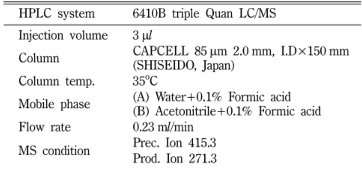
시료 건조분말 50 g을 methanol 500 ml에 넣고 상온에서 24 시간 교반한 후 Whatman 종이여과지와 0.45 µm cellulose 막여 과지에 통과시켜 불용분을 제거하고 여액을 얻었다. 여액을 rotary evaporator(EYELA)에서 감압농축한 후 동결건조하고 이들 추출 물 5 mg을 다시 5 ml methanol에 녹여 검액으로 사용하였다. 표 준품 Diosgenin은 methanol과 DMSO 3 : 1(v/v)에 녹인 후 일정 농도로 희석하여 검량선 작성하였다. Diosgenin의 함량 분석은 역상 HPLC 법으로 6410B Triple Quad LC/MS(Agilant, Santa Clara, CA)를 사용하여 Table I과 같이 실행하였다.
통계적 분석 방법
모든 실험은 3회 반복 실시하였으며, 실험 Data는 mean±S.D.
로 표시하였고, Student's t-test를 이용하여 통계학적 분석을 하 여 통계적 유의성(p<0.05)을 판정하였다.
실험결과 및 고찰
산약 발효물의 pH, 산도, 추출율 및 diosgenin 함량 변화 산약 건조분말을 3일간 젖산균 발효를 시킨 후 얻어진 발효건 조물의 pH와 발효과정에서 합성된 유기산의 산도를 측정하였다.
산약은 steroidal saponin계 화합물로 알려진 diosgenin을 함유하 고 있다. Diosgenin는 대식세포 활성 억제하는 주요성분으로 마 품종 및 산지에 따라 diosgenin의 함유량이 다소 차이가 있지만 젖산균 발효에 의한 함량 변화가 예측되므로 산약 발효물에서
diosgenin의 함량을 HPLC/MS법으로 분석하였다(Table I). 발효 직전의 산약분말(NFD) 현탁액의 pH는 6.18±0.05이었으며 유기 산 산도는 검출되지 않았다. 발효과정을 통해 얻어진 최종 발효 산물(FD)의 pH는 3.95±0.04이었고 유기산의 산도는 1.62±
0.11%를 보였다. 산약 건조분말 시료의 메탄올 추출률은 6.7±
0.3%이었으며, 발효물의 경우 미발효물에 비하여 3배 높은 20.1±
0.5% 추출율을 보였다(Table II). 이와 같이 발효물에서 용매에 의한 추출율의 변화는 생리활성물질의 함량과 밀접한 관련이 있 을 것으로 사료된다. 한편 발효물에서 산약의 지표성분으로 diosgenin의 함량에 대한 변화를 확인하고자 HPLC/MS법으로 분석하였다(Fig. 1). 산약 건조분말 및 산약발효물의 100 g당 diosgenin 함량은 각각 0.74±0.08 mg와 1.31±0.07 mg이었다. 이 와 같은 결과로부터 젖산균 발효에 의해 산약 특성이 변화하고 diosgenin의 함량이 증가하는 경향을 확인하였다. 산약에는 인체 에서 황체호르몬의 전구물질으로 알려진 dioscin 및 diosgenin을 함유하고 있음이 보고되었다.27) Diosgenin은 세포사멸신호전달 에 관여하여 암세포의 증식을 억제하는 항종양효과와28)면역억 제조절 T세포(Treg)의 활성을 자극하여 알러지등의 염증반응을 억제하는 작용이29)보고되었다. 젖산균 발효를 거친 산약시료의 Table I− HPLC/MS condition for analysis of diosgenin
HPLC system 6410B triple Quan LC/MS Injection volume 3µl
Column CAPCELL 85µm 2.0 mm, I.D×150 mm (SHISEIDO, Japan)
Column temp. 35oC
Mobile phase (A) Water+0.1% Formic acid (B) Acetonitrile+0.1% Formic acid Flow rate 0.23 ml/min
MS condition Prec. Ion 415.3 Prod. Ion 271.3
Fig. 1− The HPLC chromatograms of diosgenin in Dioscoreae batatas fermented by Lactobacillus (NFD: Non-fermented D. batatas powder, FD: Fermented D. batatas powder).
Table II− The acidity, pH, extraction yield by methanol, and diosgenin content in the fermented bark of Dioscoreae batatas
Samples Acidity
(%) pH Yield of
extraction(%)
Diosgenin contents (mg/100 g) NFD1) 0 6.18±0.05 06.7±0.3 0.74±0.08 FD2) 1.62±0.11 3.95±0.04 20.2±0.5 1.31±0.07 1) NFD: Non-fermented D. batatas powder.
2) FD: Fermented D. batatas powder.
diosgenin의 함량증가는 dioscin 등과 같은 steroidal saponin계 화합물이 발효과정을 거치며 젖산균에 의해 다당류가 대사되어 diosgenin으로 변화되는 것으로 판단된다. 따라서 산약 발효물에 서 diosgenin 함량의 증가는 약리학적인 생리활성에 변화가 예 상되며, 대식세포 활성에 미치는 효과를 조사하였다.
산약 발효물의 추출물이 J774 A.1 세포의 세포생존율에 미치 는 영향
산약 미발효물(NFD) 및 산약 발효물(FD)의 메탄올 추출물이 J774 A.1 세포의 생존 및 증식에 미치는 영향을 비교하고자 다 양한 농도로 세포에 처리하여 48시간 배양 후 미처리대조군(100%) 을 기준으로 각각의 세포생존율을 측정하였다(Fig. 2). 1~500 µg/
ml 농도범위에서 미발효물 및 발효물 메탄올추출물의 처리군 모 두에서 세포사멸은 관찰되지 않았다. 1~100 µg/ml 농도범위에 서 미발효물 메탄올추출물을 처리한 경우 세포증식에 큰 변화가 없었지만, 발효물 메탄올추출물의 경우 농도 의존적으로 세포증 식률이 증가하였으며, 100 µg/ml에서는 미발효물 메탄올추출물 에 비하여 25% 이상 세포증식율이 증가함을 보였다. 500 µg/ml 처리군에서는 세포사멸은 보이지 않았지만 세포증식을 억제하였 다. 발효물 메탄올추출물을 100 µg/ml 이하 농도에서 대식세포 에 처리한 경우 세포생존율이 증가한 점은 추출물에 세포증식을 자극하는 영양성분의 함량 증가 또는 대식세포의 활성억제 이후 보이는 Cell cycle 작동에 기인한 것으로 사료된다. 이상의 결과 로부터 산약 및 발효물 메탄올 추출물은 1~100 µg/ml 이하 농 도에서 대식세포 활성에 영향을 미치는 것으로 사료된다.
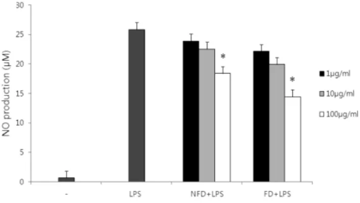
산약 발효물의 추출물이 LPS로 유도된 J774 A.1 세포의 NO 생성에 미치는 영향
LPS로 자극한 J774 A.1 세포에서 산약 미발효물 및 발효물의 메탄올추출물을 농도별로 처리하여 Nitric oxide(NO) 생성에 미
치는 영향을 비교하였다(Fig. 3). 대식세포 활성대조군으로 LPS 를 처리하였으며 24시간 배양 후 25.86 µM의 NO가 합성되었다.
NFD 및 FD 추출물들은 1, 10, 100 µg/ml 농도에서 LPS와 동시 처리한 경우 농도 의존적으로 NO 생성을 억제하였다. 특히 산 약 미발효물 및 발효물의 메탄올추출물을 100 µg/ml 처리한 경 우 LPS에 의해 유도된 NO 생성율을 각각 28.9% 및 44.2% 감 소시켰다. 이상의 결과로 산약발효물 메탄올추출물에서 LPS에 의해 유도된 대식세포 NO 생성에 대한 억제효과가 미발효물에 비해서 1.5배 유의하게 증가하는 것을 확인하였다. NO는 인체에 침입한 병원체나 암세포를 제거하는 면역반응에 중요한 세포독 성물질로 알려져 있다.30,31) NO는 대식세포가 외래 자극에 의해 iNOS 단백질에 의해 합성되며 혈관이완 및 염증반응을 조절하 는 세포간 매개체로 작용하며 조직손상에 관여하고 있다32). 현 재까지 NO 생성을 억제하는 약물의 작용기전은 NO 합성효소 iNOS의 발현이 억제되거나, 세포 밖으로 NO분비를 억제하는 것 과 관련이 있는 것으로 알려져 있다.33-35)최근 보고에서 산약의 에탄올추출물이 LPS로 처리한 대식세포주 RAW264.7에서 NF- κB 신호전달을 차단하여 NO 합성을 억제한다고 보고하였다.36) 따라서 산약 발효물 메탄올추출물의 NO 합성억제 효과는 활 성성분의 변화에 기인한 것으로 사료된다.
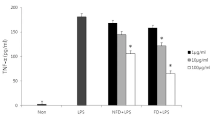
산약 발효물의 추출물이 LPS로 유도된 J774 A.1 세포의 사 이토카인 분비에 미치는 영향
산약 및 발효물 추출물이 LPS에 의해 유도된 J774 A.1 세포 의 염증성 인자 TNF-α 분비에 미치는 영향을 비교하고자 ELISA 법으로 측정하였다(Fig. 4). 세포에 LPS를 처리하고 24시간 배 양 후 181.22 pg/ml의 TNF-α가 분비되었다. 미발효물 메탄올추 출물을 LPS와 동시 처리한 경우 100 µg/ml 농도에서 105.8 pg/
Fig. 2− Cell viability by the fermentaed Dioscoreae batatas methanol extracts in J774 A.1 cells. Cells (1×10⁴cells/well) were cultured for 48 h with various concentrations of the extracts. Cell viability was measured by CCK-8 assay. Data are represented as mean from the triplicate experiments.
Fig. 3− Inhibitory effect of the fermented Dioscoreae batatas methanol extracts on nitric oxide production in LPS- stimulated J774 A.1 cells. Cells (1×10⁵cells/well) were incubated with various concentrations of the extracts in the presence of LPS (0.1µg/ml) for 24 h. NO production from the cells was determined by Griess method. Data are represented as mean from the triplicate experiments (*P<
0.05).
ml TNF-α가 분비되었으며, 발효물 메탄올추출물의 경우 65.2 pg/ml가 분비되었다. 이상의 결과로 산약 미발효물 및 발효물 메 탄올추출물이 LPS 유도 대식세포의 TNF-α 분비를 유의적으로 각각 41.6%, 64.0% 억제함을 확인하였으며, 특히 산약 발효물 메탄올추출물에서 LPS에 의해 유도된 대식세포의 TNF-α 분비 에 대한 억제효과가 미발효물에 비해서 1.5배 증가하는 것을 확 인하였다. TNF-α는 주로 LPS에 의해 자극된 macrophage가 분 비하는 사이토카인으로 알려져 있으며 항암작용 및 염증반응에 매우 중요한 역할을 나타낸다.37)최근 연구에서 Choi 등38)은 Dioscorea daemona 메탄올 추출물이 동물실험모델에서 항염증 및 type VI 알레르기 억제 효과를 나타내었으며 LPS가 유도된 Raw264.7 세포주에서 NO, TNF-α, IL-6 등 염증성 매개물의 억 제를 통하여 항염증 활성을 나타낸다고 보고하였다. 산약에서 항 염작용을 보이는 유효성분으로 Diosgenin39)와 Dioscorealide B40) 등이 알려져 있으며, 이들은 대식세포 활성에 관여하는 NF-κB, AP-1, JNK 등의 신호전달을 차단하여 염증성 사이토카인의 발 현을 억제하여 항염효과를 나타낸다. 따라서 발효과정을 거친 발 효물에서 Diosgenin 함량증가는 산약 메탄올추출물의 대식세포 활성억제능이 미발효 산약분말에 비해 증가하는 근거를 뒷받침 하고 있다. 한편 Rimbach 등41)은 발효한 구아바 잎에서 대식세 포 활성에 대한 억제능이 본 연구 결과와 유사하게 증가함을 보 고하였다. 따라서 발효기술을 활용하여 산약의 염증유발 사이토 카인 및 유발인자의 생성을 억제하는 항염효과를 향상시킬 수 있 을 것으로 사료되며 염증성 질환 개선 및 치료제로써 적용을 위 해 지속적인 연구가 필요할 것이다.
결 론
본 연구에서는 산약 분말을 젖산균 발효하여 메탄올으로 추출
한 시료에 대하여 LPS-유도된 대식세포 활성에 미치는 변화를 산약분말 메탄올추출물과 비교분석하였다. 산약을 L. fermentum 와 L. plantarum 젖산균으로 pH 4.0 이하가 될 때까지 발효시 키면 식품발효의 일반적인 특성인 유기산의 산도가 증가하였고, 산약 고유의 특성인 점액성 물질은 일반 관능검사에서 관찰되지 않았다. 한편 산약을 젖산균으로 일정조건으로 발효한 경우 메 탄올에 의한 추출률이 산약건조분말과 비교해서 3배 이상 증가 하였고, 항염작용이 알려진 steroidal saponin계 화합물 diosgenin 함량이 증가되는 것을 확인하였다. 이와 비례하여 LPS에 의해 유도된 대식세포 NO 생성과 TNF-α 분비량이 산약발효 메탄올 추출물은 농도 의존적으로 억제능이 증가하였다.
결과를 종합하면 산약을 발효함으로써 기능성원료생산이 용이 하도록 물리화학적 특성을 개선할 뿐만 아니라, diosgenin의 함 량을 증가시켜 항염증작용 활성을 증강시킬 수 있을 것으로 사 료된다. 또한 인체 섭취시 스테로이성 사포닌의 체내흡수 및 생 체이용률을 향상시킬 것으로 보이며, 항염작용, 항비만작용, 항 산화효과 등의 약리활성이 증강된 건강기능성 소재개발에 활용 될 수 있을 것으로 사료된다. 하지만 산약이 발효과정을 통하여 새로이 합성되는 화합물에 대한 정보는 전혀 알려진 바가 없으 므로 인체 섭취를 위해서는 향후 동물실험을 통하여 발효산물의 안전성, 생체이용률 및 생리활성에 관한 연구가 필요하다.
감사의 말씀
본 연구는 지식경제부 지역산업계발기술개발사업(과제번호:
70007092)의 지원에 의하여 수행되었으며 이에 감사드립니다.
참고문헌
1) Flora of Korea Editorial Committee : The Genera of Vascular Plants of Korea, Academy Publishing Co., Seoul. Korea. 1336 (2007).
2) 생약학교재편찬위원회 : 생약학. 동명사. 서울. 250 (2007).
3) Jang, S. M., Noh, S. H. and Park, S. D. : Botany of Herbal Medicine Resources. Hakmun Publishing Ltd. Seoul. Korea.
299 (1999).
4) Yang, M. H., Yoon, K. D., Chin, Y. W. and Kim, J. W. : Phytochemical and pharmacological profiles of Dioscorea species in Korea, China and Japan. Kor. J. Pharmacogn. 40, 257 (2009).
5) Sautour, M., Mitaine-Offer, A. C., Miyamoto, T., Wagner, H. and Lacaille-Dubois, M. A. : A new phenanthrene glycoside and other constituents from Dioscorea opposita. Chem. Pharm.
Bull. 52, 1235 (2004).
6) 하영득, 이삼빈, 곽연길 : 마 점질물의 중금속제거능과 ACE저해 효과. 한국식품영양과학회지 27, 751 (1998).
Fig. 4− Inhibitory effect of the fermented Dioscoreae batatas methanol extracts on TNF-α production in LPS-stimulated J774 A.1 cells. Cells (1×10⁵cells/well) were incubated with various concentrations of the extracts in the presence of LPS (0.1µg/ml) for 24 h. TNF-α production from the cells was determined by ELISA. Data are represented as mean from the triplicate experiments (*P<0.05).
7) Chung, Y. C., Chiang, B. H., Wei, J. H., Wang, C. K., Chen, P. C. and Hsu, C. K. : Effects of blanching, drying and extraction processes on the antioxidant activity of yam (Dioscorea alata). Int. J. Food Sci. Tech. 43, 859 (2008).
8) Jeon, J. R., Lee, J. S., Lee, C. H., Kim, J. Y., Kim, S. D. and Nam, D. H. : Effect of ethanol extract of dried Chinese yam (Dioscorea batatas) flour containing dioscin on gastrointestinal function in rat model. Arch. Pharm. Res. 29, 348 (2006).
9) Wang, C. H., Tsai, C. H., Lin, H. J., Wang, T. C. and Chen, H. L. : Uncooked Taiwanese yam (Dioscorea alata L. cv.
Tainung No.2) beneficially modulated the large bowel function and faecal microflora in Balb/c mice. J. Sci. Food Agr. 87, 1378 (2007).
10) Hikino, H., Konno, C., Takahashi, M., Murakami, M., Kato, Y., Karikura, M. and Hayashi, T. : Isolation and hypoglycemic activity of dioscorans A, B, C, D, E, and F; Glycans of dioscorea japonica rhizophors1. Planta Med. 52, 168 (1986).
11) Chen, H., Wang, C., Chang, C. T. and Wang, T. : Effects of Taiwanese yam (Dioscorea japonica Thunb var. pseudojaponica Yamamoto) on upper gut function and lipid metabolism in Balb/
c mice. Nutrition. 19, 646 (2003).
12) Lin, C. L., Lin, S. Y., Lin, Y. H. and Hou, W. C. : Effects of tuber storage protein of yam (Dioscorea alata cv. Tainong No.1) and its peptic hydrolyzates on spontaneously hypertensive rats. J.
Sci. Food Agr. 86, 1489 (2006).
13) Yang, M. H., Yoon, K. D., Chin, Y. W., Park, J. H., Kim, S. H., Kim, Y. C. and Kim, J. : Neuroprotective effects of Dioscorea opposita on scopolamine-induced memory impairment in vivo behavioral tests and in vitro assays. J. Ethnopharmacol. 121, 130 (2009).
14) Kwon, C. S., Sohn, H. Y., Kim, S. H., Kim, J. H., Son, K. H., Lee, J. S., Lim, J. K. and Kim, J. S. : Anti-obesity effect of Dioscorea nipponica Makino with lipase-inhibitory activity in rodents. Biosci. Biotechnol. Biochem. 67, 1451 (2003).
15) Roitt, I., Brostoff, J. and Male, D. : Immunology. 3rd Ed. Mosby, New York (1993).
16) Bosca, L., Zeini, M., Traves, P. G. and Hortelano, S. : Nitric oxide and cell viability in inflammatory cells : a role for NO in macrophage function and fate. Toxicology 208, 249 (2005).
17) Turini, M. E. and DuBois, R. N. : Cyclooxygenase-2: a therapeutic target. Annu. Rev. Med. 53, 35 (2002).
18) Jin, M., Suh, S. J., Yang, J. H., Lu, Y., Kim, S. J., Kwon, S., Jo, T. H., Kim, J. W., Park, Y. I., Ahn, G. W., Lee, C. K., Kim, C. H., Son, J. K., Son, K. H. and Chang, H. W. : Anti- inflammatory activity of bark of Dioscorea batatas DECNE through the inhibition of iNOS and COX-2 expressions in RAW264.7 cells via NF-κB and ERK1/2 inactivation. Food Chem. Toxicol. 48, 3073 (2010).
19) Jung, D. H., Park, H. J., Byun, H. E., Park, Y. M., Kim, T. W.,
Kim, B. O., Um, S. H. and Pyo, S. : Diosgenin inhibits macrophage-derived inflammatory mediators through down- regulation of CK2, JNK, NF-κB and AP-1 activation. Int.
Immunopharmacol. 10, 1047 (2010).
20) Fuller, R. : Probiotics the scientific Basis. Chapman and Hall.
London (1992).
21) Kim, D. H., Han, S. B., Park, J. S. and Han, M. J. : Fermentation of antler and its biological activity. Kor. J.
Pharmacogn. 25, 233 (1994).
22) Chi, H. and Ji, G. E. : Transformation of ginsenosides Rb1 and Re from panax ginseng by food microorganisms. Biotechnol.
Lett. 27, 765 (2005).
23) Choi, Y. M., Kim, Y. S., Ra, K. S. and Suh, H. J. : Characteristics of fermentation and bioavailability of isoflavones in Korean soybean paste (doenjang) with application of Bacillus sp. KH- 15. Int. J. Food Sci. Technol. 42, 1497 (2007).
24) Cabras, P. and Angioni, A. : Pesticide residues in grapes, wine, and their processing products. J. Agric. Food Chem. 48, 967 (2000).
25) Isolauri, E., Salminen, S. and Ouwehand, A. C. : Probiotics.
Best Prac. Res. Cl. Ga. 18, 299 (2004).
26) Kim, J. S. and Byun, G. I. : A study on the consumers recognition, preference and use of yam and yam products focused on consumers in daegu area and andon area. Kor. J.
Culinary Research 14, 441 (2008).
27) Tal, B., Tamir, I., Rokem, J. S. and Goldberg, I. : Isolation and characterization of an intermediate steroid metabolite in diosgenin biosynthesis in suspension cultures of Dioscorea deltoidea cells. Biochem. J. 219, 619 (1984).
28) Wang, L. J., Wang, Y., Chen, S. W., Ma, J. S., Fu, Q. and Wang, B. X. : The antitumor activity of Diosgenin in vivo and in vitro.
Zhongguo Zhong Yao Za Zhi. 27, 777 (2002).
29) Huang, C. H., Liu, D. Z. and Jan, T. R. : Diosgenin, a plant- derived sapogenin, enhances regulatory T-cell immunity in the intestine of mice with food allergy. J. Nat. Prod. 73, 1033 (2010).
30) Stuehr, D. J. and Nathan, C. F. : Nitric oxide. A macrophage product responsible for cytostasis and respiratory inhibition in tumor target cells. J. Exp. Med. 169, 1543 (1989).
31) Hibbs, J. B., Jr, Taintor, R. R. and Vavrin, Z. : Macrophage cytotoxicity: role for L-arginine deiminase and imino nitrogen oxidation to nitrite. Science 23, 473 (1987).
32) Bogdan, C. : Nitric oxide and the immune response. Nat.
Immun. 2, 907 (2001).
33) Cao, H., Urban, J. F. and Jr, Anderson, R. A. : Cinnamon polyphenol extract affects immune responses by regulating anti- and pro inflammatory and glucose transporter gene expression in mouse macrophages. J. Nutr. 138, 833 (2008).
34) Tao, J. Y., Zhao, L., Huang, Z. J., Zhang, X. Y., Zhang, S. L.,
Zhang, Q. G., Fei-Xiao, Zhang, B. H., Feng, Q. L. and Zheng, G. H. : Anti-inflammatory effects of ethanol extract from Kummerowia striata (Thunb.) Schindl on lps-stimulated RAW 264.7 cell. Inflammation. 31, 154 (2008).
35) Zhao, L., Zhang, S. L., Tao, J. Y., Jin, F., Pang, R., Guo, Y. J., Ye, P., Dong, J. H. and Zheng, G. H. : Anti-inflammatory mechanism of a folk herbal medicine, Duchesnea indica (Andr) Focke at RAW264.7 cell line. Immunol. Invest. 37, 339 (2008).
36) Jin, M., Suh, S. J., Yang, J. H., Lu, Y., Kim, S. J., Kwon, S., Jo, T. H., Kim, J. W., Park, Y. I., Ahn, G. W., Lee, C. K., Kim, C. H., Son, J. K., Son, K. H. and Chang, H. W. : Anti- inflammatory activity of bark of Dioscorea batatas DECNE through the inhibition of iNOS and COX-2 expressions in RAW264.7 cells via NF-κB and ERK1/2 inactivation. Food Chem. Toxicol. 48, 3073 (2010).
37) Strieter, R. M., Kunkel, S. L. and Bone, R. C. : Role of tumor necrosis factor-α in disease states and inflammation. Crit. Care Med. 21, S447 (1993).
38) Choi, E. M. and Koo, S. J. : Effects of dioscorea daemona roxb.
stem extract on the inflammatory responses, antioxidant
system and lipid levels in vivo and the production of inflammatory mediators in RAW264.7 cells. J. East. Asian Soc.
Dietary Life 15, 707 (2005).
39) Jung, D. H., Park, H. J., Byun, H. E., Park, Y. M., Kim, T. W., Kim, B. O., Um, S. H. and Pyo, S. : Diosgenin inhibits macrophage-derived inflammatory mediators through down- regulation of CK2, JNK, NF-kappaB and AP-1 activation. Int.
Immunopharmacol. 10, 1047 (2010).
40) Hiransai, P., Ratanachaiyavong, S., Itharat, A., Graidist, P., Ruengrairatanaroj, P. and Purintrapiban, J. : Dioscorealide B suppresses LPS-induced nitric oxide production and inflammatory cytokine expression in RAW 264.7 macrophages:
The inhibition of NF-kappaB and ERK1/2 activation. J. Cell.
Biochem. 109, 1057 (2010).
41) Rimbach, G., Park, Y. C., Guo, Q., Moini, H., Qureshi, N., Saliou, C., Takayama, K., Virgili, F. and Packer, L. : Nitric oxide synthesis and TNF-alpha secretion in RAW 264.7 macrophages:
mode of action of a fermented papaya preparation. Life Sci. 67, 679 (2000).