A Prospective Evaluation of Adult Men with Iron-deficiency Anemia in Korea
Gak Won Yun1, Young Joon Yang2, Ik Chan Song1, Keon Uk Park3, Seung-Woo Baek1, Hwan Jung Yun1, Samyong Kim1, Deog Yeon Jo1and Hyo Jin Lee1
Abstract
Objective Iron-deficiency anemia (IDA) is the most common nutritional deficiency worldwide. However, the information concerning various causes of IDA in adult men is still insufficient. The aim of our study was to evaluate adult men with IDA.
Methods We prospectively studied 206 adult men with IDA. All subjects had a direct history taken and un- derwent a physical examination. Esophagogastroduodenoscopy was performed in most patients, and colono- scopy was conducted if no lesion causing IDA was found or the fecal occult blood test was positive.
Results The history of prior gastrectomy and blood-letting cupping therapy that probably had caused IDA were reported in 24 (11.7%) and 11 (5.3%) patients, respectively. In terms of potential causes of IDA, 68 (33.0%) patients were found to have upper gastrointestinal disorders (34 peptic ulcers, 17 erosive gastritis, 16 gastric cancers, and one gastrointestinal stromal tumor). Colonoscopy showed 42 (20.4%) clinically relevant lesions that probably caused IDA: colon cancer (five patients), colon polyps (14 patients), ulcerative colitis (one patient), and hemorrhoids (22 patients). One small bowel tumor was detected at small bowel series.
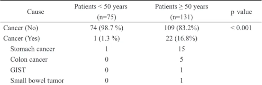
Concerning malignant lesions that were responsible for IDA, 22 malignant lesions were found in patients of 50 years or older, accounting for 16.8% (22 of 131 patients), while only one (1.3%) early gastric cancer was found in the younger patients.
Conclusion This study demonstrated that gastrointestinal blood loss is the main cause of IDA in adult men, and that there is a high rate of malignancy in men older than 50 years, emphasizing the need for a complete, rigorous gastrointestinal examination in this group of patients. Considering blood-letting cupping therapy, there is a need to consider culture-specific procedures as a possible cause of IDA.
Key words: iron deficiency anemia, adult men, gastrointestinal bleeding
(Intern Med 50: 1371-1375, 2011) (DOI: 10.2169/internalmedicine.50.5289)
Introduction
Iron deficiency is one of the most common disorders af- fecting humans, and it continues to be a major public health problem worldwide. There is an estimated 3.5 billion iron- deficient people in the world, with the majority in develop- ing countries (1-3). The estimated prevalence of iron- deficiency anemia in adults ranges from 2 to 5%, although it varies widely depending on the population investi-
gated (4, 5). It is believed that iron-deficiency anemia gener- ally results from chronic blood loss from the gastrointestinal tract or uterus and it is frequently a sign of serious underly- ing disease (6). Indeed, asymptomatic colonic and gastric cancer may present with iron-deficiency anemia and exclu- sion of these conditions is of great importance, whereas malabsorption, frequently due to celiac disease or gastrec- tomy, poor dietary intake, and non-steroidal anti- inflammatory drugs are other common possible causes (7-9).
Consequently, appropriate management requires not only the
1Department of Internal Medicine, Chungnam National University School of Medicine, Korea,2Department of Internal Medicine, Daejeon St.
Mary’s Hospital, Korea and3Department of Internal Medicine, Dongsan Medical Center, Keimyung University School of Medicine, Korea Received for publication February 8, 2011; Accepted for publication March 23, 2011
Correspondence to Dr. Hyo Jin Lee, [email protected] G.W.Y amd Y.J.Y equally contributed to this work
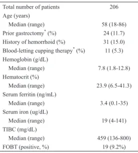
Table 1. Demographic Characteristics and Laboratory Data of the Patients with Iron De- ficiency Anemia
Total number of patients Age (years)
Median (range) Prior gastrectomy* (%) History of hemorrhoid (%) Blood-letting cupping therapy* (%) Hemoglobin (g/dL)
Median (range) Hematocrit (%) Median (range) Serum ferritin (ng/mL) Median (range) Serum iron (ug/dL) Median (range) TIBC (mg/dL) Median (range) FOBT (positive, %)
206
58 (18-86) 24 (11.7) 31 (15.0) 11 (5.3)
7.8 (1.8-12.8)
23.9 (6.5-41.3)
3.4 (0.1-35)
19 (4-141)
459 (136-800) 19 (9.2%) FOBT, fecal occult blood test.
*, that probably had caused iron deficiency anemia.
supplementation of adequate iron to replenish iron stores, but also meticulous investigations to identify the underlying cause in patients who are fit and willing to undergo further tests (6). Nevertheless, there is little information on the vari- ous causes of iron-deficiency anemia in adult men. This study prospectively evaluated adult men with iron-deficiency anemia.
Patients and Methods
Patients
Patients were referred from primary care and internal medicine clinics to the hematology section for the evaluation of anemia. The prospective study was conducted at Chung- nam National University Hospital from January 2003 to De- cember 2010.
Enrollment criteria included men !18 years old with iron-deficiency anemia giving informed written consent. The protocol was approved by the institutional review board.
Anemia was defined as hemoglobin (Hb) <13 g/dL, using the World Health Organization criteria. Iron-deficiency ane- mia was considered to be present if the serum ferritin level was ≦15 ng/mL. In patients with iron-deficiency anemia plus inflammatory conditions, a serum ferritin <50 ng/mL associated with a red blood cell (RBC) mean cell volume (MCV) of 80 fL or less with a transferrin saturation <10%
was considered diagnostic of iron-deficiency anemia.
Evaluations
All subjects had an independent, direct history taken and underwent a physical examination. The history inquiry was regarding symptoms, such as fatigue, dyspnea on exertion,
and dizziness. A history of upper gastrointestinal tract symp- toms, including dyspepsia, nausea, vomiting, early satiety, upper abdominal pain, dysphagia, heart burn, and lower gas- trointestinal symptoms, including changes in bowel habits, constipation, diarrhea, bright red blood per rectum, and lower abdominal pain, was also obtained. All patients were also asked about blood-letting cupping therapy, aspirin use, and prior gastrointestinal disorders. All patients underwent a complete physical examination, including looking for traces of blood-letting cupping therapy and a fecal occult blood test (FOBT) of three spontaneously passed stools. All pa- tients had laboratory analysis consisting of a complete blood count, serum iron and total iron-binding capacity, and a se- rum ferritin level. Most patients underwent esophagogastro- duodenoscopy. Colonoscopy was performed if no lesion causing iron-deficiency anemia was found or the FOBT was positive. As an additional test, abdominal computed to- mography (CT) or a small bowel series was performed at the clinician’s discretion. Endoscopic procedures were per- formed by experienced gastroenterologists with standard hemodynamic monitoring. Clinically relevant lesions of chronic gastrointestinal blood loss as a cause of iron- deficiency anemia included cancer, adenomatous polyps
!1.5 cm, vascular ectasia that numbered five or more or were at least 8 mm in diameter, severe erosive esophagitis or gastritis, single gastric or duodenal ulcer >1 cm in diame- ter, multiple gastric or duodenal ulcers >0.5 cm, hemor- rhoids with bleeding or a history of recurrent bleeding, ac- tive colitis, or inflammatory bowel disease. Diverticula and esophageal varices were not considered to be sources of chronic gastrointestinal blood loss based on the study of Rockey and Cello (15).
Statistical analysis
The categorical variables were analyzed using Fisher’s ex- act test. p values <0.05 were considered to indicate statisti- cal significance. All statistical analyses were conducted us- ing the SPSS 13.0 software (SPSS, Chicago, IL).
Results
We enrolled 206 patients who met the inclusion criteria.
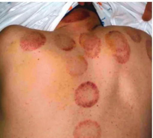
The median age was 58 (range 18-86) years. There were symptoms such as fatigue, dyspnea on exertion, dizziness, or digestive complaints in 178 of the 206 (86.4%) men with iron-deficiency anemia. Demographic and laboratory pa- rameters of the patients are provided in Table 1. Twenty-one patients used aspirin or another non-steroidal anti- inflammatory drug regularly. A history of gastrectomy and hemorrhoids was reported in 24 and 31 patients, respec- tively. Eleven patients reported a history of repeated blood- letting cupping therapy, and typical signs were seen in some patients (Fig. 1). FOBT was positive in 9.2% of the sub- jects.
Table 2 summarizes the esophagogastroduodenoscopy (EGD) findings for the 184 (89.3%) patients who underwent
Figure 1. Characteristic traces of cupping performed on the neck and back.
EGD. The most common findings at EGD were gastritis (34 erosive gastritis and 18 chronic atrophic gastritis) and peptic ulcer (22 gastric ulcers and 13 duodenal ulcers). In terms of potential causes of iron-deficiency anemia, 68 patients were found to have upper gastrointestinal disorders (22 gastric ul- cers, 17 erosive gastritis, 16 gastric cancers, 12 duodenal ul- cers, and one gastrointestinal stromal tumor). In total, 104 (50.5%) patients underwent colonoscopy, which showed 42 clinically relevant lesions that probably caused iron- deficiency anemia: colon cancer (five patients), colon polyps (14 patients), ulcerative colitis (one patient), and hemor- rhoids (22 patients; Table 3). A small bowel series and ab- dominal CT were conducted in 20 and 18 patients, respec- tively. Only one small bowel tumor was detected.
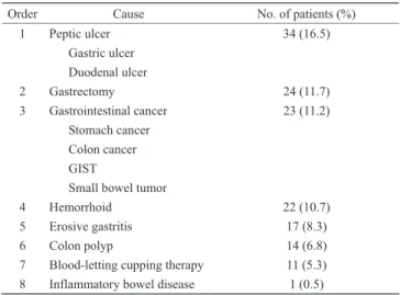
In terms of the cause of iron-deficiency anemia in this population, peptic ulcer was the most common cause of iron-deficiency anemia in adult men, followed by status of gastrectomy, gastrointestinal cancer, hemorrhoid, erosive gastritis, colon polyp, and blood-letting cupping therapy, in decreasing order (Table 4). Concerning malignant lesions that were responsible for iron-deficiency anemia, 23 (11.2%) malignant lesions were found in the population. When we separated the population into two groups using age 50 years as the cut-off, 22 malignant lesions were found in patients aged 50 years or older, accounting for 16.8% (22 of 131 pa- tients), while only one (1.3%) early gastric cancer was found in the younger patients (Table 5).
Discussion
In this prospective study, we found that chronic blood loss from the gastrointestinal tract was the main cause of iron-deficiency anemia in adult men (about 54%; 34 patients with peptic ulcer, 23 with gastrointestinal cancer, 22 with hemorrhoids, 17 with erosive gastritis, 14 with colon polyps, and one with ulcerative colitis; Table 4), and there was a higher rate of malignancy in men !50 years than in younger men (16.8% vs. 1.3%; p<0.001; Table 5), empha- sizing the importance of a complete, rigorous gastrointesti- nal examination in this group of patients. It is necessary to
consider culture-specific procedures, such as blood-letting cupping therapy, and racial or geographical differences, such as the relatively high frequency of stomach cancer in Korea versus colon cancer in Western populations, when investigat- ing possible causes of iron-deficiency anemia.
Although iron-deficiency anemia is a very common clini- cal problem, most studies have examined small, heteroge- nous cohorts of patients with varying demographic and ane- mia parameters (10). The present study focused on men
!18 years old to investigate the various causes of iron- deficiency anemia in a prospective fashion. More upper gas- trointestinal tract lesions were detected than lower upper gastrointestinal tract lesions, similar to previous re- ports (11-18). The most common lesion causing iron- deficiency anemia was a peptic ulcer (34 of 206 patients; 22 gastric ulcers and 12 duodenal ulcers, comparable with other reports) (13-15). Prior gastrectomy, gastrointestinal cancer, hemorrhoids, erosive gastritis, colon polyps, and blood- letting cupping therapy were also causes of iron-deficiency anemia, in decreasing order. Regarding cancer, 23 cases of cancer (11.2%) were detected in our population, similar to previous reports of 7-28%; however, in our series, stomach cancer (16 cases) was more frequent than colon cancer (5 cases) compared with Western studies (12, 15, 16, 18). We suggest that the difference is due to the different incidence and prevalence of each cancer type in different regions: i.e., stomach cancer is more common in East Asia, especially in Korea and Japan, suggesting special consideration of geo- graphic or racial differences when we approach the patient with iron-deficiency anemia. Additionally, when we sepa- rated the patients according to age, 22 malignant lesions were found in those !50 years, accounting for 16.8% (22 of 131 patients), while only one younger patient was found to have early gastric cancer. These results suggest that a thorough, rigorous investigation of gastrointestinal cancer is needed in iron-deficiency anemia patients older than 50 years, consistent with other reports (10, 19).
Regarding hemorrhoids, Nikpour et al (20) reported that hemorrhoids are the most common (54.2% of the popula- tion) colonoscopic lesion in patients with minimal rectal bleeding. Park et al (21) also demonstrated that hemorrhoids with or without bleeding constitute the most prevalent (18.2% of the patients) lower gastrointestinal lesion in as- ymptomatic premenopausal women with iron-deficiency ane- mia. Additionally, hemorrhoids with bleeding as a cause of chronic blood loss were reported in about 6% of patients with iron-deficiency anemia (17, 22). Kluiber and Wolff (23) studied the incidence of hemorrhoidal bleeding that caused anemia in a stable population of patients seen at the Mayo Clinic and Olmsted Community Hospital in Roch- ester, Minnesota. Furthermore, Ibrahim et al (24) reported that hemorrhoids can cause substantial bleeding over long periods of time, suggesting that hemorrhoidal bleeding should be considered in the differential diagnosis of obscure gastrointestinal bleeding. In the present study, hemorrhoids were detected in 10.7% of the patients, reemphasizing it as
Table 2. Esophagogastroduodenoscopic Finding (n=184)
Finding No. of patients No. of potential cause of IDA Reflux esophagitis
Esophageal varix Chronic atrophic gastritis Erosive gastritis Gastric ulcer Angiodysplasia Gastric polyp Gastric cancer GIST Duodenal ulcer
16 (8.7 %) 7 (3.8 %) 18 (9.8 %) 34 (18.5 %) 22 (12.0 %) 3 (1.6 %) 7 (3.8 %) 16 (8.7 %)
1 (0.5%) 13 (7.1 %)
17 (9.2 %) 22 (12.0 %)
16 (8.7 %) 1 (0.5 %) 12 (6.5 %)
Severe erosive gastritis is considered a clinically relevant lesion of chronic gastrointestinal blood loss as a cause of iron-deficiency anemia. IDA, iron deficiency anemia; GIST, gastrointestinal stromal tumor
Table 3. Colonoscopic Finding (n=104)
Finding No. of patients No. of potential cause of IDA Colon polyp
Angiodysplasia Non-specific colitis Diverticulosis Colon cancer Hemorrhoid IBD
23 (22.1 %) 1 (1.0 %) 3 (2.9 %) 2 (1.9 %) 5 (4.8 %) 24 (23.1 %)
1 (1.0 %)
14 (13.5 %)
5 (4.8 %) 22 (21.2 %)
1 (1.0 %) IDA, iron deficiency anemia; IBD, inflammatory bowel disease
Table 4. Potential Cause of Iron Deficiency Anemia in Adult Men (n=206)
Order Cause No. of patients (%)
1
2 3
4 5 6 7 8
Peptic ulcer Gastric ulcer Duodenal ulcer Gastrectomy Gastrointestinal cancer
Stomach cancer Colon cancer GIST
Small bowel tumor Hemorrhoid
Erosive gastritis Colon polyp
Blood-letting cupping therapy Inflammatory bowel disease
34 (16.5)
24 (11.7) 23 (11.2)
22 (10.7) 17 (8.3) 14 (6.8) 11 (5.3) 1 (0.5)
a potential cause of iron-deficiency anemia.
Blood-letting cupping therapy, which was detected in 5.3% of the patients, is often performed at acupuncture points, and always results in blood loss, in Eastern medi- cine. Thus, this therapy is capable of inducing iron- deficiency anemia if used over a prolonged period (25-27).
For example, Lee et al (27) reported iron-deficiency anemia and Sohn et al (28) anemia-related cardiomyopathy induced by blood-letting cupping therapy, suggesting the need to consider culture-specific practices as possible causes of iron-
deficiency anemia.
In the present study, the cause of iron-deficiency anemia remained unidentified in 29% of the study population. This rate of negative work-ups is similar to previous studies, al- though each study had different definitions of clinically rele- vant lesion as a cause of iron-deficiency anemia and was conducted in a heterogenous cohort of the subjects with varying clinical parameters (11-18). For example, in our study, diverticula and esophageal varices were not consid- ered to be sources of chronic gastrointestinal blood loss ac- cording to our predefined criteria which were based on the study of Rockey and Cello (15), although those lesions could cause chronic gastrointestinal blood loss. Additionally, those studies did not use small bowel capsule endoscopy or double-balloon enteroscopy, techniques that have revolution- ized the management of patients with obscure gastrointesti- nal bleeding. The detection of lesions such as small intesti- nal angioectasia has become possible with the advent of capsule endoscopy, and such lesions can be treated with double-balloon enteroscopy. The use of these techniques will probably decrease the unresolved cases of iron-deficiency anemia in the future (29, 30). Considering the importance of history taking, in addition, we overlooked other possible causes of iron deficiency anemia such as a vegetarian diet and regular blood donation which could cause chronic blood loss (31).
In conclusion, this study demonstrated that gastrointestinal blood loss is the main cause of iron-deficiency anemia in
Table 5. Malignant Cause of IDA in Adult Men According to Age
Cause Patients < 50 years (n=75)
Patients 50 years
(n=131) p value
Cancer (No) Cancer (Yes)
Stomach cancer Colon cancer GIST
Small bowel tumor
74 (98.7 %) 1 (1.3 %)
1 0 0 0
109 (83.2%) 22 (16.8%)
15 5 1 1
< 0.001
IDA, iron deficiency anemia; GIST, gastrointestinal stromal tumor
adult men, and that there is a high rate of malignancy in men older than 50 years, emphasizing the need for a com- plete, rigorous gastrointestinal examination in this group of patients. Considering blood-letting cupping therapy, there is a need to consider culture-specific procedures as a possible cause of iron-deficiency anemia of unknown cause.
The authors state that they have no Conflict of Interest (COI).
References
1. Alleyne M, Horne MK, Miller JL. Individualized treatment for iron-deficiency anemia in adults. Am J Med 121: 943-948, 2008.
2. Patel KV. Epidemiology of anemia in older adults. Semin Hematol 45: 210-217, 2000.
3. Killip S, Bennett JM, Chambers MD. Iron deficiency anemia. Am Fam Physician 75: 671-678, 2007.
4. Patterson RN, Johnston SD. Iron deficiency anaemia: are the Brit- ish Society of Gastroenterology guidelines being adhered to? Post- grad Med J 79: 226-228, 2003.
5. Sayer JM, Long RG. A perspective on iron deficiency anaemia.
Gut 34: 1297-1299, 1993.
6. Logan EC, Yates JM, Stewart RM, Fielding K, Kendrick D. Inves- tigation and management of iron deficiency anaemia in general practice: a cluster randomised controlled trial of a simple manage- ment prompt. Postgrad Med J 78: 533-537, 2002.
7. Goddard AF, McIntyre AS, Scott BB. Guidelines for the manage- ment of iron deficiency anaemia. British Society of Gastroenterol- ogy. Gut 46 (suppl 3-4): IV1-IV5, 2000.
8. Rasul I, Kandel GP. An approach to iron-deficiency anemia. Can J Gastroenterol 15: 739-747, 2001.
9. Pasricha SR, Flecknoe-Brown SC, Allen KJ, et al. Diagnosis and management of iron deficiency anaemia: a clinical update. Med J Aust 193: 525-532, 2010.
10. Niv E, Elis A, Zissin R, Naftali T, Novis B, Lishner M. Iron defi- ciency anemia in patients without gastrointestinal symptoms―a prospective study. Fam Pract 22: 58-61, 2005.
11. Cook IJ, Pavli P, Riley JW, Goulston KJ, Dent OF. Gastrointesti- nal investigation of iron deficiency anaemia. Br Med J (Clin Res Ed) 292: 1380-1382, 1986.
12. Zuckerman G, Benitez J. A prospective study of bidirectional en- doscopy (colonoscopy and upper endoscopy) in the evaluation of patients with occult gastrointestinal bleeding. Am J Gastroenterol 87: 62-66, 1992.
13. Hsia PC, al-Kawas FH. Yield of upper endoscopy in the evalu- ation of asymptomatic patients with Hemoccult-positive stool after a negative colonoscopy. Am J Gastroenterol 87: 1571-1574, 1992.
14. McIntyre AS, Long RG. Prospective survey of investigations in outpatients referred with iron deficiency anaemia. Gut 34: 1102- 1107, 1993.
15. Rockey DC, Cello JP. Evaluation of the gastrointestinal tract in patients with iron-deficiency anemia. N Engl J Med 329: 1691- 1695, 1993.
16. Gordon SR, Smith RE, Power GC. The role of endoscopy in the evaluation of iron deficiency anemia in patients over the age of 50. Am J Gastroenterol 89: 1963-1967, 1994.
17. Kepczyk T, Kadakia SC. Prospective evaluation of gastrointestinal tract in patients with iron-deficiency anemia. Dig Dis Sci 40:
1283-1289, 1995.
18. Reyes López A, Gómez Camacho F, Gálvez Calderón C, Miño Fugarolas G. Iron-deficiency anemia due to chronic gastrointesti- nal bleeding. Rev Esp Enferm Dig 91: 345-358, 1999 (in Span- ish).
19. Ho CH, Chau WK, Hsu HC, Gau JP, You JY, Chen CC. Predictive risk factors and prevalence of malignancy in patients with iron de- ficiency anemia in Taiwan. Am J Hematol 78: 108-112, 2005.
20. Nikpour S, Ali Asgari A. Colonoscopic evaluation of minimal rec- tal bleeding in average-risk patients for colorectal cancer. World J Gastroenterol 14: 6536-6540, 2008.
21. Park DI, Ryu SH, Oh SJ, et al. Significance of endoscopy in as- ymptomatic premenopausal women with iron deficiency anemia.
Dig Dis Sci 51: 2372-2376, 2006.
22. Park JS, Park DI, Park SK, et al. Endoscopic evaluation of signifi- cant gastrointestinal lesions in patients with iron deficiency with and without anaemia: a Korean Association for the Study of Intes- tinal Disease Study. Intern Med J 39: 441-446, 2009.
23. Kluiber RM, Wolff BG. Evaluation of anemia caused by hemor- rhoidal bleeding. Dis Colon Rectum 37: 1006-1007, 1994.
24. Ibrahim AM, Hackford AW, Lee YM, Cave DR. Hemorrhoids can be a source of obscure gastrointestinal bleeding that requires transfusion: report of five patients. Dis Colon Rectum 51: 1292- 1294, 2008.
25. Tham LM, Lee HP, Lu C. Cupping: from a biomechanical per- spective. J Biomech 39: 2183-2193, 2006.
26. Iblher N, Stark B. Cupping treatment and associated burn risk: a plastic surgeon’s perspective. J Burn Care Res 28: 355-358, 2007.
27. Lee HJ, Park NH, Yun HJ, Kim S, Jo DY. Cupping therapy- induced iron deficiency anemia in a healthy man. Am J Med 121:
e5-e6, 2008.
28. Sohn IS, Jin ES, Cho JM, et al. Bloodletting-induced cardio- myopathy: reversible cardiac hypertrophy in severe chronic anae- mia from long-term bloodletting with cupping. Eur J Echocardiogr 9: 585-586, 2008.
29. Rockey DC. Occult and obscure gastrointestinal bleeding: causes and clinical management. Nat Rev Gastroenterol Hepatol 7: 265- 279, 2010.
30. Raju GS, Gerson L, Das A, Lewis B; American Gastroenterologi- cal Association. American Gastroenterological Association (AGA) Institute technical review on obscure gastrointestinal bleeding.
Gastroenterol 133: 1697-1717, 2007.
31. Cook JD. Iron-deficiency anaemia. Baillieres Clin Haematol 7:
787-804, 1994.
Ⓒ 2011 The Japanese Society of Internal Medicine http://www.naika.or.jp/imindex.html