Protein 2 Overexpression on Endothelial Function and Apoptosis
Ki-Up Lee,* In Kyu Lee,* Jin Han, Dae-Kyu Song, Yun Mi Kim, Hai Sun Song, Hyoun Sik Kim, Woo Je Lee, Eun Hee Koh, Kee-Ho Song, Sung Min Han, Min Seon Kim,
In-Sun Park, Joong-Yeol Park
Abstract—Increased oxidative stress in vascular cells plays a key role in the development of endothelial dysfunction and atherosclerosis. Uncoupling protein 2 (UCP2) is an important regulator of intracellular reactive oxygen species (ROS) production. This study was undertaken to test the hypothesis that, UCP2 functions as an inhibitor of the atherosclerotic process in endothelial cells. Adenovirus-mediated UCP2 (Ad-UCP2) overexpression led to a significant increase in endothelial nitric oxide synthase (eNOS) and decrease in endothelin-1 mRNA expression in human aortic endothelial cells (HAECs). Moreover, UCP2 inhibited the increase in ROS production and NF- B activation, and apoptosis of HAECs induced by lysophophatidylcholine (LPC) and linoleic acid. LPC and linoleic acid caused mitochondrial calcium accumulation and transient mitochondrial membrane hyperpolarization, which was followed by depolarization.
UCP2 overexpression prevented these processes. In isolated rat aorta, Ad-UCP2 infection markedly improved impaired vascular relaxation induced by LPC. The data collectively suggest that UCP2, functions as a physiologic regulator of ROS generation in endothelial cells. Thus, measures to increase UCP2 expression in vascular endothelial cells may aid in preventing the development and progression of atherosclerosis in patients with metabolic syndrome. (Circ Res. 2005;
96:1200-1207.)
Key Words: endothelial cells 䡲 uncoupling protein 䡲 oxidative stress 䡲 vascular endothelial function 䡲 apoptosis
M etabolic syndrome is a cluster of simultaneously oc- curring vascular risk factors, such as central obesity, hypertension, dyslipidemia, and glucose intolerance.1 Im- paired endothelium-dependent vascular relaxation (endothe- lial dysfunction), generally considered a prerequisite for atherosclerosis, is a frequent finding in metabolic syndrome.
2
Subjects with metabolic syndrome frequently display ele- vated plasma concentrations of free fatty acid (FFA) and oxidized low-density lipoprotein (LDL).
1,3–5Lipid emulsion and oxidized LDL induce endothelial dysfunction similar to what is observed in metabolic syndrome.
6,7 It is therefore suggested that endothelial dysfunction in metabolic syndrome is mediated, at least in part, by elevated circulating FFA and oxidized LDL levels.
Endothelium is important in the regulation of smooth muscle cell growth, migration, and proliferation. In this regard, endothelial apoptosis is an important early event in the pathogenesis of atherosclerosis.
8Endothelium also mod- ulates vascular tone through the release of relaxing or
contracting substances, including nitric oxide (NO), prosta- cyclin, endothelium-derived hyperpolarizing factor (EDHF), and endothelin-1 (ET-1). Several lines of evidence have suggested that decreased availability of NO or increased availability of ET-1 in the vasculature are central to the pathogenesis of impaired vascular relaxation observed in the early stages of atherosclerosis.
9 –11Increased oxidative stress in vascular cells is a key mech- anism of endothelial dysfunction and atherosclerosis.
12Var- ious risk factors for atherosclerosis generate intracellular oxidative stress. A relatively high level of oxidative stress, in turn, induces vascular inflammatory “atherogenic” genes via redox-sensitive signaling pathways, and activates redox- sensitive transcription factors.
12Mitochondrial uncoupling protein (UCP)2 is a novel member of the mitochondrial anion carrier family, and displays 60% sequence identity with the well-known thermogenic UCP1 from brown adipose tissue.
13Previous studies suggest that UCP2 is involved in the control of mitochondrial membrane potential ( ⌬⌿
m)
14and reactive
Original received October 6, 2004; resubmission received April 14, 2005; revision resubmission received May 4, 2005; accepted May 5, 2005.
From the University of Ulsan College of Medicine (K.-U.L., W.J.L., E.H.K., K.-H.S., S.M.H., M.S.K., J.-Y.P.), Seoul, Korea; School of Medicine (I.K.L.), Kyungpook National University, Daegu, Korea; College of Medicine (D.-K.S.), Keimyung University, Daegu, Korea; College of Medicine (J.H.), Inje University, Pusan, Korea; Asan Institute for Life Sciences (Y.M.K., H.S.S., H.S.K.), Seoul, Korea; Inha University College of Medicine (I.S.P.), Inchon, Korea.
*Both authors contributed equally to this work.
Correspondence to Dr Joong-Yeol Park, Department of Internal Medicine, University of Ulsan College of Medicine, Asan Medical Center, Song-Pa P.O. Box 145, Seoul 138-600, Korea. E-mail [email protected]
© 2005 American Heart Association, Inc.
Circulation Research is available at http://www.circresaha.org DOI: 10.1161/01.RES.0000170075.73039.5b 1200
oxygen species (ROS) generation.
15A direct role of UCP2 as an important regulator of atherogenesis is additionally sug- gested, in view of the finding that bone marrow transplanta- tion of UCP2-deficient mice to low-density lipoprotein receptor-deficient mice markedly increased atherosclerotic lesion sizes.
16However, the function of UCP2 in vascular endothelial cells is currently unclear.
In this study, we examined the possible antiatherogenic role of UCP2 in vascular endothelial cells. For this purpose, we analyzed the effects of adenovirus-mediated UCP2 over- expression on ⌬⌿
m, ROS generation, mitochondrial Ca
2⫹flux, apoptosis, mRNA expression of endothelial nitric oxide synthase (eNOS), and ET-1 genes in cultured human aortic endothelial cells (HAECs), as well as on relaxation of isolated rat aorta.
Materials and Methods Cell Culture
HAECs were obtained from BioWhittaker Inc. (Walkersville, Md), and maintained in endothelial basal medium (EBM, BioWhittaker) supplemented with various growth factors and 2% fetal bovine serum (FBS). Cells were passaged more than 5 times before use in experiments. Cells were transferred to modified Eagle’s medium (MEM) containing 0.5% FBS, and incubated in media containing various substances for the indicated times.
Preparation of Recombinant Adenovirus
Adenoviruses containing human UCP2 (Ad-UCP2) and-galactosidase (Ad--gal) cDNA were transferred (6⫻106 p.f.u./mL) to confluent HAEC by 1 hour infection at 37°C in DMEM without serum and incubation in normal growth media for 48 hours.17Western blots in subcellular fractions showed that UCP2 was overexpressed mainly within mitochondria (see Figure I available in the online data supple- ment at http://circres.ahajournals.org). Overexpression of the UCP2 gene in the rat aortic ring was achieved by 30 minute adenoviral infection of 6⫻106p.f.u./mL at 37°C in DMEM without serum and incubation in media containing 5% BSA for 24 hours.18
Preparation of iRNA
The iRNA specific for human UCP2 (5⬘-AAC UGU UUG ACA GAA UCA UAC AGG C-3⬘) and control iRNA (5⬘- AAC ACU UGU UAG ACA GUA ACA UGG C-3⬘), which has same GC contents as target sequences and has no effect on silencing of UCP2 gene expression, were synthesized (Invitrogen, Carlsbad, Calif).
Northern Blot Analysis
ET-1 and eNOS mRNA levels were analyzed by Northern blot analysis, as described previously.19Band intensities were determined with a densitometer and corrected by that of GAPDH.
Measurement of Intracellular NO Level
The NO level in HAECs was measured in situ by using DAF-FM diacetate (Molecular Probes).20
Measurement of Intracellular ROS Production
HAECs were incubated with 10mol/mL carboxydichlorodihydroflu- orescein diacetate (DCFH2-DA, Molecular Probes, Eugene, Ore) at 37°C. After 15 minutes incubation, the increase in DCFH2oxidation was measured using FACSCalibur (Becton Dickinson).21,22
Electrophoretic Mobility Shift Assay (EMSA)
NF-B activity was determined by EMSA as previously described.23
Analysis of Apoptosis
Apotosis was measured by various methods; ELISA, DNA ladder- ing, FACScan, and caspase assay.
Confocal Fluorescence Imaging of Mitochondrial Ca
2ⴙConcentration and Membrane Potential
Mitochondrial Ca2⫹concentration ([Ca2⫹]m) and membrane potential (⌬⌿m) were measured using confocal microscope (Carl Zeiss) as described.14,24
Assessment of the Opening of Mitochondrial Permeability Transition (PT) Pores and Release of Cytochrome c
Opening of PT pores and release of cytochrome c were assessed as previously described.25
Measurement of Activity of Mitochondrial Respiratory Chain
Mitochondrial respiration was measured as previously described.26
Vascular Function Study
Vascular function study was performed using an isometric force displacement transducer (Hugo Sachs Elektronik KG D-7806) and a polygraph (Graphtec Linerecorder mark 8 WR3500) as described previously.18
Statistical Analysis
Results are expressed as means⫾SEM. Statistical significance was estimated by 1-way ANOVA and Student Newman-Keuls test for comparison of several groups. Differences were classified as signif- icant at P⬍0.05.
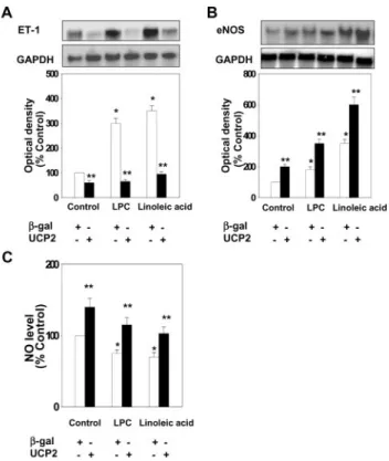
Figure 1. Effect of adenoviral overexpression of the human UCP2 gene on ET-1 (A) and eNOS (B) mRNA expression and NO production (C) in HAECs. HAECs were infected with Ad-UCP2 or Ad--gal. Two days after infection, HAECs were maintained in media containing 0.5% FBS for 12 hours, and exposed to 4g/mL LPC for 4 hours or 300 mol/L linoleic acid for 6 hours. Cells were lysed, and total RNA extracted. Total RNA (10g) was hybridized with human ET-1 and eNOS probe.
RNA quantity was normalized by GAPDH. The NO level in HAECs was measured in situ by using DAF-FM diacetate. Val- ues are presented as means⫾SEM of 4 independent experi- ments. *P⬍0.01 vs Control, **P⬍0.01 vs-gal.
An expanded Materials and Methods section is available in the online data supplement.
Results UCP2 mRNA Expression in HAECs
Cultured HAECs displayed significant levels of UCP2 mRNA (online data supplement Figure IIA). Lysophosphatidylcholine
(LPC) and linoleic acid increased UCP2 mRNA levels by 3.5 and 4.1-fold, respectively.
Effects of Adenoviral UCP2 Gene Transfer on UCP2 mRNA Expression
HAEC infected with adenovirus containing UCP2 (Ad-UCP2) cDNA at doses of 3, 6, 12, and 24 ⫻10
6p.f.u./mL exhibited 2-, 6-, 13-, and 26-fold increase in UCP2 mRNA levels, respectively, compared with cells infected with Ad- -gal (online data supple- ment Figure IIB). The level of UCP2 mRNA following adenoviral UCP2 gene transfer at the dose of 6 ⫻10
6p.f.u. was ⬇2-fold higher than that induced by LPC and linoleic acid. Accordingly, this dose of Ad-UCP2 was used in subsequent experiments.
Effects of Adenoviral UCP2 Gene Transfer on ET-1 and eNOS mRNA Expression and Intracellular NO Level
LPC and linoleic acid induced a significant increase in ET-1 mRNA expression, compared with the control (P ⬍0.01). Notably, this increased ET-1 mRNA expression was diminished to levels com- parable to that of the control, following treatment with Ad-UCP2 (Figure 1A).
Expression of eNOS mRNA was significantly increased in the presence of LPC or linoleic acid, relative to the control (P ⬍0.01, Figure 1B). However, intracellular NO level was decreased by LPC or linoleic acid (Figure 1C). UCP2 overexpression increased eNOS mRNA expression and intra- cellular NO level (Figure 1B and 1C).
Effects of Adenoviral UCP2 Gene Transfer on Intracellular ROS Generation and
NF- B Activation
Incubation with LPC and linoleic acid significantly increased intracellular ROS production. On UCP2 overexpression, the
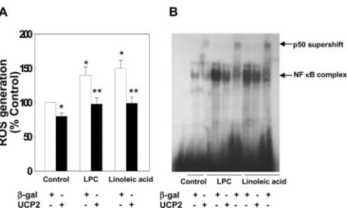
Figure 2. Effect of adenoviral overexpression of UCP2 on ROS gener-ation (A) and NF-B activation (B). Quiescent HAECs were infected with Ad-UCP2 or Ad--gal. Two days after infection, cells were main- tained in media containing 0.5% FBS for 12 hours, and exposed to 4
g/mL LPC for 4 hours or 300 mol/L linoleic acid for 6 hours. (A) After 15 minutes incubation with 10mol/mL carboxydichlorodihydro- fluorescein diacetate (DCFH2-DA), the increase in DCFH2oxidation was measured using FACSCalibur. Values are presented as means⫾SEM of 4 independent experiments. *P⬍0.01 vs Control,
**P⬍0.01 vs-gal. (B) NF-B binding activity was determined by EMSA. Nuclear extracts were prepared from HAECs. For the antibody supershift assay, nuclear extracts were incubated with 10g of anti- p50 polyclonal antibody for 15 minutes at room temperature before the addition of the32P-labeled oligonucleotides. The upper arrow indi- cates the p50 supershift, and the lower arrow specifies the position of the NF-B-specific complex. The bottom depicts the free probe.
Figure 3. Effect of adenoviral overex- pression of UCP2 on LPC- and linoleic acid-induced apoptosis and caspase activation. HAECs were infected with Ad-UCP2 or Ad--gal. Two days after infection, cells were maintained in media containing 0.5% FBS for 12 hours and exposed to 4g/mL LPC for 8 hours or 300mol/L linoleic acid for 16 hours.
After treatment, apoptosis was analyzed by measuring the levels of cytosolic histone-bound DNA fragments using a cell death ELISA assay kit (A) and visual- ization of DNA fragmentation using Southern blot (B). Caspase-3 activation was analyzed by measuring activities using a caspase fluorescence assay kit (C) and immunoblotting for active subunit of caspase-3 (D). The values are present- ed as means⫾SEM of 5 independent experiments. *P⬍0.01 vs Control,
**P⬍0.01 vs-gal.
increase in ROS production induced by LPC and linoleic acid was suppressed (Figure 2A). Catalase and uric acid, which scavenges hydrogen peroxide and peroxynitrite respectively, inhibited linoleic acid/LPC-increased DCF fluorescence, in- dicating that linoleic acid and LPC increased both hydrogen peroxide and peroxynitrite production (online data supple- ment Figure III). LPC and linoleic acid activated NF- B in HAECs. Ad-UCP2 infection markedly decreased activation of NF- B induced by these agents (Figure 2B).
Effects of Adenoviral UCP2 Gene Transfer on Apoptosis and Caspase Activation
LPC and linoleic acid increased endothelial apoptosis by 2.3- and 2.5-fold, respectively. UCP2 overexpression protected HAEC from LPC- and linoleic acid-induced apoptosis by 38% and 40%, respectively (Figure 3A). Southern blotting data additionally revealed that UCP2 decreased linoleic acid-induced DNA fragmentation (Figure 3B). FACScan revealed that UCP2 overexpression inhibited both apoptosis and necrosis of HAECs (online data supplement Figure IV).
LPC and linoleic acid induced a significant increase in caspase-3 (Figure 3C), -8, and -9 activities (online data supplement Figure VA and B) in HAECs. Following UCP2 overexpression, this lipid-induced increase in caspase activity was inhibited. The generation of active caspase-3 from the proenzyme was enhanced in the presence of linoleic acid (Figure 3D). Linoleic acid-induced cleavage of caspase-3 into active subunits was prevented in the presence of UCP2.
Effects of UCP2 iRNA on Apoptosis and Caspase Activation
UCP2 iRNA could effectively reduce the endogenous level of UCP2 mRNA at doses higher than 10 pmol/L compared with control iRNA (Figure 4A). The UCP2 iRNA activated caspase 3 to a significant extent, whereas the control iRNA did not (Figure 4B). UCP2 iRNA exaggerated LPC or linoleic
acid-induced apoptosis compared with control cells (Figure 4C). UCP1 and UCP3 iRNA did not affect apoptosis in control and UCP2 overexpressed cells (data not shown), indicating that the effect of UCP2 is specific. These data
Figure 4. Effect of UCP2 iRNA transfection on caspase activation and apoptosis.HAECs were transfected with UCP2 or con- trol iRNA. Two days after transfection, UCP2 mRNA expression was examined by RT-PCR (A). Transfected cells were main- tained in media containing 0.5% FBS for 12 hours, and 2g/mL LPC for 8 hours or 200mol/L linoleic acid for 16 hours. Fol- lowing treatment, caspase-3 activation and apoptosis were analyzed by measuring activities using a caspase fluorescence assay kit (B) and the cell death ELISA assay kit that measures the levels of cytosolic histone-bound DNA fragments (C). The val- ues are presented as means⫾SEM of 4 independent experiments. *P⬍0.01 vs Con- trol iRNA, **P⬍0.01 vs LPC or linoleic acid⫹Control iRNA.
Figure 5. Effects of adenoviral overexpression of UCP2 on LPC- and linoleic acid-induced change in⌬⌿mand [Ca2⫹]m. UCP2 overexpres- sion attenuated the 4g/mL LPC (A)- and 300 mol/L linoleic acid (B)-induced change in TMRE fluorescence. Vertical axis was expressed as the arbitrary units of TMRE fluorescence. UCP2 overex- pression attenuated the 4g/mL LPC (C)- and 300 mol/L linoleic acid (D)-induced increase in Rhod-2 fluorescence. Pretreatment with 100mol/L ruthenium red inhibits the 4 g/mL LPC (E)- and 300mol/L linoleic acid (F)-induced increase in Rhod-2 fluorescence.
Vertical axis was expressed as the ratio of the baseline value. F is the measured Rhod-2 fluorescence and Fois that at the beginning of the experiment.
demonstrate that endogenous UCP2 expression is important for preventing apoptosis in HAECs.
Effects of UCP2 Overexpression on the LPC- and Linoleic Acid-Induced Changes in ⌬⌿
mand [Ca
2ⴙ]
mIn the Ad-UCP2-infected HAECs, the intensity of TMRE fluorescence at baseline was lower than that in the Ad- -gal- infected HAECs (Figure 5A and 5B), indicating that the mitochondria are partially depolarized. LPC and linoleic acid caused transient hyperpolarization, which was followed by a gradual decrease in ⌬⌿
m.In the Ad-UCP2-infected HAECs, LPC and linoleic acid did not cause these changes (Figure 5A and 5B, and online data supplement Figure VIA).
Mitochondrial Ca
2⫹uptake, particularly in association with oxidative stress, was shown to trigger cell death.
27A reduc- tion of ⌬⌿
mwas shown to limit intramitochondrial Ca
2⫹accumulation.
27LPC and linoleic acid increased, and UCP2 overexpression prevented Ca
2⫹accumulation (Figure 5C and 5D, and online data supplement Figure VIB and VIC).
Pretreatment with ruthenium red, the mitochondrial Ca
2⫹uniporter blocker, blocked the LPC- and linoleic acid-induced increase in [Ca
2⫹]
m(Figure 5E and 5F). These data show that the cytoprotective effect of UCP2 overexpression is related to the inhibition of mitochondrial Ca
2⫹overload.
Effects of UCP2 Overexpression on the Activity of Mitochondrial Respiratory Chain
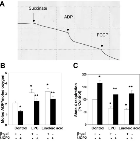
Linoleic acid and LPC significantly decreased state 3 respiration (increased ADP/O ratio; less oxygen consumed
per amount of ADP converted to ATP) and state 4 respiration, as measured by O
2consumption rate in the presence of oligomycin to inhibit ATP synthase (Figure 6B and 6C). Adenoviral overexpression of UCP2 significantly increased both state 3 and state 4 respiration (Figure 6B and 6C).
Inhibition of the Opening of PT Pores in UCP2 Overexpressed Cells
The fluorescence because of calcein in isolated mitochondria from LPC or linoleic acid-treated cells was significantly lower than that of control cells (Figure 7A), indicating an opening of PT pores (PTP). In contrast, UCP2 overexpression inhibited the opening of PTP (Figure 7A). In addition, treatment with linoleic acid resulted in the liberation of cytochorome c from mitochondria to cytosol (Figure 7B). The liberation of cytochrome c by linoleic acid was blocked in cells overexpressing UCP2 (Figure 7B).
Effects of Adenoviral UCP2 Gene Transfer on LPC-Induced Vascular Dysfunction
To determine whether UCP2 overexpression in aortic tissue improves vascular function, we infected the aortic ring with Ad-UCP2 ex vivo, and measured endothelium-dependent and independent vasorelaxation. Immunohistochemical staining for UCP2 protein in Ad-UCP2-treated vessel preparations showed that UCP2 protein was found mainly in endothelium and adventitia in Ad-UCP2-treated aortas (online data sup- plement Figure VII).
Figure 6. Effects of adenoviral overex- pression of UCP2 on mitochondrial oxygen consumption. A, Representative recording of oxygen consumption by mitochondria isolated from HAECs.
Mitochondria isolated from Ad-UCP2 or Ad--gal infected HAECs treated with LPC or linoleic acid were suspended at 60% (vol/vol) in incubat ion buffer.
Additions of succinate, ADP, and car- bonyl cyanide p-[trifluoromethoxy]- phenyl-hydrazone (FCCP) were as shown. The ADP/O ratio (B) is calcu- lated from the amount of oxygen con- sumed during ADP consumption and the known amount of ADP added.
State 4 respiration (C) was measured in the presence of oligomycin (2mol/L) to inhibit ATP synthase. The values are presented as means⫾SEM of 4 inde- pendent experiments. *P⬍0.01 vs Con- trol, **P⬍0.01 vs-gal.
Endothelium-dependent (acetylcholine-induced) and inde- pendent (nitroprusside-induced) relaxation was not affected by Ad-UCP2 alone (Figure 8A and 8B). LPC significantly decreased endothelium-dependent relaxation of the aortic ring infected with Ad- -gal (Figure 8A), but did not affect endothelium-independent relaxation. Ad-UCP2 infection sig- nificantly improved impaired endothelium-dependent relax- ation induced by LPC.
Discussion
In this study, we have shown that UCP2 is expressed in HAECs, and upregulated by LPC and linoleic acid.
Adenovirus-mediated transfer of the UCP2 gene into HAECs profoundly suppressed ROS generation, NF- B activation, and mRNA expression of ET-1, and enhanced eNOS tran- scription. UCP2 additionally inhibited caspase activation and endothelial apoptosis in response to LPC and linoleic acid.
LPC and linoleic acid caused mitochondrial calcium accumu- lation, transient mitochondrial membrane hyperpolarization (followed by depolarization), decreased oxygen consumption,
and cytochrome c release. UCP2 overexpression prevented all these processes and improved impaired endothelium- dependent vascular relaxation induced by LPC. Suppression of endogenous expression of UCP2 exaggerated linoleic acid- and LPC-induced caspase activation and apoptosis.
UCPs are located in the mitochondrial inner membrane, and partially dissipate the ⌬⌿
m. Previous studies have shown that UCP2 is beneficial to cells and organisms, because it reduces ROS formation.
15,28 –30For example, UCP2 prevents the apoptosis of cardiomyocytes and neuronal cells by de- creasing ROS.
15,31,32The mitochondrial respiratory chain generate ROS when the electrochemical gradient across the mitochondrial inner membrane is high and the rate of electron transport is limited.
15In agreement with previous studies in other cells,
26we found that adenoviral UCP2 overexpression decreased coupling of mitochondrial oxidation and phosphor- ylation, as evidenced by a decrease in the ADP/O ratio and increased state 4 respiration. These results suggest that UCP2 decreases ROS generation by reducing ⌬⌿
mand increasing the rate of electron transport.
Linoleic acid and LPC stimulate ROS generation in cells and activate the redox-sensitive transcription factors, NF B and AP-1.
33,34The promoter region of human UCP2 contains AP-1 binding sites near the transcription initiation site.
35This finding strongly suggests that transcription of the UCP2 gene is induced by intracellular ROS and the consequent activation of AP-1. Consistent with this theory, recent studies demon- strated that superoxide activates UCPs in various tissues.
36It has been proposed that induction of UCP2 by ROS is a compensatory mechanism to counteract increased intracellu- lar oxidative stress.
37In this study, UCP2 mRNA expression increased 3- to 4-fold by LPC or linoleic acid. However, this increase in UCP2 may not be sufficient to protect against LPC/linoleic acid-induced increases in ROS. Supporting this idea, UCP2 overexpression at a sufficient dose (6 ⫻10
6p.f.u.) almost completely suppressed the increase in ROS production induced by LPC and linoleic acid, whereas at a lower dose (3 ⫻10
6p.f.u.), UCP2 incompletely suppressed ROS genera- tion (online data supplement Figure IX).
Endothelium acts not only as a barrier, but also as a regulator of vascular tone and smooth muscle cell growth, migration, and proliferation. Vascular tone is modulated through the release of relaxing and contracting substances.
Among them, NO and ET-1 are the physiologically most important regulators of vascular tone. NO produced by eNOS has anti-inflammatory effects on the vascular wall, and inhibits the migration and proliferation of vascular smooth muscle cells.
11On the other hand, ET-1 displays vasocon- strictive and cell-proliferating effects.
38In agreement with previous studies,
39,40linoleic acid and LPC increased the expression of both eNOS and ET-1 in HAECs. This finding prompts the question of why LPC and linoleic acid decrease vascular relaxation yet increase eNOS levels? The bioavail- ability of NO is determined by the balance between produc- tion and degradation. NO is quenched by superoxide to form peroxynitrite. In this study, both catalase and uric acid, which scavenge hydrogen peroxide and peroxynitrite respectively, significantly inhibited linoleic acid/LPC-increased DCF fluo- rescence, indicating that linoleic acid and LPC increase both
Figure 7. Effects of adenoviral overexpression of UCP2 on opening ofpermeability transition (PT) pores and cytochrome c release. Mito- chondria (1 mg of protein/mL) were isolated from Ad-UCP2 or Ad-- gal infected HAECs treated with LPC or linoleic acid. To measure the release of calcein from PT pores (A), isolated mitochondria were sus- pended in a mitochondrion-isolation buffer. Calcein was introduced into the mitochondrial matrix by incubation with 1mol/L calcein-AM.
The mitochondria were then washed and fluorescence was measured with microplate spectrofluorometer. For analysis of the release of cytochrome c from mitochondria into the cytosol (B), proteins in cyto- solic and mitochondrial fractions were separated by SDS/PAGE. Cyto- chrome c was detected with an enhanced chemiluminescence sys- tem with specific monoclonal antibodies. The values are presented as means⫾SEM of 5 independent experiments. *P⬍0.01 vs Control
hydrogen peroxide and peroxynitrite production. In addition, direct measurement of NO in the cells by using DAF-FM diacetate showed that NO level is decreased by linoleic acid and LPC. These results suggest that increased quenching by superoxide decreases NO availability, and that eNOS expres- sion is increased to overcome this decrease in NO availabil- ity. UCP2 suppressed ET-1 expression, and increased eNOS expression induced by LPC and linoleic acid. Increased eNOS and decreased ET-1 expression in the presence of UCP2 may contribute to prevention of atherosclerosis.
Endothelial apoptosis is an important early event in the pathogenesis of atherosclerosis,
8and increased intracellular oxidative stress plays an important causative role in the pathogenesis of endothelial apoptosis.
8,12In agreement with previous data on other cell types,
14,31overexpression of UCP2 inhibited LPC- and linoleic acid-induced endothelial apopto- sis and caspase activation. In addition, suppression of UCP2 expression using UCP2-specific iRNA exaggerated caspase activation and apoptosis induced by LPC and linoleic acid.
Furthermore, UCP2 overexpression prevented the changes in
⌬⌿
mand reduced mitochondrial Ca
2⫹accumulation, ROS generation, PTP opening, and cytochrome c release induced by LPC or linoleic acid. It has long been thought that a decrease of ⌬⌿
mis a marker of apoptotic cells.
41More recently, it was proposed that a significant increase in ⌬⌿
m(hyperpolarization) represents an earlier prerequisite for cell death.
42,43Consistent with these studies, we found that LPC and linoleic acid caused transient hyperpolarizaion that was followed by depolarization, and that UCP2 overexpression prevented these processes. A reduction in ⌬⌿
mwas shown to limit the mitochondrial accumulation of Ca
2⫹.
44The accumu- lation of mitochondrial Ca
2⫹is associated with the production of ROS
14,37and leads to the increased mitochondrial perme- ability to cytochrome c and other molecules (through the PT pores), which are known to initiate a cascade of events culminating in cell death. Taken together, the present results indicate that lowering ⌬⌿
mby UCP2 is the primary mecha- nism preventing the cascade of other events leading to apoptosis.
Finally, we examined whether UCP2 overexpression af- fects vascular function in isolated rat aorta. As reported previously,
45LPC impaired endothelium-dependent but not endothelium-independent vascular relaxation. UCP2 partially
reversed the endothelium-dependent vascular dysfunction induced by LPC. Defective endothelium-dependent vasodila- tion is an important early event in the development of atherosclerosis.
10These ex vivo results confirm the anti- atherogenic role of UCP2 in vascular endothelial cells.
In summary, UCP2 overexpression reduced ROS genera- tion and led to a significant increase in eNOS and decrease in endothelin-1 mRNA expression in HAECs. UCP2 inhibited endothelial apoptosis in response to LPC and linoleic acid, and improved impaired endothelium-dependent vascular re- laxation induced by LPC. The data collectively suggest that UCP2, functions as a physiologic regulator of ROS genera- tion in endothelial cells. Measures to increase UCP2 expres- sion in vascular cells may aid in preventing the development and progression of atherosclerosis in patients with metabolic syndrome.
Acknowledgments
This study was supported by an NRL grant
(M1040000000804J000000810) from the Ministry of Science and Technology, grants from Ministry of Health and Welfare (02-PJ1- PG10-20708-0007) and Asan Institute for Life Sciences (01-122, 01-279, 05-006), Republic of Korea.
References
1. Reaven GM. Role of insulin resistance in human disease. Diabetes. 1988;
37:1595–1607.
2. Steinberg HO, Chaker H, Leaming R, Johnson A, Brechtel G, Baron AD.
Obesity/insulin resistance is associated with endothelial dysfunction.
Implications for the syndrome of insulin resistance. J Clin Invest. 1996;
97:2601–2610.
3. Egan BM, Lu G, Greene EL. Vascular effects of non-esterified fatty acids: implications for the cardiovascular risk factor cluster. Prostaglan- dins Leukot Essent Fatty Acids. 1999;60:411– 420.
4. Reaven GM, Hollenbeck C, Jeng CY, Wu MS, Chen YD. Measurement of plasma glucose, free fatty acid, lactate, and insulin for 24 h in patients with NIDDM. Diabetes. 1988;37:1020 –1024.
5. Sigurdardottir V, Fagerberg B, Hulthe J. Circulating oxidized low-density lipoprotein (LDL) is associated with risk factors of the metabolic syndrome and LDL size in clinically healthy 58-year-old men (AIR study). J Intern Med. 2002;252:440 – 447.
6. Steinberg HO, Tarshoby M, Monestel R, Hook G, Cronin J, Johnson A, Bayazeed B, Baron AD. Elevated circulating free fatty acid levels impair endothelium-dependent vasodilation. J Clin Invest. 1997;100:1230 –1239.
7. Li D, Mehta JL. 3-hydroxy-3-methylglutaryl coenzyme A reductase in- hibitors protect against oxidized low-density lipoprotein-induced endo- thelial dysfunction. Endothelium. 2003;10:17–21.
Figure 8. Effects of adenoviral overexpres- sion of UCP2 on LPC-induced impairment of vasodilatory response. UCP2 gene was overexpressed in the rat aortic ring by a 30 minutes adenoviral infection (6⫻106 p.f.u./mL) at 37°C in DMEM without serum and 24 hours incubation with medium con- taining 5% BSA. After exposure to 4g/mL LPC, contraction of aortic rings was intro- duced by treatment with 300mol/L phenyl- ephrine. Acetylcholine (from 10⫺9to 10⫺5 mol/L) and sodium nitroprusside (from 10⫺11 to 10⫺7mol/L) were added serially to the bath to induce endothelium-dependent vasorelaxation (A) and endothelium- independent vasorelaxation (B), respec- tively. Values are presented as means⫾SEM of 6 independent experi- ments. *P⬍0.01 vs-gal.
8. Choy JC, Granville DJ, Hunt DW, McManus BM. Endothelial cell apo- ptosis: biochemical characteristics and potential implications for athero- sclerosis. J Mol Cell Cardiol. 2001;33:1673–1690.
9. Mombouli JV, Vanhoutte PM. Endothelial dysfunction: from physiology to therapy. J Mol Cell Cardiol. 1999;1:61–74.
10. Ross R. Atherosclerosis-an inflammatory disease. N Engl J Med. 1999;
340:115–126.
11. Williams IL, Wheatcroft SB, Shah AM, Kearney MT. Obesity, athero- sclerosis and the vascular endothelium: mechanisms of reduced nitric oxide bioavailability in obese humans. Int J Obes Relat Metab Disord.
2002;26:754 –764.
12. Kunsch C, Medford RM. Oxidative stress as a regulator of gene expression in the vasculature. Circ Res. 1999;85:753–766.
13. Boss O, Hagen T, Lowell BB. Uncoupling proteins 2 and 3: potential regulators of mitochondrial energy metabolism. Diabetes. 2000;49:
143–156.
14. Teshima Y, Akao M, Jones SP, Marban E. Uncoupling protein-2 over- expression inhibits mitochondrial death pathway in cardiomyocytes. Circ Res. 2003;93:192–200.
15. Arsenijevic D, Onuma H, Pecqueur C, Raimbault S, Manning BS, Miroux B, Couplan E, Alves-Guerra MC, Goubern M, Surwit R, Bouillaud F, Richard D, Collins S, Ricquier D. Disruption of the uncoupling protein-2 gene in mice reveals a role in immunity and reactive oxygen species production. Nat Genet. 2000;26:435– 439.
16. Blanc J, Alves-Guerra MC, Esposito B, Rousset S, Gourdy P, Ricquier D, Tedgui A, Miroux B, Mallat Z. Protective role of uncoupling protein 2 in atherosclerosis. Circulation. 2003;107:388 –390.
17. Becker TC, BeltrandelRio H, Noel RJ, Johnson JH, Newgard CB. Over- expression of hexokinase I in isolated islet of Langerhans via recombinant adenovirus. Enhancement of glucose metabolism and insulin secretion at basal but not stimulatory glucose levels. J Biol Chem. 1994;269:
21234 –21238.
18. Zanetti M, Sato J, Jost CJ, Gloviczki P, Katusic ZS, O’Brien T. Gene transfer of manganese superoxide dismutase reverses vascular dys- function in the absence but not in the presence of atherosclerotic plaque.
Hum Gene Ther. 2001;12:1407–1416.
19. Park JY, Takahara N, Gabriele A, Chou E, Naruse K, Suzuma K, Yamauchi T, Ha SW, Meier M, Rhodes CJ, King GL. Induction of endothelin-1 expression by glucose: an effect of protein kinase C acti- vation. Diabetes. 2000;49:1239 –1248.
20. Shao C, Stewart V, Melvyn Folkard M, Michael BD, Prise KM. Nitric Oxide-Mediated Signaling in the Bystander Response of Individually Targeted Glioma Cells. Cancer Res. 2003;63:8437– 8442.
21. van Reyk DM, King NJ, Dinauer MC, Hunt NH. The intracellular oxi- dation of 2⬘,7⬘-dichlorofluorescin in murine T lymphocytes. Free Radic Biol Med. 2001;30:82– 88.
22. Bass DA, Parce JW, Dechatelet LR, Szejda P, Seeds MC, Thomas M.
Flow cytometric studies of oxidative product formation by neutrophils: a graded response to membrane stimulation. J Immunol. 1983;130:
1910 –1917.
23. Ahn JD, Morishita R, Kaneda Y, Lee KU, Park JY, Jeon YJ, Song HS, Lee IK. Transcription factor decoy for activator protein-1 (AP-1) inhibits high glucose- and angiotensin II-induced type 1 plasminogen activator inhibitor (PAI-1) gene expression in cultured human vascular smooth muscle cells. Diabetologia. 2001;44:713–720.
24. Trollinger DR, Cascio WE, Lemasters JJ. Mitochondrial calcium tran- sients in adult rabbit cardiac myocytes: inhibition by ruthenium red and artifacts caused by lysosomal loading of Ca2⫹-indicating fluorophores.
Biophys J. 2000;79:39 –50.
25. Imai H, Koumura T, Nakajima R, Nomura K, Nakagawa Y. Protection from inactivation of the adenine nucleotide translocator during hypoglycaemia-induced apoptosis by mitochondrial phospholipid hydroperoxide glutathione peroxidase. Biochem J. 2003;371:799 – 809.
26. Hong Y, Fink BD, Dillon JS, Sivitz WI. Effects of adenoviral overex- pression of UCP-2 and -3 on mitochondrial respiration in insulinoma cells. Endocrinology. 2001;142:249 –256.
27. Duchen MR. Mitochondria and calcium: from cell signaling to cell death.
J of Physiol. 2000;529:57– 68.
28. Skulachev VP. Uncoupling: new approaches to an old problem of bioen- ergetics. Biochim Biophys Acta. 1998;1363:100 –124.
29. Lee F-YJ, Li Y, Yang EK, Yang SQ, Lin HZ, Trush MA, Dannenberg AJ, Diehl AM. Phenotypic abnormalities in macrophages from leptin- deficient, obese mice. Am J Physiol. 1999;276:C386 –C394.
30. Li LX, Skorpen F, Egenberg K, Jorgensen IH, Grill V. Uncoupling protein 2 participates in cellular defence against oxidative stress in clonal beta-cells. Biochem Biophys Res Commun. 2001;282:273–277.
31. Mattiasson G, Shamloo M, Gido G, Mathi K, Tomasevic G, Yi S, Warden CH, Castilho RF, Melcher T, Gonzalez-Zulueta M, Nikolich K, Wieloch T. Uncoupling protein-2 prevents neuronal death and diminishes brain dysfunction after stroke and brain trauma. Nat Med. 2003;9:1062–1068.
32. Vincent AM, Olzmann JA, Brownlee M, Sivitz WI, Russell JW.
Uncoupling proteins prevent glucose-induced neuronal oxidative stress and programmed cell death. Diabetes. 2004;53:726 –734.
33. Masamune A, Sakai Y, Yoshida M, Satoh A, Satoh K, Shimosegawa T.
Lysophosphatidylcholine activates transcription factor NF-kappaB and AP-1 in AR42J cells. Dig Dis Sci. 2001;46:1871–1881.
34. Park JY, Kim YM, Song HS, Park KY, Kim YM, Kim MS, Pak YK, Lee IK, Lee JD, Park SJ, Lee KU. Oleic acid induces endothelin-1 expression through activation of protein kinase C and NF-kappa B. Biochem Biophys Res Commun. 2003;303:891– 895.
35. Tu N, Chen H, Winnikes U, Reinert I, Marmann G, Pirke KM, Lentes KU. Molecular cloning and functional characterization of the promoter region of the human uncoupling protein-2 gene. Biochem Biophys Res Commun. 1999;265:326 –334.
36. Echtay KS, Roussel D, St-Pierre J, Jekabsons MB, Cadenas S, Stuart JA, Harper JA, Roebuck SJ, Morrison A, Pickering S, Clapham JC, Brand MD. Superoxide activates mitochondrial uncoupling proteins. Nature.
2002;415:96 –99.
37. Paradis E, Clavel S, Bouillaud F, Ricquier D, Richard D. Uncoupling protein 2: a novel player in neuroprotection. Trends Mol Med. 2003;9:
522–525.
38. Schiffrin EL. Role of Endothelin-1 in Hypertension and Vascular Disease. Am J Hypertens. 2001;14:83S– 89S.
39. Ramasamy S, Parthasarathy S, Harrison DG. Regulation of endothelial nitric oxide synthase gene expression by oxidized linoleic acid. J Lipid Res. 1998;39:268 –276.
40. Hirata K, Miki N, Kuroda Y, Sakoda T, Kawashima S, Yokoyama M.
Low concentration of oxidized low-density lipoprotein and lysophos- phatidylcholine upregulate constitutive nitric oxide synthase mRNA expression in bovine aortic endothelial cells. Circ Res. 1995;76:958 –962.
41. Zamzami N, Marchetti P, Castedo M, Zanin C, Vayssiere JL, Petit PX, Kroemer G. Reduction in mitochondrial potential constitutes an early irreversible step of programmed lymphocyte death in vivo. J Exp Med.
1995;181:1661–1672.
42. Russell JW, Golovoy D, Vincent AM, Mahendru P, Olzmann JA, Mentzer A, Feldman EL. High glucose-induced oxidative stress and mitochondrial dysfunction in neurons. FASEB J. 2002;16:1738 –1748.
43. Matarrese P, Gambardella L, Cassone A, Vella S, Cauda R, Malorni W.
Mitochondrial membrane hyperpolarization hijacks activated T lym- phocytes toward the apoptotic-prone phenotype: homeostatic mechanisms of HIV protease inhibitors. J Immunol. 2003;170:6006 – 6015.
44. Krieger C, Duchen MR. Mitochondria, Ca2⫹ and neurodegenerative disease. Eur J Pharmacol. 2002;447:177–188.
45. Murohara T, Kugiyama K, Ohgushi M, Sugiyama S, Ohta Y, Yasue H.
LPC in oxidized LDL elicits vasocontraction and inhibits endothelium- dependent relaxation. Am J Physiol. 1994;267:H2441–H2449.
![Figure 5. Effects of adenoviral overexpression of UCP2 on LPC- and linoleic acid-induced change in ⌬⌿ m and [Ca 2⫹ ] m](https://thumb-ap.123doks.com/thumbv2/123dokinfo/5012838.306747/4.877.73.566.99.463/figure-effects-adenoviral-overexpression-ucp-linoleic-induced-change.webp)