교신저자 : 김 재 문
대전시 중구 대사동 640
충남대학교 의과대학 신경과학교실
TEL) 042-863-9920, FAX) 042-252-8654, e-mail) [email protected]
*본 연구는 보건복지부 보건의료 기술연구개발사업의 지원에 의하여 이루어진 것임(HMP-98-N-2-0004).
흰쥐에서 고용량의 P i l o c a r p i n e 에 의하여 유발된 간질중첩증의 양상
충남대학교 의과대학 신경과학교실
이경목・정기영・김재문
EEG Patterns of High dose Pilocarpine-Induced Status Epilepticus in Rats
Kyung-Mok Lee, Ki-Young Jung, Jae-Moon Kim
Department of Neurology, Chungnam National University Hospital, Taejon, Korea
- Abstract -
B a c k g r o u n d : We studied EEG changes during pilocarpine-induced status epilepticus(SE), a widely used model whose EEG characteristics have not been fully described previously. M e t h o d s : Male Sprague-Dawley rats weighing 250-350 grams were used as subjects. SE was induced 5-7 days after placement of chronic epidural electrodes, using 360-380 mg/Kg pilocarpine IP. Rats were observed with continuous EEG recording following pilocarpine injection until end of the SE episode. R e s u l t s : SE occurred in 10/12 rats studied. SE began with a series of discrete seizures 11.1±3.93 minutes after pilocarpine injection. 5.2±2.71 seizures occurred over 10.9±4.62 minutes, until the EEG converted to a waxing and waning pattern, during which the amplitude and frequency of epileptiform activity increased. After 1.4±1.82 minutes, a pattern of continuous high amplitude rapid spiking was established. Continuous spiking continued for 3.4±0.48 hours with a very gradual decline in amplitude and frequency, until periodic epileptiform discharges(PEDs) began to occur. The EEG consisted primarily of PEDs for another 7.4±3.09 hours, until electrographic generalized seizures began to occur. These continued for 5.8±4.82 hours until death. Duration of SE was 17.0±5.88 hours. Flat periods were a prominent feature during all EEG patterns in this model. C o n c l u s i o n : EEG features distinctive in pilocarpine SE(but not unique to it) include flat periods during all patterns and resumption of continuous spik- ing episodes after the onset of PEDs. The sequence of discrete seizures to waxing and waning to continuous spiking to PEDs was identical to that which has been described in humans and other animal models.
Key Words : Status epilepticus(SE), Pilocarpine, Electroencephalography(EEG)
I N T R O D U C T I O N
Treiman et. al.1 , 2staged SE according to the evo-
lution of EEG characteristics in human and sever- al animal models. These five distinct electroen- cephalographic patterns and their behavioral fea- tures in human and several epilepsy models were characteristic and stereotypic. They reported the
initial discrete seizures were followed by merging stages with wax and waning of frequencies and amplitudes. Later, continuous spiking follows and the continuous spiking progressed into continuous with intermittently punctuated flat periods and the finally, periodic epileptiform discharges(PEDs) ensued. Not only in human SE, with only minimal variance, these patterns were found in lithium- pilocarpine, cobalt-homocysteine, kainic acid- induced SE models and in the electrical kindling m o d e l1 - 4. Recently even in the in vivo h i p p o c a m p a l slice model, these EEG patterns were suggested5 , 6. These EEG patterns have been expected to have significant meaning in the SE evolution as well as physiologic alteration and therapeutic results.
Since introduction of pilocarpine as a chemical convulsant, high-dose pilocarpine-induced status epilepticus(PISE) model is widely used in epilepsy r e s e a r c h7. This model shows initial complex partial seizures originating from the limbic structures and following convulsive SE8 - 1 2. Although the behav- ioral and EEG changes of high-dose PISE model had been reported and the results were very con- sistent and stereotypic, interpretation of their EEG recordings with reference to the SE patterns are lacking, and EEG recordings were not stan- d a r d i z e d1 3 , 1 4.
To verify whether these distinct EEG patterns are sequentially exist in PISE model, we record- ed and analyzed the EEG of PISE rats.
M E T H O D S
Twelve male Sprague-Dawley rats weighing 150~200 grams were used in this experiment. Rats were anesthetized by i.p. injections of 87 mg/Kg ketamine plus 13 mg/Kg xylazine and mounted in a stereotaxic frame. After the skull is exposed, epidural recording electrodes(0-80 * 1/8 inch stain- less steel screw and wire) were placed at the follow- ing stereotaxic coordinates: A-P+1.5 mm, -2 . 5 mm and lateral±2.0 mm) from the bregma.
Electrodes were anchored to the skull using dental acrylic. After the operation, the rats were housed separately and allowed to recover at least five days prior to SE induction. All the rats had food and water available ad lib. And maintained in a 12 hour
light/dark cycle.
EEG was recorded as follows: paper speed 15~30 mm/sec, high frequency filter 60 Hz and low fre- quency filter 1 Hz, sensitivity were variable accord- ing to the EEG amplitude and every change of sen- sitivity were written on the EEG. Montage was bipolar: F4-P4, P4-P3, P3-F3, F3-F4.
Definition of each EEG stages were as follows:
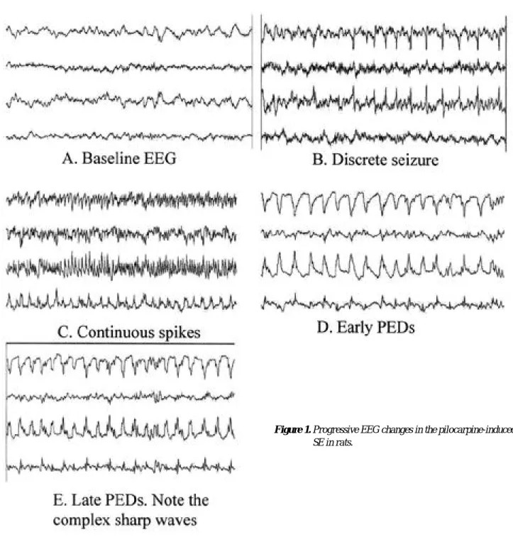
Discrete seizure is an onset of SE. It is defined as a clearly identifiable generalized but may be asym- metric epileptiform discharge that is first seen on the EEG with behavioral seizure of forelimb clonus.
Merging stage is characterized by waxing and waning ictal discharges. Rhythmic but frequently asymmetric sharp or spike/wave patterns are seen with recurrent build-up and then slowing of fre- quency and waxing and waning of the amplitudes.
Continuous ictal discharges are defined as the EEG of rhythmic, relatively constant, sharp or spike/wave discharges those are usually symmet- ric. This pattern is usually associated with either continuous generalized clonic jerks or subtle clonic m o v e m e n t s .
Continuous ictal discharges with flat period are similar to the continuous ictal discharge in the EEG aside from the intermittently intervening flat period.
They usually tend to be slower in frequency and lower in amplitude.
PEDs on a flat background is characterized by bilateral, high voltage, monomorphic, sharp waves superimposed on a relatively flat background. PEDs were defined as monotonous spike/wave with flat background, less than 2 Hz in frequencies, lasts at least 10 seconds.
Before 10 minutes of baseline EEG recording, rats were allowed to adapt in the recording cage for 20 minutes with electrode connected state.
After the baseline EEG recording, scopolamine methyl nitrate 1 mg/Kg was injected S.C. to min- imize peripheral cholinergic effects. 360~380 mg/Kg of pilocarpine hydrochloride was freshly dissolved in saline to make 100 mg/ml and inject- ed by i.p. Every rat was continuously observed and recorded EEG until the SE stops or the rats die. Video recording was performed simultaneous- ly. No supportive care or treatment was done.
R E S U LT S
SE was induced in 10 out of 12 rats. With 380 mg/Kg of pilocarpine injection, all the five rats showed SE, whereas only 5/7 of 360 mg/Kg pilo- carpine injected rats developed SE. Every rat was died of SE.
1. Behavioral Changes
Within less than one to two minutes after pilo- carpine injection, rats showed initial akinesia.
Behavior of rat was changed after akinesia.
Common behaviors following akinesia were; facial automatism, sniffing, ataxic lurching, ear trem- bling, and masticatory movements and other limbic seizures. Head nodding, rearing, and falling occa- sionally followed. These behaviors persisted until the first discrete seizure occurs and also in between the discrete seizures. Nature of discrete seizure was variable from mild forelimb clonus to jump/running fit. During the early continuous spiking stage, most rats showed cholinergic effects of piloerection and red eye, severe foaming and drooling. At this stage, severe convulsive movements involved bilateral forelimb clonus, rearing and falling. After 30~60 minutes of vigorous seizure, these convulsive move- ments were replaced by mild, rhythmic and slow head movements(so called ‘subtle SE’). But occa- sionally during this stage and during the PEDs, usually after a long flat period, occasional big jerk or jump occurred. In the stage of PEDs, epileptic movements were weaker or even absent. In the very late PEDs, as the frequency of epileptic dis- charges in the EEG decrease and slow wave replace the flat period, rats were awaking and walk around the cage and responded to the external stimulation.
Wet dog shakes were not only observed just after discrete seizure but were more prominent feature during the electrical seizure of later PEDs.
During the electrographic generalized seizure in the later PED, rat was nonresponsive without dis- cernible epileptic movement. But, after this elec- trographic seizure, rats frequently moved around the cage.
2. EEG Changes
Just after the pilocarpine injection, when rat is akinetic or in the limbic behavior, predrug EEG backgrounds were replaced by low amplitude fast activities. Four to eight minutes later, they are changed with relatively rhythmic theta waves.
Later, these theta waves occasionally superim- posed with isolated random spikes or progressed into discrete seizures.
Every rat started its SE with discrete seizure.
First discrete seizure occurred 11.1±3.93 minutes after the pilocarpine injection and number of dis- crete seizures were 5.2±2.71(2~11 seizures).
Characteristically, even in the discrete seizure, punctuated flat periods and postictal depression were frequently seen in this model. Discrete seizures repeated until the EEG converted to a waxing and waning pattern, during which the amplitude and frequency of epileptiform activity increased to a pat- tern of continuous high amplitude rapid spiking was established. Generalized tonic-clonic seizure was attained in half of the rats during the first discrete seizure although the initiation of first discrete seizure was invariably limbic seizure. No single dis- crete seizure exceeds 2 minutes, and if it last more than 2 minutes, it invariably entered into continu- ous ictal discharges.
Merging(waxing and waning) period was very brief or sometimes absent. This period occurred 2 2 . 2±7.85 min. from the injection and lasted 1 . 4±1.82 min only.
Continuous spiking continued for 3.4±0 . 4 8 hours with a very gradual decline in amplitude and frequency after a brief build-up. The maxi- mum EEG frequency was 10.4±1.11/sec at 14.4
±18.88 min from the beginning of this stage.
And highest amplitude was established in 35.4±
15.78 minutes(1290±203.0 mV). As the punc- tuated flat period was evident from the onset of SE, continuous ictal discharges with flat period were not identified.
Transition from the continuous spiking to the PEDs was very gradual. During the late contin- uous ictal discharge and early PEDs, both con- tinuous spiking and periodic sharp/spike were mixed for hours. The EEG consisted primarily of
PEDs continued 7.4±3.09 hours, until electro- graphic generalized seizures began to occur.
These late PEDs continued 5.8±4.82 hours until death. These electrographic seizures were generally brief and frequent(4~19 times), and mostly followed by WDS. EEGs of this seizure showed low amplitude fast spikes with rather rhythmic intervening flat periods. During the mid- to late PEDs, occasional very low voltage fast rhythms mimicking alpha wave followed spike/sharp wave. Overall duration of SE until death was 17.0±5.88 hours. And no rat recov- ered to the pre-drug EEG.
D I S C U S S I O N
SE was very fatal in this model without any supportive care and treatment as all rat was died of SE. Comparing 20%~70% mortality rates of previous reports, it was exceptionally high in this experiment and it may be resulted from minimal supportive care only.
Behavioral patterns were not different from previous reports1 5 , 1 6. Every rat showed stereotyp- ic behavior with reliable progression.
Injection of 1 ml/Kg of scopolamine did not influ-
Figure 1. Progressive EEG changes in the pilocarpine-induced SE in rats.
ence much in their behavior and EEG but rats are tend to be more explorative and EEG was disorga- nized and amplitude was increasing. Regarding the initial effect of pilocarpine, Turski et. al.1 3 reported early appearance(2~5 min) of theta rhythms in hippocampus after pilocarpine injection and at the same time cortical EEG was replaced by fast activities. Extradural recording in this experiment was almost same to the reported corti- cal recording, but the appearance of fast wave was sometimes within a minute after injection.
Prominent flat periods from the initiation of SE were a prominent feature during all EEG pat- terns in this model. Also postictal depression was very frequent findings. Due to the long postictal suppression, initiation of merging stage was not easy to determine. The highest amplitude in EEG usually precedes maximum frequency. But, the earlier the highest amplitude occurs, maxi- mal frequency was established earlier.
PEDs can be divided into early and late PEDs in this model. Early PEDs are mainly comprised of monotonous spike/sharp waves with the typi- cal flat background, whereas late PEDs are com- prised of occasional electrographic seizures and low amplitude fast waves following relatively rhythmic spike/sharp waves. This difference in the PEDs were suggested by Reiher et. al.1 7 a s
‘PLEDs proper(uniform PLEDs with periodicity)’
and ‘PLEDs plus(complex morphology PLEDs).
In the very late PEDs or recovery from PEDs, slow waves started to replace flat period. The electrographic seizures were conspicuous finding during the late PEDs and the numbers of them were variable.
Previous reports that EEG rhythms gradually normalized and became indistinguishable from the pre-drug activity by 48~72 hours. Until death(up to 36 hours), we could not find so called “silent period”1 2 , 1 7 , 1 8or normalized pattern in EEG. The possible explanation may be ; 1) as the amplitude of electrical seizures in the late PEDs are too low, only proper EEG recording identifies the epileptic discharge; 2) silent peri- od may develops more than 36 hours after the SE. But as isolated high voltage spikes were recorded up to 5 days in systemic pilocarpine injection and saline injection in substantia
nigra, true silent period may be developed later.
C O N C L U S I O N
PISE model follows the SE stages suggested by Treiman et al., aside from the absence of contin- uous ictal discharge with intermittent flat peri- od. Each stage showed its own characteristic EEG patterns.
참고문헌
01. Treiman DM, Walton NY, Kendrick C. A progressive sequence of electroencephalographic changes during generalized convul- sive status epilepticus. Epilepsy Res 1 9 9 0 ; 5 : 4 9 - 6 0 .
02. Treiman DM. Electroclinical features of status epilepticus. J of Clin Neurophysiol 1995;12:343-362.
03. Walton NY, Treiman DM, Gunnawan S. Brain amino acid con- centrations during status epilepticus induced by lithium and pilocarpine. Exp Neurol 1 9 9 0 ; 1 0 8 : 6 1 - 7 0 .
04. Walton NY, Treiman DM. Experimental secondarily general- ized convulsive status epilepticus induced by D,L-homocys- teine thiolactone. Epilepsy Res 1988;2:79-86.
05. Rafiq A, Zhang YF, DeLorenzo RJ, Coulter DA. Long-duration self-sustained epileptiform activity in the hippocampal-parahip- pocampal slice: a model of status epilepticus. J Neurophysiol 1 9 9 5 ; 1 7 : 2 0 2 8 - 2 0 4 2 .
06. Lothman EW, Bertram EH, Bekenstein JW, Perlin JB. Self- sustaining limbic status epilepticus induced by ‘continuous’
hippocampal stimulation:electrographic and behavioral char- acteristics. Epilepsy Res 1989;3:107-119.
07. Grossman SP. Chemically induced epileptiform seizures in the cat. Science 1963;142:409-411.
08. Turski WA, Cavalheiro EA, Schwarz M, Czuczwar SJ, Klein- rok Z, Turski L. Limbic seizures produced by pilocarpine in rats: Behavioral, electroencephalographic and neuropathologic study. Behavioral Brain Research 1983;9:315-335.
09. Turski WA, Cavalheiro EA, Bortolotto ZA, Mello LM, Sch- warz M, Turski L. Seizures produced by pilocarpine in mice:
A behavioral, electroencephalographic and morphological analysis. Brain Research 1984;321:237-253.
10. Turski WA, Cavalheiro EA, Ikonomidou C, Mello LEA, Bortolotto ZA, Turski L. Effects of aminophylline and 2-chlo- radenosine on seizures produced by pilocarpine in rats:
Morphological and electroencephalographic correlates. B r a i n Res 1985;361:309-323.
11. Turski LW, Cavalheiro EA, Schwarz M, Turski WA, Mello LEA, Bortolotto ZA, Klokgether T, Sontag K-H. Susceptibility to seizures produced by pilocarpine in rats after microinjection of Isoniazid or gamma-Vinyl-GABA into the substantia nigra.
Brain Res 1 9 8 6 ; 3 7 0 : 2 9 4 - 3 0 9 .
12. Turski L, Cavalheiro EA, Sieklucka-Dziuba M, Ikonomidou- Turski C, Czuczwar SJ, Turski WA. Seizures produced by pilo- carpine: Neuropathological sequelae and activity of glutamate decarboxylase in the rat forebrain. Brain Res 1 9 8 6 ; 3 9 8 : 3 7 - 4 8 . 13. Turski L, Cavalheiro EA, Czuczwar SJ, Turski WA, Kleinrok
Z. The seizures induced by pilocarpine: behavioral, electroen- cephalographic and neuropathological studies in rodents. Pol J Pharmacol Pharm 1987;39:545-555.
14. Turski L, Ikonomidou C, Turski WA, Bortolotto ZA, Caval- heiro EA. Review: Cholinergic mechanisms and epileptogene- sis. The seizure induced by pilocarpine: A novel experimental model of intractable epilepsy. Synapse 1989;3:154-171.
15. Cavalheiro EA, Leite JP, Bortolott ZA, Turski WA, Ikonomi- dou C, Turski L. Long-term effects of pilocarpine in rats: Struc- tural damage of the brain triggers kindling and spontaneous recurrent seizures. E p i l e p s i a 1 9 9 1 ; 3 2 : 7 7 8 - 7 8 2 .
16. Cavalheiro EA, Santos NF, Priel MR. The pilocarpine model of epilepsy in mice. Epilepsia 1996;37:1015-1019.
17. Butterbaugh GG, Michelson HB, Keyser DO. Status epilepti- cus facilitated by pilocarpine in amygdala-kindled rats. E x p Neurol 1986;94:91-102.
18. Reiher J, Rivest J, Grand’Maison F, Leduc CP. Periodic later- alized epileptiform discharges with transitional rhythmic dis- charges: associated with seizures. Electroencephalogr Clin Neurophysiol 1991:78:11-17.
19. Liu Z, Nagao T, Desjardins, Gloor, Avoli M. Quantitative evaluation of neuronal loss in the dorsal hippocampus in rats with long-term pilocarpine seizure. Epilepsy Res 1994;17:237- 247.
20. Mello LEAM, Cavalheiro EA, Tan AM, Kupfer WR, Pretorius JK, Babb TL, Finch DM. Circuit mechanisms of seizures in the pilocarpine model of chronic epilepsy: cell loss and mossy fiber sprouting. Epilepsia 1993;34:985-995.