서 론
난치성 간질의 수술 전 검사에서 발작 시작 부위를 정확히 확인하는 것은 중요하다. 간질의 가장 흔한 유형인 측두엽간 질의 경우 전방 내측두엽이 발작 시작 부위로 흔한데 국제표 준 10-20 전극계(international 10-20 electrodes sys- t e m )1에 의한 두피 뇌파는 전방 내측두엽과 거리가 있어 만
족할 만한 결과를 얻지 못하는 경우가 많다. 그래서 내측-기 저부 측두엽의 발작파를 좀더 정확히 찾아내기 위해 비전형전 극들이 필요하게 되었는데 이러한 전극으로는 관골( z y g o- m a t i c )전극,2 비인두( n a s o p h a r y n g e a l )전극,3 고실( t y m- p a n i c )전극,4 하악( m a n d i b u l a r )전극,5 표면 나비뼈( s u r- face sphenoidal)전극,5및 나비뼈전극,6등이 있다. 이러한 비전형전극들 사이의 유용성을 비교하는 연구7 - 1 0은 여러 가지 있었으며 이중 나비뼈 전극이 해부학적으로 내측-기저부 측두 엽에 가장 가까이 위치하여 임상적으로 유용하다고 알려져 있
다.1 1 - 1 3또한 나비뼈전극은 선단부 위치에 따라 검사 결과의
민감도에 차이가 있어서 투시기(fluoroscope) 안내하에 정 확히 나비뼈전극을 삽입한 경우에 유용성이 더 높다고 하였
다.1 4 , 1 5하지만 나비뼈 전극은 부분적으로 침습적인 방법으로
시술하기가 간편하지 않고 환자에게 고통과 불편감을 주며 드 물게는 출혈, 감염, 전극의 내부 분절 등의 다양한 부작용이 있을 수 있다.1 0 , 1 6 , 1 7그래서 비침습적인 방법으로 유용한 전극
발작 시작 파의 확인에 있어서 뺨전극과 나비뼈전극의 비교
계명대학교 의과대학 신경과학교실
조용원
Comparison of Cheek Electrode with
Sphenoidal Electrodes for Identification of Ictal Onset Activity
Yong-Won Cho, M.D.
Department of Neurology, Keimyung University, School of Medicine
B a c k g r o u n d : The sphenoidal electrodes are used to localize epileptiform discharge in temporal lobe epilepsy.
However, the insertion of the sphenoidal electrodes is a semi-invasive procedure that is painful and uncomfortable. The sensitivity of sphenoidal electrodes varies depending on the tip position of the wire electrode. We investigated the use- fulness of cheek electrodes for the identification of the ictal onset activity in temporal lobe epilepsy, and then compared it with that of sphenoidal electrodes. Methods : Both the cheek electrodes and the sphenoidal electrodes were posi- tioned and seizure monitoring was performed on 17 patients suffering from complex partial seizures. Remontaging the EEG using the sphenoidal and cheek electrodes produced EEG printouts for each seizure, alternatively. Two neurolo- gists interpreted all of the records independently. The EEGs were used to lateralize and localize the ictal onset activity and time of onset of ictal activity. Results : There were a total of 95 seizures in the 17 patients. The overall amplitude recorded by cheek electrodes was slightly lower than sphenoidal electrodes. But there were no significant differences between these two types of electrodes in detection of ictal onset. Conclusions : The cheek electrodes are comparable with the sphenoidal electrodes in its effectiveness for the localization of ictal activity in patients with complex partial seizures. It is a relatively comfortable technique. It may replace sphenoidal electrodes for the identification of ictal onset activity in complex partial seizures.
J Korean Neurol Assoc 20(4):373~378, 2002
Key Words : Cheek electrodes, Sphenoidal electrodes, Ictal EEG pattern
Manuscript received April 15, 2002.
Accepted in final form June 21, 2002.
* Address for correspondence Yong-Won Cho, M.D.
Department of Neurology, Keimyung University School of Medicine, 194 Dongsan-dong, Daegu, 700-310, Korea
Tel : +82-53-250-7831 Fax : +82-53-250-7840 E-mail : [email protected]
본 연구는 2 0 0 1년도 계명대학교 비사연구기금으로 이루어졌음
의 필요성이 대두되어, 이러한 전극으로 전방 측두( a n t e r i o r temporal) 전극이나 뺨( c h e e k )전극이 비침습적인 방법으로 나비뼈전극과 임상적으로 차이가 없이 측두엽간질에서 발작 시작 파(ictal onset activity)를 확인하였다는 연구들이 있
다.1 0 , 1 8 , 1 9하지만 국내에서는 측두엽간질에서 다른 비전형전극
들사이의 효율성 비교 연구7 , 9는 있었지만 나비뼈전극과 직접 비교하는 연구가 없었고 두피 뺨전극에 관하여 익숙하지 않은 간질 센터가 많아 발작 시작 파를 확인함에 있어 두피 뺨전극 과 나비뼈전극을 직접 비교하여 뺨전극이 임상적으로 충분히 나비뼈전극을 대신 할 수 있는지를 알아 보았다.
대상과 방법 1. 대상
난치성 간질로 간질센터집중 검사실에 입원하여 비디오-뇌
파검사를 받은 환자 중 임상적으로 측두엽간질이 의심되는 연 속적인 1 7명의 환자를 대상으로 하였다. 임상적으로 측두엽 간질이 의심되었지만 비디오-뇌파검사에서 측두엽간질이 아 닌 경우는 대상에서 제외 하였다.
2. 방법
모든 환자는 본 센터 간질 환자 등록 프로그램에 따라 자세 한 병력 청취와 신경학적검사 및 고해상도 M R I를 실시하였 다. 비디오-뇌파검사시 항견련제는 점차 감량하여 중단하였으 며 국제 표준 10-20 전극계에 의해 부착한 두피전극과 함께 나비뼈전극과 뺨 전극을 동시에 부착하여 뇌파를 기록하였다.
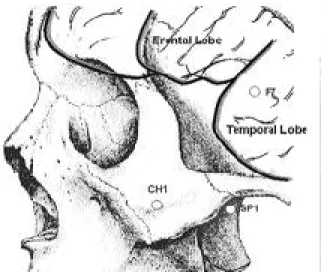
나비뼈전극은 하악절흔(mandibular notch), 즉 이주( t r a- gus) 2~3 cm 전방에서 외측익돌근판(lateral pterygoid p l a t e )에 인접하여 반대쪽 이주 방향을 향해 4~6 cm 깊이 로 삽입하였다. 뺨전극은 접형전극 삽입부의 전방 약 1 cm 부위로서 관골안면공(zygomaticofacial foramen)의 직하 방 부위에 접지하였다(Fig. 1).
장비는 T e l e f a c t o r사의 비디오-뇌파 감시장치( V i d e o - EEG monitoring system, West Conshohocken, USA) 를 이용하였고 뇌파는 여러 가지 몽타주를 사용하여 디지털 평가 장비를 이용하여 분석하다가 필요한 경우 종이로 출력하 여 판독하였다.
뇌파기록의 판독
발작시 뇌파는 나비뼈전극 혹은 뺨전극을 포함하는 종축 ( l o n g i t u d i n a l )과 횡축( t r a n s v e r s e )의 양극 몽타주( b i p o- lar montage) 혹은 기준 몽타주(reference montage)로 출력하여 분석하였는데 양극 몽타주에서 나비뼈전극( S p )과 뺨전극( C h )을 F7-Sp1(or Ch1)-T3-T5/F8-Sp2(or Ch2)- T4-T6 혹은 Cz-Sp2(or Ch2)-Sp1(or Ch1)-Cz로 배열하 Figure 1. Approximate placement of sphenoidal and cheek
electrodes in the left temporal lobe. SP; sphenoidal electrode, CH; cheek electrode
Table 1. Clinical, imaging and EEG findings
Cases Age(years)/sex No. of Seizure MRI Ictal onset patterns Localization by SE Localization by CE
01 34/M 5 HA RTA Left temporal Left temporal
02 43/M 7 HA RTA Right temporal Right temporal
03 16/F 3 HA RTA Left temporal Left temporal-suspicious
04 23/F 4 Tumor RTA Right temporal Right temporal
05 21/M 5 HA RTA Right temporal Right temporal
06 22/M 1 HA RTA Left temporal Left temporal
07 26/M 7 CM RTA Right temporal Right temporal
08 41/F 5 HA RTA Right temporal Right temporal
09 26/F 4 HA RTA Left temporal Left temporal
10 20/F 1 HA RTA Left temporal Left temporal
11 28/M 8 Normal RTA Left temporal Left temporal
12 31/F 12 HA Arrhythmic activity Bilateral Bilateral
13 21/M 10 HA RTA Right temporal Right temporal
14 9/M 9 HA RTA Left temporal (+ noise) Left temporal
15 26/F 3 CM RAA Bilateral Bilateral
16 41/M 4 HA RTA Left temporal Left temporal
17 35/F 7 Normal RDA Right temporal Right temporal
SE; sphenoidal electrode, CE; cheek electrode, HA; hippocampal atrophy, CM; cortical malformation, RTA; rhythmic theta activity, RAA; rhythmic alpha activity, RDA; rhythmic delta activity, Tumor; pilocytic astrocytoma
여 비교 분석하였다. 두피 뇌파기록은 2인의 간질 전문의가 독립적으로 판독하였다.
발작시 뇌파는 배경 뇌파와 뚜렷이 구분되는 r h y t h m i c activity (alpha, theta or delta frequencies), parox- ysmal fast (≥13 Hz), suppression (≤10 ㎶ i n amplitude), repetitive epileptiform activity (3 or more discharges in sequence), arrhythmic activity, obscured 로 나누어 분석하였으며, 각 몽타주에서 발작 시작 파가 보이는 위치를 기록하고, 발작 시작 시간을 기록하였다.
결 과
측두엽간질이 의심되는 일련의 대상 환자 1 7명 중 남자가 9명, 여자는 8명이었다. 연령은 9세부터 4 3세까지 평균 연 령은 2 7±9세였다. 발작의 원인은 특발성 1 3예, 피질 기형 2예, 외상 1예, 뇌종양 1예였다. 발작회수는 한 환자 당 1회 에서 1 2회까지 총 9 5회였다(Table 1).
1. 발작시 뇌파 소견
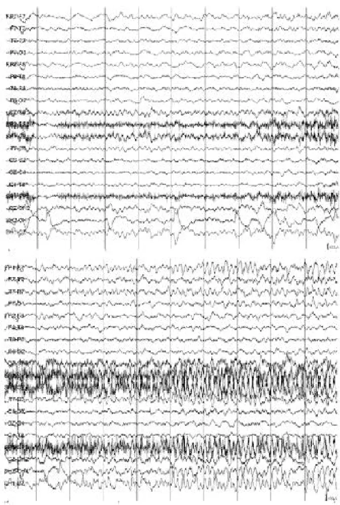
발작파의 모양은 rhythmic theta activity가 대부분으로 나비뼈전극이나 뺨전극을 포함한 몽타주에서 현저히 비슷한 모양이었으나 진폭은 뺨전극에서 조금 낮은 경향을 보였다 (Fig. 2). 발작 시작 파의 위치와 분포에 대하여 각각의 전극 몽타주를 이용한 판독에서 두 명의 판독자가 모두 일치하게 동일한 부위로 판독한 경우는 1 7명 중 1 6명으로 9 4 . 1 %의 일치도(agreement rate)를 보였으며 나머지 한 환자(증례
3 )의 경우에서도 뺨전극에서 잡파가 섞여 분명하지 않았지만 뇌파 판독자 2명의 의견을 조율하였을 때 뺨전극 몽타주에서 나비뼈전극 몽타주와 동측으로 발작파를 판독하여 전체적으 로 거의 1 0 0 %의 일치도를 보였다. 어느 환자도 나비뼈전극 이나 뺨전극을 포함한 몽타주에서 발작 시작 부위가 서로 반 대측으로 상이하게 판독된 경우는 없었다. 나비뼈전극 몽타주 에서 발작파의 분포는 뺨전극 몽타주에서 역시 동일하였다.
발작 시작 시간은 각 전극을 포함한 몽타주에서 거의 동일하 며 나비뼈전극 몽타주에서 뺨전극 몽타주에 비해 평균 0 . 4 9
±0.3 초 빨리 감지 되었지만 임상적 의미는 없었다. 대상 환자 가운데 발작 중 나비뼈전극이 밖으로 빠진 경우 그 이후 로 일련의 발작시 뺨전극을 통해 발작 시작 부위를 확인하는 데 도움이 된 경우도 있었으며 드물게는 발작시 나비뼈전극에 서 잡파가 섞여 보이는 경우 뺨전극에서 발작파를 더 분명히 보이는 경우도 있었다(Fig. 3). 대체로 나비뼈전극 뇌파는 씹는(chewing) 동작에, 뺨전극 뇌파는 안구운동에 영향을 많이 받았다.
고 찰
내측 측두엽간질은 수술적 치료를 요하는 간질 중 비교적 흔한 종류이다. 측두엽간질이 의심되는 환자에서 비디오-뇌파 검사시 국제 표준 10-20 전극계에 의한 전극과 더불어 비전 형전극을 많이 이용한다.2 - 6이중 나비뼈전극은 해부학적으로 내측-기저부 측두엽에 가장 가까이 위치하며, 전극과 뇌파를 생성하는 신경 조직 사이에 전도를 방해하는 뼈 등의 물질이 Figure 2. Representative EEG shows nearly identical patterns when either sphenoidal or cheek electrodes are substituted in bipolar montages. The amplitude of recorded epileptiform activity is little higher with sphenoidal electrodes compared to check electrodes.
SP; sphenoidal electrode, CH; cheek electrode
Figure 3. An example This is more clear ictal pattern with the use of cheek electrodes compared with sphenoidal electrodes. SP;
sphenoidal electrode, CH; cheek electrode
없어 내측 측두엽의 진폭이 낮은 발작파를 빨리 감지 할 수 있고, 잡파가 적어서 내측 측두엽간질과 외측 측두엽간질의 감별에 도움이 된다고 알려져 왔다.1 3 - 1 5하지만 최근 나비뼈에 인접한 다른 두피전극들이 이를 대신할 수 있다는 연구도 있 어 나비뼈전극의 임상적 필요성에 관하여 논란이 있다.1 0 , 1 8 - 2 2
측두엽간질 환자에서 비록 투시기 안내 하에 나비뼈 전극을 삽입한 경우라도 발작 시작 부위를 확인하는데 있어 두피전극 보다 단지 5 . 4 ~ 7 %만 더 도움이 된다는 연구가 있고,1 8측두 엽간질이 의심되는 환자들을 대상으로 발작파를 관찰한 결과 나비뼈 전극과 뺨전극에서 거의 동일하게 발작파를 확인할 수 있었으며 뺨전극에서 뇌파의 진폭이 나비뼈전극에 비해 약 10~20% 정도 작았지만 이러한 차이는 발작 시작 파의 위 치 결정에 있어 영향을 주지 않는다는 연구도 있었다.1 9본 연 구에서도 측두엽간질 환자 대부분에서 뺨전극과 나비뼈전극 뇌파에서 발작 시간, 위치 결정에 관하여 동일한 임상적 정보 를 제공해 주어 Krauss 등1 9의 결과와 일치 하였다. 본 연구 에서 비록 1 7명의 측두엽간질이 의심되는 환자의 9 5회 발작 을 대상으로 하였지만 모두 본원을 방문한 연속적인 환자들을 대상으로 하여 거의 1 0 0 %에 가까운 일치도를 보였기에 상당 히 의미가 있다고 생각되며 더욱이 나비뼈전극에서는 씹는 동 작에 대한 잡파가, 뺨전극에는 안구운동에 관한 잡파가 많이 포함되는 경향을 보여 드물지만 나비뼈전극에 잡파가 포함된 경우 뺨전극에서 더 분명히 발작파를 확인할 수 있었던 경우 도 있어 뺨전극의 유용성이 강조되었다.
두피 뺨전극이 발작 기시부인 내측-기저부 측두엽에 인접한 나비뼈전극보다 거리가 있음에도 불구하고 동일하게 발작파 를 감지한 것은 발작파가 용적 전도(volume conduction) 를 하며 난원공(foramen ovale)과 상안와열( s u p e r i o r orbital fissure) 같은 열공을 통해 두피에 도달하여 두개강 외로 넓게 분포한다는 연구를 통하여 설명할 수 있다.2 3 , 2 4이 것은 또한 측두엽간질에 있어 발작파를 측두엽에 가까이 위치 한 T3/T4 전극보다 전방 측두전극에서 더 민감하게 감지할 수 있다는 연구2 3나, 발작파가 두피에 일정한 양상으로 전파 되며 관골 안면공 주위에서 가장 집중적으로 나타난다는 연구
2 4도 이러한 결과를 뒷받침 한다. 즉 본 연구에 사용된 뺨전극 이 이러한 부위에 위치하여 나비뼈전극과 거의 동일하게 발작 파를 감지 할 수 있었다고 생각된다. 그런데 나비뼈전극 뇌파 와 뺨전극 뇌파 사이에 진폭이 차이가 나는 것은 발작 기시부 로부터 전극까지의 거리가 다르기 때문으로 생각된다. 이러한 나비뼈전극 뇌파가 두피전극 뇌파에 비해 작은 차이지만 진폭 이 커서 진폭이 낮은 발작파는 두피전극까지 도달하지 않거나 도달하더라도 나비뼈전극보다 늦게 감지할 것이라고 이론적 으로 생각할 수 있다. 하지만 본 연구와 다른 연구에서 보듯 이 이러한 차이는 실지 임상에서 크게 문제가 되지 않는
다.1 0 , 1 8 - 2 0부분 발작의 경우 발작이 진행함에 따라 발작파의
진폭은 증가하고 주파수는 감소하나2 5 이러한 뇌파를 감지하 는데 있어 나비뼈전극과 두피전극은 서로 시간적이 차이가 나 더라도 3 . 7초 이내로 크지 않고 또한 서로 상반되게 편측화 되지 않아서 임상적인 차이가 없다고 하며1 8 실제로 본 연구 에서도 약간의 시간적 차이가 나지만 이러한 시간적 차이는
뇌파의 진폭 차이에 의해 생겼다고 생각되며 발작 시작 부위 를 확인하는데 문제가 되지는 않았다.
임상에서 내측 측두엽간질의 경우 나비뼈전극에서 먼저 발 작파가 국한하여 나타나다가 다른 두피전극으로 전파되는 양 상을 보이는 경우가 있다는 보고1 3가 있으며 측두엽간질의 경 우에도 드물게는 나비뼈전극이나 뺨전극 등 두피 뇌파에 보이 지 않을 수 있고, 임상 양상과 뇌영상 소견에 따라서 선택적 으로 나비뼈전극이나 두개강내전극이 필요로 할 때가 있다는 것을 기억해야 한다. 또한 Pacia 등1 3은 두개강내전극을 이용 하여 신피질 측두엽간질로 확인된 2 0명을 대상으로 발작 뇌 파를 분석한 결과 5 1회 발작에서 두피전극과 나비뼈전극에서 동시에 발작파를 보였다고 하였고, Marks 등2 6은 나비뼈전극 과 두개강내전극 등으로 동시에 기록된 극파를 비교한 결과 내측두엽뿐 아니라 측두엽외 부위에서 발생하는 극파도 나비 뼈전극에서 최대 진폭의 전위를 보일 수 있기 때문에, 나비뼈 전극에서 최대 진폭을 보이는 극파가 모두 내측두엽간질 발작 에 특이 소견은 아니라고 하였다. 따라서 본 연구에서도 두개 강내전극을 삽입하여 동시에 검사를 하지 않았으므로 나비뼈 전극이나 뺨전극에서 보이는 발작파 소견이 모두 내측 측두엽 발작파라고 단정할 수는 없으므로 발작파 분석에 있어 이점을 유념하여야 하겠다.
나비뼈전극은 부분적으로 침습적인 방법으로 환자에게 상 당한 고통과 불편감을 주며 드물지만 출혈, 감염, 전극의 내 부 분절, 전극에 의한 타액선이나 타액관 관통, 일시적인 안 면신경 마비, 입 주위에 이상 감각, 뇌막염 및 지주막하출혈 등의 다양한 부작용이 보고되어 있다.1 0 , 1 6 , 1 7그래서 본 연구를 통하여 볼 때 나비뼈전극을 일상적으로 사용하는 것은 바람직 하지 않으며 많은 경우에서 두피 뺨전극이 나비뼈전극을 대신 할 수 있을 것으로 생각된다.
결론적으로, 뺨전극이 측두엽에서 발생하는 간질파만을 기 록하는 것은 아니고 전두엽 등의 간질파를 기록할 가능성도 있으나 뺨전극은 측두엽간질이 의심되는 환자에서 발작의 시 작 부위를 확인하는데 나비뼈전극과 거의 동일하게 발작 시작 부위를 확인할 수 있었다. 이러한 결과는 향후 측두엽간질 환 자들을 검사함에 있어 뺨전극이 나비뼈전극을 대신할 수 있는 하나의 이론적 근거가 될 수 있다. 그리고 나아가 만약 나비 뼈전극이 꼭 필요한 경우라도 뺨전극을 함께 부착하면 발작파 를 하나 이상의 전극에서 감지할 수 있고 잡파가 생기더라도 두 전극 동시에 나타나지 않을 가능성도 있어 발작 시작 부위 를 확인하는데 더 많은 도움이 될 것으로 생각되며 향후 뺨전 극의 진단율의 민감도와 특이도에 관하여 연구해 보는 것도 의미 있을 것으로 생각된다.
R E F E R E N C E S
01. Jasper HH. Report of the committee on methods of clinical examination in electroencephalography. E l e c t r o e n c e p h a l - ogr Clin Neurophysiol 1958;10:370-375.
02. S i n d r u p E , T h y g e s e n N , K r i s t e n s e n O , A l v i n g J . Zygomatic electrodes: their use and value in complex par- tial epilepsy. In: M.Dam, I. Gram and J.K. Penry(Eds.),
Advances in epileptology . New York: Reven Press, 1981;313.
03. Sunwoo IN, Kim KW, Kim SM. Clinical significance of nasopharyngeal EEG in patients with psychomotor seizure. J Korean Neurol Assoc 1987;5:13-23.
04. Arellano AP. A tympanic lead. Electroenceph clin Neurophysiol 1949;1:112-113.
05. Sadler RM. Goodwin J. Multiple electrodes for detecting spikes in partial complex seizures. Can J Neurol Sci 1989;16:326-329.
06. Jones DP. Recording of the basal electroencephalogram with sphenoidal needles electrodes. E l e c t r o e n c e p h a l o g r Clin Neurophysiol 1951;3:100.
07. Choi SM, Byun YJ, Kim SM, Lee J, Kim MJ, Park MY, Hah JS. Efficacy of Non-standard Surface Electrodes for Detecting Epileptiform Discharges in Patients with Temporal Lobe epilepsies. J Korean Neurol Assoc 1995;13:233-238.
08. Kim OJ, Lee BI. A New Approach in Clinical Usefulness of Foramen Ovale Electrode in Epilepsy Surgery. J Korean Neurol Assoc 1999;17:505-513.
09. Lee NS, Lee JH, Lee KW, Myung HJ. A comparison of anterior temporal and nasopharyngeal electrodes in the detection of epileptiform activities from temporal Lobes, a preliminary report. J Korean Neurol Assoc 1 9 9 1 ; 9 : 4 5 1 - 455.
10. Wilkus RJ, Vossler DG, Thompson PM. Comparison of EEG derived from sphenoidal, infrazygomatic, anterior temporal, and midtemproal electrodes during complex par- tial seizures. J Epilepsy 1993;6:152-161.
11. Kristensen O, Sindrup EH. Sphenoidal electrodes: their use and value in the electroencephalographic investigation of complex partial epilepsy. Acta Neurol Scand 1978;58:157-166.
12. Pampiglione G, Kerridge J. EEG abnormalities from the temporal lobe studied with sphenoidal electrodes. J Neurol Neurosurg Psychiatry 1956;19:117-129.
13. Pacia SV, Jung WJ, Devinsky O. Localization of mesial temporal lobe seizures with sphenoidal electrodes. J Clin Neurophysiol 1998;15:256-261.
14. Kanner AM, Ramirez L, Jones JC. The utility of placing sphenoidal electrodes under the foramen ovale with fluo- roscopic guidance. J. Clin Neurophysiol 1995;12:72-81.
15. Kanner AM, Jones JC. When do sphenoidal electrodes yield additional data to that obtained with antero-temporal electrodes? Electroencephalogr Clin Neurophysiol
1997;102:12-19.
16. Iriarte J, Parra J, Kanner AM. Transient facial palsy in sphenoidal electrode placement. E p i l e p s i a 1 9 9 6 ; 3 7 : 1 2 3 9 - 1241.
17. Wilkus RJ, Vossler DG, Rudd TG. Salivary ductal pene- tration during insertion of a sphenoidal electrode.
Electroencephalogr Clin Neurophysiol 1990;76:36.
18. Kissani N, Alarcon G, Dad M, Binnie CD, Polkey CE.
Sensitivity of recordings at sphenoidal electrode site for detecting seizure onset: evidence from scalp, superficial and deep foramen ovale recordings. E l e c t r o e n c e p h a l o g r Clin Neurophysiol 2001;112:232-240.
19. Krauss GL, Lesser RP, Fisher RS, Arroyo S. Anterior
“cheek” electrodes are comparable to sphenoidal elec- trodes for the identification of ictal activity.
Electroencephalogr Clin Neurophysiol 1992;83:333-338.
20. Homan RW, Jones MC, Rawat S. Anterior temporal elec- trodes in complex partial seizures. E l e c t r o e n c e p h a l o g r Clin Neurophysiol 1988;70:105-109.
21. Fernandez Torre JL, Alarcon G, Binnie CD, Polkey CE.
Comparison of sphenoidal, foramen ovale and anterior temporal placements for detecting interictal epileptiform discharges in presurgical assessment for temporal lobe epilepsy. E l e c t r o e n c e p h a l o g r Clin Neurophysiol 1999;110:895-904.
22. Binnie CD, Marston D, Polkey CE, Amin D. Distribution of temporal spikes in relation to the sphenoidal electrode.
Electroencephalogr Clin Neurophysiol 1989;73:403-409.
23. Fernandez Torre JL, Alarcon G, Binnie CD, Seoane JJ, Juler J, Guy CN, Polkey CE. Generation of scalp dis- charges in temporal lobe epilepsy as suggested by intraop- erative electrocorticographic recordings. J Neurol Neurosurg Psychiatry 1999;67;51-58.
24. Fisch BJ. EEG Primer. Basic principles of digital and analog EEG. 3rd ed. Amsterdam: Elsevier Science BV, 1999;22-24.
25. Alarcon G, Binnie CD, Elwes RD, Polkey CE. Power spectrum and intracranial EEG patterns at seizure onset in partial epilepsy. Electroencephalogr Clin Neurophysiol 1995;94:326-337.
26. Marks DA, Katz A, Booke J, Spencer DD, Spencer SS.
Comparison and correlation of surface and sphenoidal electrodes with simultaneous intracranial recording: an interictal study. Electroencephalogr Clin Neurophysiol 1992;82:23-29.