Korean J Intern Med 2016;31:54-64 http://dx.doi.org/10.3904/kjim.2016.31.1.54
INTRODUCTION
Nonvariceal upper gastrointestinal bleeding (NVUGIB) is the most common medical emergency, and is a con- siderable clinical and economic burden [1]. Although the mortality rate in patients with NVUGIB has declined dramatically in recent years due to the development of proton pump inhibitors and endoscopic therapy, it remains high at 5% to 10% [2,3]. A recent internation- al consensus on NVUGIB emphasized the importance
of early risk stratification for rebleeding and mortality [4]. Several risk factors for this condition have been pro- posed, and of these, rebleeding was a significant predic- tor of mortality [5-7]. Other predictive factors include advanced age, liver cirrhosis, chronic kidney disease, ad- vanced neoplasia, low hemoglobin level, cardiac failure, and hemodynamic instability [1,8]. Early recognition of the risk of death would facilitate differentiation between high- and low-risk patients. Close monitoring and care- ful management of high-risk patients could improve
1Division of Gastroenterology and Hepatology, Department of Internal Medicine, Keimyung University School of Medicine, Daegu; 2Division of Gastroenterology and Hepatology, Department of Internal Medicine, Kyungpook National University School of Medicine, Daegu, Korea
Received : April 3, 2014 Revised : July 28, 2014 Accepted : November 13, 2014 Correspondence to
Eun Soo Kim, M.D.
Division of Gastroenterology and Hepatology, Department of Internal Medicine, Keimyung University School of Medicine, 56 Dalseong-ro, Jung-gu, Daegu 41931, Korea
Tel: +82-53-250-8096 Fax: +82-53-250-7442
E-mail: [email protected]
*These authors contributed equally to this work.
Background/Aims: Nonvariceal upper gastrointestinal bleeding (NVUGIB) is a common medical emergency that can be life threatening. This study evaluated predictive factors of 30-day mortality in patients with this condition.
Methods: A prospective observational study was conducted at a single hospital be- tween April 2010 and November 2012, and 336 patients with symptoms and signs of gastrointestinal bleeding were consecutively enrolled. Clinical characteristics and endoscopic findings were reviewed to identify potential factors associated with 30-day mortality.
Results: Overall, 184 patients were included in the study (men, 79.3%; mean age, 59.81 years), and 16 patients died within 30 days (8.7%). Multivariate analyses re- vealed that comorbidity of diabetes mellitus (DM) or metastatic malignancy, age
≥ 65 years, and hypotension (systolic pressure < 90 mmHg) during hospitalization were significant predictive factors of 30-day mortality.
Conclusions: Comorbidity of DM or metastatic malignancy, age ≥ 65 years, and hemodynamic instability during hospitalization were predictors of 30-day mor- tality in patients with NVUGIB. These results will help guide the management of patients with this condition.
Keywords: Gastrointestinal hemorrhage; Mortality; Prognosis; Comorbidity
Predictive factors of mortality within 30 days in patients with nonvariceal upper gastrointestinal bleeding
Yoo Jin Lee1,*, Bo Ram Min1,*, Eun Soo Kim1, Kyung Sik Park1, Kwang Bum Cho1, Byoung Kuk Jang1, Woo Jin Chung1, Jae Seok Hwang1, and Seong Woo Jeon2
their prognosis. Therefore, predictors of mortality in patients with NVUGIB are clinically valuable. The aim of this prospective study was to investigate potential predictors associated with 30-day mortality in patients admitted to the emergency unit for this condition.
METHODS
PatientsThis prospective, single-center, observational study was conducted at a tertiary hospital in Daegu, Korea (Keimyung University Dongsan Hospital), and was approved by the institutional review board of the university (No. 11-294).
Written informed consent was obtained from all of the subjects enrolled in the study. This study is registered in the World Health Organization International Clinical Trials Registry Platform (KCT0000514).
Adult patients (> 18 years), clinically diagnosed with gastrointestinal bleeding between April 2010 and No- vember 2012, were enrolled. Patients were excluded if they did not complete at least 30 days of follow-up, if the bleeding source was varices or gastric cancer, if bleed- ing was associated with endoscopic procedures such as endoscopic mucosal resection, or if bleeding occurred in the lower gastrointestinal tract. The following infor- mation was documented prospectively: patient data (age, sex, date of admission, and endoscopy results); historical data (presenting symptoms, previous history of gastro- intestinal bleeding, or peptic ulcer disease); social histo- ry such as current smoking status, alcohol consumption (heavy alcoholics were defined as women and men who consumed more than 40 and 60 g alcohol per day, re- spectively, for more than 5 years) [9]; physical findings (results of nasogastric tube aspiration, results of rec- tal examination, and initial hemodynamic status); ini- tial laboratory data (hemoglobin and blood urea levels, platelet count, and nitrogen prothrombin time); and co- morbidities including hypertension, diabetes mellitus (DM), ischemic heart disease, cerebrovascular disease, heart failure, liver cirrhosis (liver cirrhosis was defined according to clinical, laboratory, and radiologic data but not liver biopsy results because biopsy was not per- formed) [10], chronic kidney disease (defined as patients with an estimated glomerular filtration rate < 60 mL/
min for at least 3 months calculated using the four-vari-
able Modification of Diet in Renal Disease Study equa- tion) [11], metastatic malignancy, and peripheral vascu- lar disease. Hemodynamic instability was defined as tachycardia with a heart rate > 100 beats per minute and hypotension with a systolic pressure < 90 mmHg upon admission and during hospitalization. Medication his- tory (antiplatelet agents, vitamin K antagonists, nonste- roidal anti-inflammatory drugs [NSAIDs], steroids, and proton pump inhibitors) was also recorded if patients had taken the aforementioned drugs 1 week before the first bleeding event. Patients were managed according to recent consensus recommendations, including en- doscopic and pharmacologic management as well as transfusion [4]. All of the patients were intravenously administered proton pump inhibitors. In addition, each patient’s risk of bleeding was assessed by a Rockall score and a Blatchford score. The total Rockall score includes both endoscopic and nonendoscopic variables, where- as the Blatchford score is only based on nonendoscopic variables [6,7]. High-risk patients were defined as those with a total Rockall score ≥ 5, indicating an estimated mortality rate of 40% [6].
Endoscopic procedures
All of the endoscopic procedures for acute upper gas- trointestinal bleeding (UGIB) were performed within 24 hours after arrival at the hospital. Procedures were per- formed by experienced endoscopists who had previously conducted > 1,000 endoscopies. Urgent endoscopy was defined as an endoscopy procedure conducted within 12 hours of admission [4]. High-risk patients underwent urgent endoscopy according to a strict hospital proto- col. However, in patients who showed unfavorable clin- ical conditions for endoscopy or in those who arrived at night or during the weekend, the decision to perform this procedure was made by the individual endoscopist.
Endoscopic variables included the cause of UGIB and location of the lesion. Lesions are either located in the body of the stomach, where the risk of bleeding is high due to numerous supplying vessels, or in other parts of the stomach [12]. Unidentified causes of bleeding were defined as the presence of blood in the stomach without any identifiable source of bleeding [3]. In patients with ulcers, bleeding activity at the ulcer base was classified according to the Forrest classification [13]. Forrest I, IIa, and IIb represent high-risk bleeding stigmata (HRBS)
[3,6]. Endoscopic findings were reviewed and adjusted by two expert endoscopists (YJL and ESK) to improve interobserver agreement.
Outcomes
The primary outcome was mortality within 30 days of admission. Patients were advised to return for an exam- ination at 4 and 12 weeks after discharge. For discharged or deceased patients, the relevant data from hospital records were extracted by investigators. Patients with hospital record data that did not satisfy the inclusion criteria were excluded. A 30-day rebleeding episode was defined as the onset of new hematemesis or hematoche- zia with hypovolemic shock, or a greater than 2 g/dL de- crease in blood hemoglobin levels after a 24-hour period of stable vital signs within 30 days of admission [3,14]. All of the rebleeding episodes were confirmed with endos- copy. Bleeding-related 30-day mortality included death from irreversible hypovolemic shock during surgery for uncontrolled bleeding or endoscopy-related mortality occurring within 30 days of the index-bleeding episode.
Nonbleeding-related 30-day mortality was defined as death from cardiac, pulmonary or cerebrovascular dis- ease, liver or kidney failure, and malignant disease [15].
Statistical analysis
Statistical analyses were performed using SPSS version 18.0 (SPSS Inc., Chicago, IL, USA). The Student t test was used for comparison of continuous variables. Categor- ical variables were compared using Fisher exact test or a chi-square test. Independent risk factors for 30-day mortality were assessed by multivariate logistic regres- sion analysis. An odds ratio (OR) and 95% confidence interval (CI) was calculated for each independent factor.
A two-tailed p < 0.05 was considered statistically signif- icant.
RESULTS
Patient characteristics
During the study period, 336 patients with gastrointes- tinal bleeding were admitted, and 184 (54.8%) met the inclusion criteria (Fig. 1). A total of 144 patients were ex- cluded from the study because the source of bleeding was varices (82 patients), the lower gastrointestinal tract
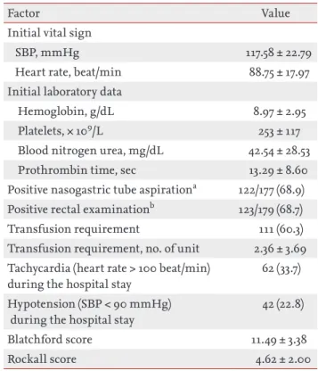
(59 patients), or gastric cancer (3 patients). Eight patients were lost to follow-up. Patient characteristics are shown in Table 1. The mean age was 59.81 years, and 79.3% of patients were male. The most frequently presenting symptom was hematemesis (50.0%). A total of 48 patients (26.1%) had a previous history of gastrointestinal bleed- ing, and 60 (32.6%) had a previous history of peptic ulcer disease. The most common comorbidity was hyperten- sion (82 patients, 44.6%), followed by DM (44 patients, 23.9%). With regard to concomitant use of drugs that could have been related to bleeding, 53 patients (28.8%) used antiplatelet agents including aspirin, clopidogrel, or cilostazol, and 37 patients (20.1%) used NSAIDs. As outlined in Table 2, the mean serum level of hemo- globin upon admission was 8.97 g/dL. The percentage of patients with tachycardia (heart rate > 100 beats per minute) and hypotension (systolic pressure < 90 mmHg) during hospitalization was 33.7% and 22.8%, respective- ly. The percentage of patients with positive nasogas- tric tube aspiration and digital rectal examination was 68.9% (122/177) and 68.7% (123/179), respectively. Packed red blood cells were transfused in 111 patients (60.3%), and the mean number of units transfused was 2.36 ± 3.69. The mean total Rockall score was 4.62 ± 2.00, and 96 patients (52.2%) had a score ≥ 5, indicating a high risk of mortality. The mean Blatchford score was 11.49 ± 3.38.
An urgent endoscopy (< 12 hours after admission) was conducted in 121 patients (65.8%), whereas the remain- ing patients (63, 34.2%) underwent endoscopy 12 to 24
336 Patients suspected UGIB (from 2010~2012)
3 Gastric cancer bleeding
8 Lost to follow-up
184 Included patients
82 Varix bleeding 59 Lower gastrointestinal bleeding
Figure 1. Flow chart of patient selection in the study. UGIB, upper gastrointestinal bleeding.
hours after admission. Peptic ulcer was the main cause of bleeding (78.3%); other causes of bleeding were Mal- lory-Weiss syndrome (12.5%), angiodysplasia (1.6%), and hemorrhagic gastritis (0.5%). The cause of bleeding in 13 patients (7.1%) could not be identified despite the pres- ence of blood in the stomach. Of the 144 patients with peptic ulcer, 78 (54.2%) showed HRBS (Forrest I, 22.2%;
IIa, 20.8%; and IIb, 11.1%) (Table 3).
Clinical results
The median follow-up period was 196 days (interquar-
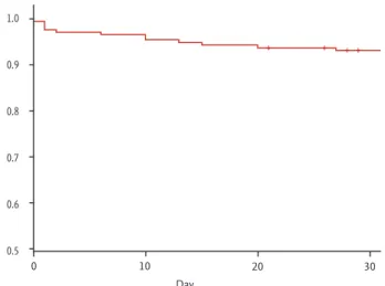
tile range, 77 to 404), and the overall number of deaths during this period was 38 (20.7%). The number of deaths within 30 days was 16 (8.7%) (Table 4). Fig. 2 shows the survival curve of patients with NVUGIB over a follow-up period of 30 days. Among patients who died within 30 days, half (8/16, 50.0%) died during the first 7 days; the 7-day survival probability was estimated to be 96.6%.
The causes of 30-day mortality are presented in Table 5. Bleeding-related death within 30 days occurred in five patients (31.2%), and all of them died within the first 7 days. The deaths of the remaining 11 patients (68.8%) were associated with their comorbidities. Cardiovascu- lar events including heart failure or acute myocardial infarction were the most frequent nonbleeding-related causes of 30-day mortality (seven patients, 43.8%), fol- lowed by liver failure (two patients, 12.5%), brain hemor- rhage (one patient, 6.3%), and metastatic cancer progres- sion (one patient, 6.3%). Overall, rebleeding occurred in 38 patients (20.7%) during the follow-up period, and rebleeding within 30 days of admission occurred in 27 Table 1. General characteristics of patients with nonvariceal
upper gastrointestinal bleeding (n = 184)
Factor Value
Male sex 146 (79.3)
Age, yr 59.81 ± 15.80
Bleeding related symptoms
Hematemesis 92 (50.0)
Tarry stool 77 (41.8)
Hematochezia 12 (6.5)
Acute onset anemia 3 (1.6)
Heavy alcoholics 72 (39.1)
Current smoker 68 (37.0)
Past history of gastrointestinal bleeding 48 (26.1) Past history of peptic ulcer disease 60 (32.6) Comorbidities
Hypertension 82 (44.6)
Diabetes mellitus 44 (23.9)
Liver cirrhosis 30 (16.3)
Chronic kidney disease 28 (15.2)
Cerebrovascular disease 26 (14.1)
Heart failure 27 (14.7)
Cardiovascular disease 22 (12.0)
Metastatic malignancy 8 (4.3)
Peripheral vascular disease 4 (2.2) Use of medication
Antiplatelet agents 53 (28.8)
NSAIDs 37 (20.1)
Vitamin K antagonist 8 (4.3)
PPI co-medication 7 (3.8)
Values are presented as number (%) or mean ± SD.
NSAID, nonsteroidal anti-inflammatory drug; PPI, proton pump inhibitor.
Table 2. Clinical data at the time of admission to the hospi- tal for nonvariceal upper gastrointestinal bleeding (n = 184)
Factor Value
Initial vital sign
SBP, mmHg 117.58 ± 22.79
Heart rate, beat/min 88.75 ± 17.97
Initial laboratory data
Hemoglobin, g/dL 8.97 ± 2.95
Platelets, × 109/L 253 ± 117
Blood nitrogen urea, mg/dL 42.54 ± 28.53
Prothrombin time, sec 13.29 ± 8.60
Positive nasogastric tube aspirationa 122/177 (68.9) Positive rectal examinationb 123/179 (68.7)
Transfusion requirement 111 (60.3)
Transfusion requirement, no. of unit 2.36 ± 3.69 Tachycardia (heart rate > 100 beat/min)
during the hospital stay
62 (33.7)
Hypotension (SBP < 90 mmHg) during the hospital stay
42 (22.8)
Blatchford score 11.49 ± 3.38
Rockall score 4.62 ± 2.00
Values are presented as mean ± SD or number (%).
SBP, systolic blood pressure.
aNasogastric tube was performed in 177 patients.
bRectal examination was performed in 179 patients.
patients (14.7%). The median length of hospitalization was 6 days.
Predictive factors for 30-day mortality in patients with NVUGIB
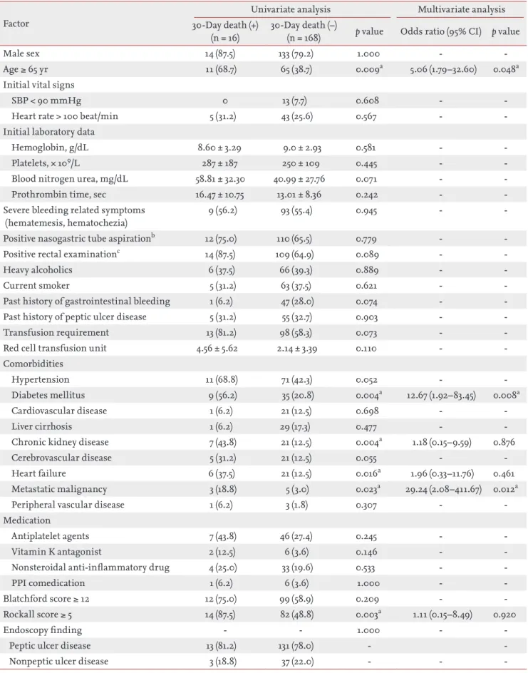
According to univariate analysis, age ≥ 65 years (p = 0.009), DM (p = 0.004), chronic kidney disease (p = 0.004), metastatic malignancy (p = 0.023), heart failure (p = 0.016), Rockall score ≥ 5 (p = 0.003), tachycardia (heart rate > 100 beats per minute) during hospitalization (p < 0.001), hy- potension (systolic pressure < 90 mmHg) during hos- pitalization (p < 0.001), and rebleeding within 30 days (p
< 0.001) were significant risk factors for 30-day mortal- ity. Multivariate logistic regression analysis identified the following variables as independent predictors of increased 30-day mortality in patients with NVUGIB:
comorbidity of DM (OR, 12.67; 95% CI, 1.92 to 83.45;
p = 0.008) or metastatic malignancy (OR, 29.24; 95% CI, 2.08 to 411.67; p = 0.012), age ≥ 65 years (OR, 5.06; 95% CI, 1.79 to 32.60; p = 0.048), and hypotension (systolic pres- sure < 90 mmHg) during hospitalization (OR, 16.63; 95%
CI, 2.56 to 107.90; p = 0.003) (Table 6). Predictors for 30- day mortality were also analyzed after dividing patients into high- and low-risk bleeding stigmata groups. How- ever, there were no independent risk factors for 30-day mortality according to risk stratification by Forrest clas- sification (Supplementary Tables 1 and 2).
Table 3. Endoscopic features of patients with nonvariceal upper gastrointestinal bleeding (n = 184)
Factor No. (%)
Urgent endoscopya 121 (65.8)
Endoscopy finding
Peptic ulcer disease 144 (78.3)
Mallory-Weiss syndrome 23 (12.5)
Angiodysplasia 3 (1.6)
Hemorrhagic gastritis 1 (0.5)
No evidence of upper gastrointestinal bleeding
13 (7.1)
Endoscopy lesion location
Cardia, angle, antrum 89 (48.4)
Body 46 (25.0)
Duodenum 35 (19.0)
No specific lesion 14 (7.6)
Forrest classificationb
I 32/144 (22.2)
IIa 30/144 (20.8)
IIb 16/144 (11.1)
IIc 40/144 (27.8)
III 26/144 (18.1)
aEndoscopy which was performed within 12 hours of admis- sion.
bClassified in 144 patients who had ulcers.
Table 4. Clinical outcomes of patients with nonvariceal upper gastrointestinal bleeding (n = 184)
Factor Value
Rebleeding
During follow-up perioda 38 (20.7)
Within 30 days 27 (14.7)
Death
During follow-up perioda 38 (20.7)
Within 30 days 16 (8.7)
Hospital stay, day 5.90 ± 5.80
Values are presented as number (%) or mean ± SD.
aMedian follow-up period of 196 days (interquartile range, 77–404 days).
1.0 0.9 0.8 0.7 0.6
0.5
0 10 20 30
Day
Figure 2. Kaplan-Meier survival curve of 30-day mortality in patients with nonvariceal upper gastrointestinal bleed- ing. Eight patients died during the first 7 days (cumulative survival, 96.6%).
DISCUSSION
In our study, we found that age (≥ 65 years), comorbid- ity of DM or metastatic malignancy, and hypotension (systolic pressure < 90 mmHg) during hospitalization were independently associated with mortality within 30 days. The 30-day mortality rate was 8.7%, which was slightly higher than the rates of 5.4% and 4.5% reported by the Canadian registry [16] and Italian database study [1], respectively. Although the reason for the higher mor-
tality rate in our study was not clear, it may have been due to differences in the study populations. This study was conducted at a tertiary hospital; therefore, patients’
conditions may have been more severe, which may have contributed to poor patient outcomes and a higher mor- tality rate. On the other hand, the mortality rate was low- er than that in a recent study from England that report- ed a 28-day mortality rate of 13.1% [17].
It is notable that half of all 30-day deaths occurred within the first 7 days, and all rebleeding-related 30-day Table 5. Characteristics of the 16 patients who died within 30 days
Patient Age/Sex Rockall score
Blatchford score
Forrest class
Endoscopy finding
Bleeding
lesion Comorbidities
Days from admission to death
Cause of death
1 63/M 6 13 2A GU Antrum HTN, CKD 1 Rebleeding
2 86/M 6 14 1B GU UB CKD, HF 1 Rebleeding
3 87/M 9 12 1B GU & DU 2nd HTN, DM, CKD,
CVA
1 Rebleeding
4 79/F 7 13 1B DU Bulb HTN, HF 1 Rebleeding
5 49/M 4 10 - No lesion - HTN, DM, HF,
CHB
1 Liver failure
6 86/M 7 13 2B GU Angle DM 2 AMI
7 72/F 5 13 3 GU Antrum HTN, DM 2 Brain
hemorrhage
8 69/M 11 8 2A DU Bulb HTN, CVA,
metastatic malignancy
6 Rebleeding
9 76/F 4 14 2C GU Antrum HTN, DM, CVA,
HF
10 HF
10 80/F 5 12 2B DU 2nd HTN 10 HF
11 63/M 5 7 - No lesion - Metastatic
malignancy
13 Cancer
progression
12 71/M 7 12 2A GU MB HTN, DM, CKD,
CVA
15 AMI
13 42/M 6 10 1B MWS Cardia LC, CKD,
metastatic malignancy
20 Liver failure
14 72/M 7 12 3 GU Antrum HTN, DM, CKD
CVD, HF
24 AMI
15 69/M 5 13 2C GU LB DM, CVA 27 HF
16 69/M 6 15 2C GU Cardia HTN, DM, HF,
CKD
30 HF
GU, gastric ulcer; HTN, hypertension; CKD, chronic kidney disease; UB, upper body; HF, heart failure; DU, duodenal ulcer;
2nd, duodenum 2nd portion; DM, diabetes mellitus; CVA, cerebrovascular attack; CHB, chronic hepatitis B; AMI, acute myo- cardial infarction; MB, mid body; MWS, Mallory-Weiss syndrome; LC, liver cirrhosis; LB, lower body.
Table 6. Predictive factors for 30-day mortality (n = 184)
Factor
Univariate analysis Multivariate analysis 30-Day death (+)
(n = 16)
30-Day death (–)
(n = 168) p value Odds ratio (95% CI) p value
Male sex 14 (87.5) 133 (79.2) 1.000 - -
Age ≥ 65 yr 11 (68.7) 65 (38.7) 0.009a 5.06 (1.79–32.60) 0.048a
Initial vital signs
SBP < 90 mmHg 0 13 (7.7) 0.608 - -
Heart rate > 100 beat/min 5 (31.2) 43 (25.6) 0.567 - -
Initial laboratory data
Hemoglobin, g/dL 8.60 ± 3.29 9.0 ± 2.93 0.581 - -
Platelets, × 109/L 287 ± 187 250 ± 109 0.445 - -
Blood nitrogen urea, mg/dL 58.81 ± 32.30 40.99 ± 27.76 0.071 - -
Prothrombin time, sec 16.47 ± 10.75 13.01 ± 8.36 0.242 - -
Severe bleeding related symptoms (hematemesis, hematochezia)
9 (56.2) 93 (55.4) 0.945 - -
Positive nasogastric tube aspirationb 12 (75.0) 110 (65.5) 0.779 - -
Positive rectal examinationc 14 (87.5) 109 (64.9) 0.089 - -
Heavy alcoholics 6 (37.5) 66 (39.3) 0.889 - -
Current smoker 5 (31.2) 63 (37.5) 0.621 - -
Past history of gastrointestinal bleeding 1 (6.2) 47 (28.0) 0.074 - -
Past history of peptic ulcer disease 5 (31.2) 55 (32.7) 0.903 - -
Transfusion requirement 13 (81.2) 98 (58.3) 0.073 - -
Red cell transfusion unit 4.56 ± 5.62 2.14 ± 3.39 0.110 - -
Comorbidities
Hypertension 11 (68.8) 71 (42.3) 0.052 - -
Diabetes mellitus 9 (56.2) 35 (20.8) 0.004a 12.67 (1.92–83.45) 0.008a
Cardiovascular disease 1 (6.2) 21 (12.5) 0.698 - -
Liver cirrhosis 1 (6.2) 29 (17.3) 0.477 - -
Chronic kidney disease 7 (43.8) 21 (12.5) 0.004a 1.18 (0.15–9.59) 0.876
Cerebrovascular disease 5 (31.2) 21 (12.5) 0.055 - -
Heart failure 6 (37.5) 21 (12.5) 0.016a 1.96 (0.33–11.76) 0.461
Metastatic malignancy 3 (18.8) 5 (3.0) 0.023a 29.24 (2.08–411.67) 0.012a
Peripheral vascular disease 1 (6.2) 3 (1.8) 0.307 - -
Medication
Antiplatelet agents 7 (43.8) 46 (27.4) 0.245 - -
Vitamin K antagonist 2 (12.5) 6 (3.6) 0.146 - -
Nonsteroidal anti-inflammatory drug 4 (25.0) 33 (19.6) 0.533 - -
PPI comedication 1 (6.2) 6 (3.6) 1.000 - -
Blatchford score ≥ 12 12 (75.0) 99 (58.9) 0.209 - -
Rockall score ≥ 5 14 (87.5) 82 (48.8) 0.003a 1.11 (0.15–8.49) 0.920
Endoscopy finding - - 1.000 - -
Peptic ulcer disease 13 (81.2) 131 (78.0) - -
Nonpeptic ulcer disease 3 (18.8) 37 (22.0) - - -
deaths also occurred during this period, suggesting that rebleeding was the cause of early death (within 7 days), whereas comorbidities were responsible for later deaths.
These findings are consistent with those from the Ital- ian database study in which early death was mainly re- lated to hemorrhage and late death was associated with patients’ comorbidities [18].
In our study, underlying comorbidities were the cause of death in more than half of patients (11/16, 68.8%). In- terestingly, 30-day rebleeding was not an independent risk factor of 30-day mortality on multivariate analysis after adjusting for confounding factors, which is in ac- cordance with previous findings [19-21]. Sung et al. [21]
reported that 10,429 patients with NVUGIB had 6.2%
mortality, and bleeding-related death was observed in only 29.2%; in the remaining patients (70.8%), comorbid- ities were the cause of death. Therefore, early monitor- ing of rebleeding in patients with NVUGIB is essential for reducing early death. In patients with comorbidities, long-term monitoring of rebleeding after discharge is an appropriate strategy to reduce mortality.
Advanced age is associated with adverse outcomes such as rebleeding or mortality in UGIB [2,22]. Consis- tent with previous studies, we found that age (≥ 65 years)
was an important predictor of 30-day mortality regard- less of severity and source of bleeding. It is unclear why the mortality rate in elderly patients with UGIB is so high, but it is likely due to multiple complex factors. For example, elderly patients are more likely to have diverse and complex comorbidities and be more susceptible to physiological changes associated with acute bleed- ing events than younger patients. Therefore, careful follow-up with intensive monitoring is needed in this population following a bleeding episode.
In addition to older age, certain comorbidities such as metastatic malignancies, renal failure, hepatic dis- ease, and heart failure are associated with a greater risk of rebleeding and mortality in patients with UGIB [6,7].
In our study, patients with DM (OR, 12.67; 95% CI, 1.92 to 83.45; p = 0.008) had a high risk of 30-day mortality.
There is limited information on how DM contributes to a higher mortality rate in patients with NVUGIB.
However, there are several possible explanations. First, although cardiovascular disease was not a predictive factor of 30-day mortality, it accounted for a large pro- portion of the leading cause of death (7/16 patients) in this study. In general, patients with DM have elevated rates of cardiovascular complications and mortality due Table 6. Continued
Factor
Univariate analysis Multivariate analysis 30-Day death (+)
(n = 16)
30-Day death (–)
(n = 168) p value Odds ratio (95% CI) p value
Endoscopy lesion location - - 0.765 - -
Body 3 (18.8) 44 (26.2) - - -
Other than body 13 (81.2) 124 (73.8) - - -
High risk endoscopic stigmata (Forrest I, IIa, and IIb)d
8 (61.5) 70 (53.4) 0.576 - -
Tachycardia (heart rate > 100 beat/min) during the admission
12 (75.0) 50 (29.8) <0.001a 3.59 (0.60–21.40) 0.161
Hypotension (SBP < 90 mmHg) during the admission
11 (68.8) 31 (18.5) <0.001a 16.63 (2.56–107.90) 0.003a
Rebleeding within 30 days 8 (50.0) 19 (11.3) <0.001a 6.40 (0.80–51.07) 0.080
Hospital stay, day 6.75 ± 6.66 5.88 ± 5.74 0.566 - -
Values are presented as number (%) or mean ± SD.
CI, confidence interval; SBP, systolic blood pressure; PPI, proton pump inhibitor.
aSignificant values with p < 0.05.
bNasogastric tube was performed in 177 patients.
cRectal examination was performed in 179 patients.
dClassified in 144 patients who had ulcers.
to impaired coronary microvascular function [23,24].
Second, diabetic angiopathy impairs mucosal integrity, leading to more severe ulcers [25]. Lastly, DM increases susceptibility to acute gastrointestinal injury and affects mucosal healing [26]. In addition to DM, metastatic ma- lignancy was associated with a higher 30-day mortality rate (OR, 29.24; 95% CI, 2.08 to 411.67; p = 0.012). Critically ill patients with cancer have an overall 30-day mortality rate of up to 50% and a high risk of severe sepsis related to immunosuppression caused by the malignancy and its treatment [27]. We hypothesized that patients with metastatic malignancy may have been in poor physio- logical condition and more vulnerable to acute illness.
In our study, hypotension (systolic pressure < 90 mmHg) during hospitalization was associated with a high risk of 30-day mortality after adjustment for confounding fac- tors (OR, 16.63; 95% CI, 2.56 to 107.90; p = 0.003). These findings are in agreement with those from prior reports showing an association between hemodynamic instabili- ty from UGIB and mortality [5-7].
In our study, both Rockall and Blatchford scores were calculated in all of the patients on the basis of clinical and endoscopic index variables. Although 30-day mor- tality rates tended to be higher in patients with a high Rockall score (≥ 5), this tendency was not observed with the Blatchford score in univariate analysis. However, the Blatchford score was originally designed to assess the need for clinical intervention to control bleeding rather than to predict mortality [7]. Notwithstanding, in the multivariate analysis, the Rockall score also failed to show a significant association with 30-day mortality.
To clarify predictive factors of NVUGIB-related death, it would be necessary to compare the predictive factors of 30-day mortality according to endoscopic risk strat- ification. However, our results showed that no factors were independently associated with 30-day mortality ac- cording to Forrest classification, probably due to small sample size. Therefore additional large-scale studies are needed to identify predictors associated with 30-day mortality in patients with NVUGIB.
The strengths of this study include its prospective design and inclusion of many factors potentially pre- dictive of mortality in NVUGIB. In addition, the effects of various debated factors on clinical outcomes of pa- tients with NVUGIB were analyzed by examining the data from high-risk patients after adjusting for severity
of bleeding. We also attempted to solve the colineari- ty problem by including Rockall scores ≥ 5 rather than continuous Rockall scores in the multivariate analysis.
This study had several limitations. First, it was a sin- gle-center study with a relatively small sample size, which could lead to sampling bias. Second, the causes of death may have been, to some extent, subjective. How- ever, we assumed that the cause of death, as determined on the basis of imaging findings, clinical symptoms and signs, might have been clinically relevant. Third, although Helicobacter pylori eradication reduces rebleed- ing in patients with peptic ulcers, we did not test for its presence in our patients [28]. Despite these limitations, our findings provide valuable insight on the outcomes of patients with NVUGIB.
In conclusion, the risk of 30-day mortality in patients with UGIB was significantly higher in patients with ad- vanced age, comorbidity of DM or metastatic malignan- cy, and hemodynamic instability during hospitalization.
These findings suggest that aggressive management and careful monitoring according to specific guidelines should be provided for high-risk patients. Additional prospective studies with a larger number of subjects are needed to support these findings.
Conflict of interest
No potential conflict of interest relevant to this article was reported.
REFERENCES
1. Marmo R, Koch M, Cipolletta L, et al. Predictive factors
KEY MESSAGE
1. This study demonstrates that comorbidity of diabetes mellitus or metastatic malignancy, age
≥ 65 years and hemodynamic instability during hospitalization are risk factors of 30-day mor- tality in patients with nonvariceal upper gastro- intestinal bleeding.
2. Early death was mainly caused by hemorrhage, whereas death after hospital discharge was mainly caused by patients’ comorbidities.
of mortality from nonvariceal upper gastrointestinal hemorrhage: a multicenter study. Am J Gastroenterol 2008;103:1639-1647.
2. Holster IL, Kuipers EJ. Management of acute nonvarice- al upper gastrointestinal bleeding: current policies and future perspectives. World J Gastroenterol 2012;18:1202- 1207.
3. Gonzalez-Gonzalez JA, Vazquez-Elizondo G, Garcia-Com- pean D, et al. Predictors of in-hospital mortality in pa- tients with non-variceal upper gastrointestinal bleeding.
Rev Esp Enferm Dig 2011;103:196-203.
4. Barkun AN, Bardou M, Kuipers EJ, et al. International consensus recommendations on the management of pa- tients with nonvariceal upper gastrointestinal bleeding.
Ann Intern Med 2010;152:101-113.
5. Saltzman JR, Tabak YP, Hyett BH, Sun X, Travis AC, Jo- hannes RS. A simple risk score accurately predicts in-hos- pital mortality, length of stay, and cost in acute upper GI bleeding. Gastrointest Endosc 2011;74:1215-1224.
6. Rockall TA, Logan RF, Devlin HB, Northfield TC. Risk as- sessment after acute upper gastrointestinal haemorrhage.
Gut 1996;38:316-321.
7. Blatchford O, Murray WR, Blatchford M. A risk score to predict need for treatment for upper-gastrointestinal haemorrhage. Lancet 2000;356:1318-1321.
8. Romagnuolo J, Barkun AN, Enns R, Armstrong D, Gregor J.
Simple clinical predictors may obviate urgent endoscopy in selected patients with nonvariceal upper gastrointesti- nal tract bleeding. Arch Intern Med 2007;167:265-270.
9. Wiley TE, McCarthy M, Breidi L, McCarthy M, Layden TJ.
Impact of alcohol on the histological and clinical pro- gression of hepatitis C infection. Hepatology 1998;28:805- 809.
10. Marin-Gabriel JC, Solis-Herruzo JA. Noninvasive assess- ment of liver fibrosis: serum markers and transient elas- tography (FibroScan). Rev Esp Enferm Dig 2009;101:787- 799.
11. The 86th Congress of the Japanese Society of Legal Med- icine. Okayama, Japan. April 17-19, 2002. Abstracts. Nihon Hoigaku Zasshi 2002;56:1-203.
12. Jeon SW, Jung MK, Cho CM, et al. Predictors of immedi- ate bleeding during endoscopic submucosal dissection in gastric lesions. Surg Endosc 2009;23:1974-1979.
13. Forrest JA, Finlayson ND, Shearman DJ. Endoscopy in gastrointestinal bleeding. Lancet 1974;2:394-397.
14. Xu HW, Wang JH, Tsai MS, et al. The effects of cefazolin
on cirrhotic patients with acute variceal hemorrhage after endoscopic interventions. Surg Endosc 2011;25:2911-2918.
15. Mungan Z. An observational European study on clinical outcomes associated with current management strategies for non-variceal upper gastrointestinal bleeding (ENERG- IB-Turkey). Turk J Gastroenterol 2012;23:463-477.
16. Barkun A, Sabbah S, Enns R, et al. The Canadian Registry on Nonvariceal Upper Gastrointestinal Bleeding and Endoscopy (RUGBE): endoscopic hemostasis and proton pump inhibition are associated with improved outcomes in a real-life setting. Am J Gastroenterol 2004;99:1238- 1246.
17. Crooks C, Card T, West J. Reductions in 28-day mortality following hospital admission for upper gastrointestinal hemorrhage. Gastroenterology 2011;141:62-70.
18. Manguso F, Riccio E, Bennato R, et al. In-hospital mortal- ity in non-variceal upper gastrointestinal bleeding For- rest 1 patients. Scand J Gastroenterol 2008;43:1432-1441.
19. Leontiadis GI, Molloy-Bland M, Moayyedi P, Howden CW. Effect of comorbidity on mortality in patients with peptic ulcer bleeding: systematic review and meta-analy- sis. Am J Gastroenterol 2013;108:331-345.
20. Nahon S, Hagege H, Latrive JP, et al. Epidemiological and prognostic factors involved in upper gastrointestinal bleeding: results of a French prospective multicenter study. Endoscopy 2012;44:998-1008.
21. Sung JJ, Tsoi KK, Ma TK, Yung MY, Lau JY, Chiu PW.
Causes of mortality in patients with peptic ulcer bleed- ing: a prospective cohort study of 10,428 cases. Am J Gas- troenterol 2010;105:84-89.
22. Chiu PW, Ng EK, Cheung FK, et al. Predicting mortality in patients with bleeding peptic ulcers after therapeutic endoscopy. Clin Gastroenterol Hepatol 2009;7:311-316.
23. Ishihara M, Kagawa E, Inoue I, et al. Impact of admission hyperglycemia and diabetes mellitus on short- and long- term mortality after acute myocardial infarction in the coronary intervention era. Am J Cardiol 2007;99:1674- 1679.
24. Cox AJ, Hugenschmidt CE, Wang PT, et al. Usefulness of biventricular volume as a predictor of mortality in patients with diabetes mellitus (from the Diabetes Heart Study). Am J Cardiol 2013;111:1152-1158.
25. Thomsen RW, Riis A, Christensen S, Norgaard M, So- rensen HT. Diabetes and 30-day mortality from peptic ul- cer bleeding and perforation: a Danish population-based cohort study. Diabetes Care 2006;29:805-810.
26. Murata A, Matsuda S, Kuwabara K, Ichimiya Y, Fujino Y, Kubo T. The influence of diabetes mellitus on short-term outcomes of patients with bleeding peptic ulcers. Yonsei Med J 2012;53:701-707.
27. Larche J, Azoulay E, Fieux F, et al. Improved survival of critically ill cancer patients with septic shock. Intensive
Care Med 2003;29:1688-1695.
28. Gisbert JP, Khorrami S, Carballo F, Calvet X, Gene E, Dominguez-Munoz E. Meta-analysis: Helicobacter pylo- ri eradication therapy vs. antisecretory non-eradication therapy for the prevention of recurrent bleeding from peptic ulcer. Aliment Pharmacol Ther 2004;19:617-629.
Supplementary Table 1. Predictive factors for 30-day mortality in patients with low risk endoscopic stigmata (n = 66)
Factor Univariate analysis Multivariate analysis
30-Day death (+) (n = 5)
30-Day death (–)
(n = 61) p value Odds ratio (95% CI) p value
Male sex 3 (60.0) 45 (73.8) 0.608 - -
Age ≥ 65 yr 5 (100.0) 28 (45.9) 0.053 - -
Initial vital signs
SBP < 90 mmHg 0 2 (3.3) 1.000 - -
Heart rate > 100 beat/min 0 15 (24.6) 0.581 - -
Initial laboratory data
Hemoglobin, g/dL 6.40 ± 2.89 8.04 ± 2.56 0.177 - -
Platelets, × 109/L 248 ± 178 246 ± 113 0.965 - -
Blood nitrogen urea, mg/dL 84.60 ± 31.92 40.64 ± 29.19 0.002a 1.03 (0.99–1.08) 0.174
Prothrombin time, sec 16.02 ± 10.32 12.43 ± 3.86 0.481 - -
Severe bleeding related symptoms (hematemesis, hematochezia)
2 (40.0) 25 (41.0) 1.000 - -
Positive nasogastric tube aspirationb 3 (60.0) 29 (47.5) 1.000 - -
Positive rectal examinationc 5 (100.0) 47 (77.0) 0.576 - -
Heavy alcoholics 2 (40.0) 23 (37.7) 1.000 - -
Current smoker 1 (20.0) 23 (37.7) 0.645 - -
Past history of gastrointestinal bleeding 0 17 (27.9) 0.317 - -
Past history of peptic ulcer disease 1 (20.0) 19 (31.1) 1.000 - -
Transfusion requirement 5 (100.0) 40 (65.6) 0.169 - -
Red cell transfusion unit 2.60 ± 0.89 1.72 ± 1.69 0.258 - -
Comorbidities
Hypertension 4 (80.0) 30 (49.2) 0.357 - -
Diabetes mellitus 5 (100.0) 17 (27.9) 0.003a 24.79 (0.12–81.98) 0.107
Cardiovascular disease 1 (20.0) 13 (21.3) 1.000 - -
Liver cirrhosis 0 14 (23.0) 0.576 - -
Chronic kidney disease 2 (40.0) 6 (9.8) 0.047a 2.34 (0.82–66.62) 0.619
Cerebrovascular disease 2 (40.0) 8 (13.1) 0.162 - -
Heart failure 3 (60.0) 11 (18.0) 0.027a 2.81 (0.29–27.21) 0.373
Metastatic malignancy 0 3 (4.9) 1.000 - -
Peripheral vascular disease 1 (20.0) 1 (1.6) 0.147 - -
Medication
Antiplatelet agents 2 (40.0) 22 (36.1) 1.000 - -
Vitamin K antagonist 1 (20.0) 0 0.076 - -
Nonsteroidal anti-inflammatory drug 2 (40.0) 13 (21.3) 0.318 - -
PPI comedication 0 2 (3.3) 1.000 - -
Blatchford score ≥ 12 5 (100.0) 43 (70.5) 0.312 - -
Rockall score ≥ 5 4 (80.0) 25 (41.0) 0.160 - -
Endoscopy lesion location - - 1.000 - -
Body 1 (20.0) 10 (16.4) - - -
Other than body 4 (80.0) 51 (83.6) - - -
Tachycardia (heart rate > 100 beat/min) during the admission
2 (40.0) 17 (27.9) 0.621 - -
Hypotension (SBP < 90 mmHg) during the admission
2 (40.0) 6 (9.8) 0.107 - -
Rebleeding within 30 days 1 (20.0) 2 (3.3) 0.214 - -
Hospital stay, day 9.80 ± 8.44 4.82 ± 4.95 0.259 - -
Values are presented as number (%) or mean ± SD.
CI, confidence interval; SBP, systolic blood pressure; PPI, proton pump inhibitor.
aSignificant values with p < 0.05.
bNasogastric tube was performed in 64 patients.
cRectal examination was performed in 66 patients.
Supplementary Table 2. Predictive factors for 30-day mortality in patients with high-risk endoscopic stigmata (n = 78)
Factor
Univariate analysis Multivariate analysis 30-Day death (+),
(n = 8)
30-Day death (–)
(n = 70) p value Odds ratio (95% CI) p value
Male sex 7 (87.5) 58 (82.9) 1.000 - -
Age ≥ 65 yr 6 (75.0) 25 (35.7) 0.015a 39.15 (0.72–890.47) 0.121
Initial vital signs
SBP < 90 mmHg 0 8 (11.4) 0.591 - -
Heart rate > 100 beat/min 3 (37.5) 18 (25.7) 0.675 - -
Initial laboratory data
Hemoglobin, g/dL 8.04 ± 1.47 8.82 ± 2.89 0.456 - -
Platelets, × 109/L 381 ± 177 267 ± 110 0.115 - -
Blood nitrogen urea, mg/dL 48.38 ± 26.84 46.77 ± 27.75 0.877 - -
Prothrombin time, sec 16.98 ± 13.01 13.30 ± 9.12 0.306 - -
Severe bleeding related symptoms (hematemesis, hematochezia)
4 (50.0) 40 (57.1) 0.723 - -
Positive nasogastric tube aspirationb 7 (87.5) 56 (80.0) 1.000 - -
Positive rectal examinationc 8 (100.0) 51 (72.9) 0.187 - -
Heavy alcoholics 3 (37.5) 26 (37.1) 1.000 - -
Current smoker 3 (37.5) 26 (37.1) 1.000 - -
Past history of gastrointestinal bleeding 1 (12.5) 19 (27.1) 0.672 - -
Past history of peptic ulcer disease 3 (37.5) 30 (42.9) 1.000 - -
Transfusion requirement 7 (87.5) 43 (61.4) 0.247 - -
Red cell transfusion unit 7.25 ± 7.03 2.90 ± 4.53 0.127 - -
Comorbidities
Hypertension 6 (75.0) 25 (35.7) 0.040a 13.85 (0.55–347.77) 0.110
Diabetes mellitus 3 (37.5) 12 (17.1) 0.177 - -
Cardiovascular disease 0 6 (8.6) 1.000 - -
Liver cirrhosis 0 8 (11.4) 0.591 - -
Chronic kidney disease 4 (50.0) 11 (15.7) 0.040a 1.66 (0.36–75.75) 0.796
Cerebrovascular disease 3 (37.5) 6 (8.6) 0.051 - -
Heart failure 2 (25.0) 6 (8.6) 0.188 - -
Metastatic malignancy 1 (12.5) 2 (2.9) 0.280 - -
Peripheral vascular disease 0 2 (2.9) 1.000 - -
Medication
Antiplatelet agents 4 (50.0) 18 (25.7) 0.212 - -
Vitamin K antagonist 0 4 (5.7) 1.000 - -
Nonsteroidal anti-inflammatory drug 2 (25.0) 16 (22.9) 1.000 - -
PPI comedication 0 3 (4.3) 1.000 - -
Blatchford score ≥ 12 7 (87.5) 45 (64.3) 0.257 - -
Rockall score ≥ 5 8 (100.0) 50 (71.4) 0.105 - -
Endoscopy lesion location - - 0.275 - -
Body 2 (25.0) 34 (48.6) - - -
Other than body 6 (75.0) 36 (51.4) - - -
Tachycardia (heart rate > 100 beat/min) during the admission
7 (87.5) 22 (31.4) 0.003a 15.80 (0.50–500.38) 0.117 Hypotension (SBP < 90 mmHg)
during the admission
7 (87.5) 21 (30.0) 0.003a 39.01 (0.46–3,310.03) 0.106
Rebleeding within 30 days 6 (75.0) 16 (22.9) 0.005a 13.32 (0.25–714.42) 0.202
Hospital stay, day 4.50 ± 5.43 7.80 ± 6.19 0.153 - -
Values are presented as number (%) or mean ± SD.
CI, confidence interval; SBP, systolic blood pressure; PPI, proton pump inhibitor.
aSignificant values with p < 0.05.
bNasogastric tube was performed in 76 patients.
cRectal examination was performed in 76 patients.