• 검색 결과가 없습니다.

A Study on the Effect of Chitin, Chitosan and Dithiocarbamate Chitosan on the Nickel Toxicity in Rat liver

N/A
N/A
Protected

Academic year: 2021

Share "A Study on the Effect of Chitin, Chitosan and Dithiocarbamate Chitosan on the Nickel Toxicity in Rat liver"

Copied!
7
0
0

로드 중.... (전체 텍스트 보기)

전체 글

(1)

A Study on the Effect of Chitin, Chitosan and Dithiocarbamate Chitosan on the Nickel Toxicity in Rat liver

Il-sou Yoo

·Kyung-soon Choi*·Mun-hee Ryu**

Department of Polymer-Nano Science and Technology, Chonbuk National University

*Department of Food and Nutrition Sahm Yook University

**Department of Food Science and Biotechnology Chonbuk National University (Received June 10, 2008/Revised July 10, 2008/Accepted July 25, 2008)

ABSTRACTS

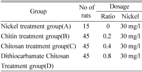
This study was performed to investigate the effects of Chitosan on the nickel poisoning in rats. In the study, 150 male Sprague-Dawley were used. The experimental groups were divided into four: A (30 mg/L nickel), B (30 mg/

L nickel+0.2% Chitin, Chitosan and Dithiocarbamate Chitosan), C (30 mg/L nickel+0.4% Chitin, Chitosan and Dithiocarbamate Chitosan), D (30 mg/L nickel+0.8%Chitin, Chitosan and Dithiocarbamate Chitosan).

The results were as flows;

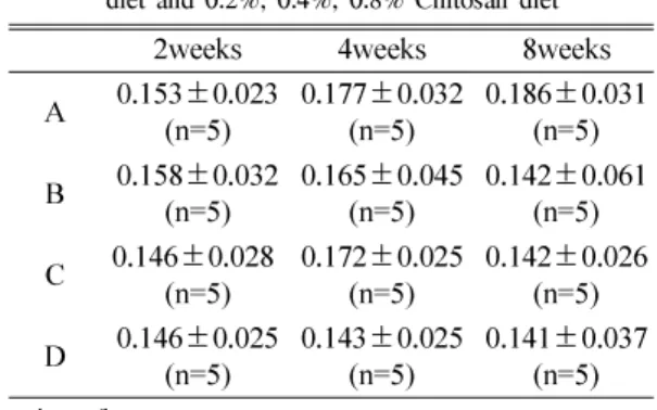
1. The nickel concentration in the livers of the control group (A) was 0.153~0.186 mg/kg but the nickel concentration in the livers of the experimental decreased during the experimental period (P<0.05).

2. Metallothionenin levels in rat liver were 2.77~3.25 ug/g wet,wt in control group (A), but were 2.89~3.51 ug/g wet,wt (B), 2.97~3.62 ug/g wet,wt (C), 2.68~3.68 ug/g wet,wt (D). Resectively in the experimental groups. The experimental groups were inclined to increase compare to the control group (P<0.05).

In conclusion, this study revealed a preventive effect of Chitin, Chitosan and Dithiocarbamate Chitosan against nickel toxicity.

Keywords: nickel, preventive effect, toxicity, chitin, chitosan, Dithiocarbamate Chitosan, liver

I. Introduction

Nickel, a chemical catalyst for electroplating, is used in the process of the stainless steel production, as a coloring matter for ceramic ware and in the production of nickel cadmium batteries.

It is reported that, if a person is exposed to nickel compound, it causes contact dermatitis, pulmonary fibrosis, cardiovascular diseases and renal diseases, and is known to be absorbed mainly to respiratory or digestive organs.

1)

In an in vivo experiment of administering nickel into the abdominal cavity of rats, increase of AST (Alamine Amino Transferase) and ALT (Aspartate Amino Transferase) was verified from the liver tissues of the exposed subjects, which suggested

hepatotoxicity of nickel. It is reported that, if nickel compound is administered through mouth, development of lung adenoma and intestinal cancer increases.

2)

The carcinogenesis proceeds as cohesion of nickel with DNA molecules of cells causes deformation and variation by disturbing the phosphorylation process.

3)

As an antidote of heavy metal, Ca

2

Na

2

EDTA is used. It is a chelating agent very friendly with heavy metal but it creates kidney toxicity.

4,5)

Also while the antidote Penicillamine has a merit of being smoothly absorbed in the stomach and intestine, it is known to probably cause leukopenia, aplastic anemia.

6,7)

But Chitin and Chitosan are considered to be harmless to human bodies and are being much studied as new medical material because these antidotes do not form any antibody and have no side effects.

8,9,10,11)

Chitin and Chitosan can be applied as new medical material for human bodies and, for these

Corresponding author : Department of Polymer-Nano Sci-

ence and Technology, Chonbuk National University

Tel:82-63-270-2335, Fax: 82-63-270-2335

E-mail: [email protected]

(2)

are material which directly contact with tissues of human bodies, Chitin and Chitosan are a subject of interest as material for artificial internal organs such as artificial kidneys or hearts, which are under development.

12)

These natural polymers are used for waste water treatment, food industry, textile industry, cosmetics and medical supplies.

Chitosan is produced by deacetylating Chitin and is reported to have an excellent absorption capacity for heavy metals favored by the increased free first amino group.

13,14)

Kim et al . produced Diehiocarbamate Chitosan and then tetracycline (Tc), an antibiotic, which can be used as a metal complexing agent, and investigated the effect of these on the medicine.

15,16)

In this experiment, the detoxification effect of Chitosan and Dithiocarbamate Chitosan was measured by checking the result after admini- stering them to a ratte which was exposed to and contaminated by nickel. The Dithiocarbamate Chitosan was obtained by treating Chitosan with carbon disulfide in alkali, which were obtained through deacetylation of Chitin and Chitosan came from crabs treated by acid and alkali.

II. Materials and Methods

1. Materials

1) Animals and Method

The subject of this experiment used was a male of Sprague Dawley rats 3 to 4 weeks old which was adapted to the environment of the laboratory for 2 weeks. The environment of the breeding cage was maintained with the temperature between 18 and 24

o

C and the humidity between 40 and 70%.

2) Production of chitin

The crab shells purchased from the market were cleaned using water to remove impurities and were deposited in the 2N HCl liquid for 12 hours to elute calcium carbonate, which was then grinded and treated in the normal temperature for 24 hours. After fully cleaning the same, the work of separating protein using 4% NaOH solution for 24 hours under the temperature of 15

o

C was repeated for 5 times. It was then treated by 3%

H

2

O

2

-1NHCl liquid for 6 hours to oxidize the pigment, treated by alkali, cleaned by distilled

water, ethanol, and by ether in order and dried to obtain pure white Chitin, which was grinded to produce Chitin of 80 to 100 mesh.

3) Production of chitosan

The Chitin of 80 to 100 mesh was deacetylated by treating with 47% NaOH solution at the temperature of 110

o

C for an hour, which was repeated for 5 times in order to increase the rate of deacetylation. It was then fully cleaned by distilled water, ethanol and ether in its order and dried in vacuum at the temperature of 70

o

C.

4) Composition of Dithiocarbamate Chitosan 60 g of Chitin powder was poured into 1 liter of 40% sodium hydroxide solution, which was then heated to 110

o

C for 8 hours, filtered and fully cleaned by distilled water. Then 500 ml of methanol and 100 ml of ammonia were dispersed and 60 ml of carbon disulfide was added before it is left in the normal temperature condition for 2 days. It was filtered, cleaned by distilled water repeatedly for 7 times, dried and stored in a desiccator.

2. Method

1) Administering of Chitin, Chitosan and Dithio- carbamate Chitosan to a rats

Chitin, Chitosan and Dithiocarbamate Chitosan produced were mixed with animal feed as per the Table 1, which the rats was allowed to eat without restriction.

2) Administering nickel to the rats.

Water with nickel concentration of 30 mg/L was given to the rats to freely drink during the period of the experiment.

Table 1. Experimental dosage with rats treated Chitin, Chitosan and Dithiocarbamate Chitosan and nickel

Group No of

rats Dosage

Ratio Nickel

Nickel treatment group(A) 15 0 30 mg/l

Chitin treatment group(B) 45 0.2 30 mg/l

Chitosan treatment group(C) 45 0.4 30 mg/l

Dithiocarbamate Chitosan 45 0.8 30 mg/l

Treatment group(D)

(3)

3) Measurement of nickel concentration rate in the livers of the rats.

The liver of the rats was taken out by 0.1 g and was put into a Kjeldahl flask where 5 ml of concentrated HNO

3

and 10 ml of concentrated H

2

SO

4

were added. From this, organic matters were decomposed on a hot plate heated up to 100

o

C. At this time, concentrated HNO

3

was added until the color of the decomposed solution becomes yellow- colorless. After heavy metals were extracted using the extraction method, DDTC-MIBK, from such sample of the completed decomposition of organic matters, the quantity of heavy metals contained was measured by an atomic absorption spectro- photometer(Varian specta. AA-30) with the wave length 232 nm and slit 0.2 nm.

4) Measurement of Metallothionein in the liver of the rats

In order to measure the quantity of Metallo- thionein in the liver of the rats, 0.1 g of the liver of a rats was taken out and the tissue was homogenized using Teflon glass homogenizer adding sugar solution (sucrose, Sigma) of 0.25 M after cleaned by physiological saline, and it was centrifuged (Beckman) for 20 minutes at the temperature of 4

o

C to get cytosol. After 0.2 ml of the cytosol was put into the buffer solution of 0.03 M tris-HCl (pH=8.0), it was saturated by 1 ml of CdCl

2

(standard solution) of 10 mg/kg and was cultivated for 5 minutes in the normal temperature. After all the bioligands except the excessive Cd and MT were removed from this by adding 0.2 ml of rat RBC hemolysate and Cd-bound hemoglobinit was denaturalized by depositing it in the water of 100

o

C for 1 minute, the liquid of the upper layer was taken after it was centrifuged by 1,000 g (Beckman, room temperature).

The sample obtained by repeating addition of rat RBC hemolysate, heat treatment and centrifugal process three times was used in measurement of the concentration of cadmium and the final calculation of the concentration of MT was made by converting 6 g of cadmium atom to 1 M of MT (molecular weight 6.050) which combines with each other to get the concentration of ugMT per g of the tissue.

III. Results and Discussion Nickel degrades the function of immune system, promotes bacillus and virus infection, degenerates the ability to form antibodies, deteriorates the phagocytic activity of macrophage and lower the function of apoptosis of cells. Nickel absorbed to human bodies through air, water and food is combined with plasma protein and absorbed to and the nerve system, liver, kidney and lung. It accumulates and also threatens human lives by causing structural, genetic and biochemical changes as well as physiological changes. It was reported that Nickel accumulates in human bodies, which are the final consumers in the food chain and causes toxication as synthesis of Metallothionein does not take place.

17)

Also, in the metabolism in human bodies, Cadmium with toxicity combines with thiol (SH) which has a high appetence with heavy metals among proteins which form the cell walls, to alleviate its toxicity. It is reported that heavy metals have effect on the formation of Metallothionein and, especially, divalent metals like Cadmium function as an important factor for formation of Metallothionein, which, having cysteine, is known to alleviate the toxicity of Cadmium by combining with it. Thus, in this experiment, we tried to find out what effect there is on the formation of Metallothionein by the toxicity of nickel.

Though the resources of Chitin and Chitosan,

which are abundant as they are contained in

crustacea and higher vegetation in big quantity,

were almost not used in the past, the quantity used

at present is fast increasing Chitin and Chitosan

are widely used because of their antimicrobial

activity, antitumoral activity and the effect of

lowering cholesterol for food industries, cosmetics,

textile industries and waste water treatment.

18)

Chitosan which is obtained by deacetylation of

Chitin has better adhesion to ions of heavy metals

than that of Chitin because of the increased free

first amino group and the change of the adhesion

degree to Hg and Cu as the deacetylation of Chitin

increases was once measured and reported.

19)

Also, Choi et al once produced separation material

of revitalized Chitosan system and examined its

effect of metal ion separation.

(4)

1) Concentration of nickel in the liver of the rat.

Water with Nickel concentration of 30 mg/L was given to the rats to freely drink. As a result, the quantity of nickel accumulation in the livers of the group to which only nickel was administered increased as the duration of the exposure gets longer from 0.153 mg/kg (2 weeks) to 0.177 mg/kg (4 weeks), 0.179 mg/kg (6 weeks) and to 0.186 mg/kg (8 weeks)(Table1). In the experiment of measuring the concentration of nickel in the livers of the rats administering nickel and Chitin at the same time, when the Chitin contained in the feed for the rats was 0.2%, the nickel concentration measured after two weeks was found to be 0.151mg/

kg which showed no effect of detoxification of Chitin. When the concentration of the Chitin administered was increased to 0.4% and 0.8%, it did not show almost any decrease in the quantify of nickel accumulated in the livers of the rats.

Though, it partially showed decrease of the concentration of nickel in the livers of the rats by administering Chitin, it was an insignificant value.

And, in the 8 week experiment of feeding the rats with increased quantity of Chitin mixed in the feed to 0.4% and to 0.8% to check the quantity of nickel detoxification, it only showed a little decrease in the nickel quantity than when the rats were exposed to nickel only (Table 1). When 0.2%, 0.4% and 0.8% of Chitosan mixed in the feeds was fed to the rats classified into 2 week, 4 seek and 8 week group, the result showed that, in the 2 week group, there was no difference from the group which was exposed only to nickel but in the 4 week group and 8 week group, some effect could be expected. (Table 2). When 0.2%, 0.4%

and 0.8% of Dithiocarbamate Chitosan mixed in the feeds was fed to the rats classified into 2 week, 4 seek and 8 week group exposing them to nickel in drinking water, the quantity of nickel contained in the livers of the rats of 4 week group which was exposed only to nickel was measured as 0.177 mg/

kg while that of the 4 week group to which 0.8%

of Dithiocarbamate Chitosan was administered was 0.138 mg/kg showing the biggest effect but the decrease of nickel quantity contained in the livers of the 2 week group and 8 week group shown was small (Table 3).

2) Quantity of Metallothionein in the livers of the rats

The quantity of Metallothionein in the livers of the rats for which only nickel was administered showed

Table 1. Concentration of nickel in rat liver consuming water containing 30 mg/L nickel chloride with basal diet and 0.2%, 0.4%, 0.8% Chitin diet

2weeks 4weeks 8weeks A 0.153 ± 0.023

(n=5) 0.177 ± 0.032

(n=5) 0.186 ± 0.031 (n=5) B 0.151 ± 0.031

(n=5) 0.169 ± 0.029

(n=5) 0.182 ± 0.029 (n=5) C 0.154 ± 0.029

(n=5) 0.147 ± 0.023

(n=5) 0.185 ± 0.019 (n=5) D 0.147 ± 0.038

(n=5) 0.151 ± 0.035

(n=5) 0.175 ± 0.052 (n=5)

unit:mg/kg

Table 2. Concentration of nickel in rats liver consuming water containing 30 mg/L nickel chloride with basal diet and 0.2%, 0.4%, 0.8% Chitosan diet

2weeks 4weeks 8weeks

A 0.153 ± 0.023

(n=5) 0.177 ± 0.032

(n=5) 0.186 ± 0.031 (n=5) B 0.158 ± 0.032

(n=5) 0.165 ± 0.045

(n=5) 0.142 ± 0.061 (n=5) C 0.146 ± 0.028

(n=5) 0.172 ± 0.025

(n=5) 0.142 ± 0.026 (n=5) D 0.146 ± 0.025

(n=5) 0.143 ± 0.025

(n=5) 0.141 ± 0.037 (n=5)

unit:mg/kg

Table 3. Concentration of nickel in rats liver consuming water containing 30 mg/L nickel chloride with basal diet and 0.2%, 0.4%, 0.8% Dithiocabarmate Chitosan diet

2 weeks 4weeks 8weeks A 0.153 ± 0.023

(n=5) 0.177 ± 0.032

(n=5) 0.186 ± 0.031 (n=5) B 0.146 ± 0.021

(n=5) 0.153 ± 0.072

(n=5) 0.163 ± 0.037 (n=5) C 0.149 ± 0.028

(n=5) 0.141 ± 0.052

(n=5) 0.151 ± 0.051 (n=5) D 0.152 ± 0.016

(n=5) 0.138 ± 0.065

(n=5) 0.162 ± 0.034 (n=5)

unit:mg/kg

(5)

decrease as the period of exposure to nickel increased, from 3.25 ug/g wet,wt for 2 weeks to 3.09 ug/g wet,wt for 4 weeks and to 2.77 ug/g wet,wt for 8 weeks. In the group for which nickel and 0.2%

Chitin was fed, it was 3.41 ug/g wet,wt after 2 weeks, 3.18 ug/g wet,wt after 4 weeks and 2.89 ug/g wet,wt after 8 weeks. And when the quantity of Metallothionein in the livers was checked after increasing the quantity of Chitin to 0.4% and 0.8%, it was 3.23 ug/g wet,wt and 3.51 ug/g wet,wt for 2 week group, which was an inconsistent result, 3.42 ug/g wet,wt and 3.39 ug/g wet,wt for 4 week group and 3.32 ug/g wet,wt and 3.21 ug/g wet,wt (Table 4). In the group to which Chitosan was administered, the quantity of Metall- othionein in the livers of the rats measured increasing the quantity of Chitosan from 0.2% to 0.4% and 0.8%, the quantities shown for the 2 week group were 3.29 ug/g wet,wt, 3.62 ug/g wet,wt and 3.52 ug/g wet,wt respectively and a tendency of increase in the quantity of Metallothionein in the livers as the quantity of Chitosan increased could be seen (Table 5).

The quantities of Metallothinein measured after feeding Dithiocarbamate Chitosan mixed into the feed at the rates of 0.2%, 0.4% and 0.8% were 3.68 ug/g wet,wt (2 week group)-2.68 ug/g wet,wt (8 week group), which showed that it decreased as the period of the feeding is longer (Table 6).

IV. Recommendations and Conclusions

Chitin obtained from crabs treated by acid and

alkali, Chitosan obtained by deacetylation of Chitin and Dithiocarbamate Chitosan obtained by treating Chitosan with carbon disulfide in alkali were mixed into the feed for the rats and, after feeding it, the concentration of nickel and the quantity of accumulated Metallothionein in the livers of the rattes were measured to conclude as follows.

1. Concentration of nickel

1) The accumulated quantity of nickel in the livers of the group of the rats to which only nickel was fed was 0.153 mg/kg (2 week group)~0.186 mg/kg (8 week group).

2) The concentration of nickel in the livers of the group of the rats which was fed with 0.2% Chitin~

0.8% Chitin mixed in the feed showed 0.151~

0.147 mg/kg (2 week group), 0.169~0.151 mg/kg

Table 4. Metallothionenin levels in rats liver consuming water containing 30 mg/L nickel chloride with basal diet and 0.2%, 0.4%, 0.8% Chitin diet

2 weeks 4weeks 8weeks A 3.25 ± 0.27

(n=5) 3.09 ± 0.38

(n=5) 2.77 ± 0.36 (n=5) B 3.41 ± 0.21

(n=5) 3.18 ± 0.42

(n=5) 2.89 ± 0.41 (n=5) C 3.23 ± 0.37

(n=5) 3.42 ± 0.36

(n=5) 3.32 ± 0.32 (n=5) D 3.51 ± 0.31

(n=5) 3.39 ± 0.29

(n=5) 3.21 ± 0.20 (n=5)

unit:ug/g wet,wt

Table 5. Metallothionenin levels in rats liver consuming water containing 30 mg/L nickel chloride with basal diet and 0.2%, 0.4%, 0.8% Chitosan diet

2 weeks 4weeks 8weeks A 3.25 ± 0.27

(n=5) 3.09 ± 0.38

(n=5) 2.77 ± 0.36 (n=5) B 3.29 ± 0.43

(n=5) 2.97 ± 0.56

(n=5) 2.98 ± 0.47 (n=5) C 3.62 ± 0.87

(n=5) 3.45 ± 0.36

(n=5) 3.51 ± 0.58 (n=5) D 3.52 ± 0.46

(n=5) 3.37 ± 0.39

(n=5) 3.38 ± 0.30 (n=5)

unit:ug/g wet,wt

Table 6. Metallothionenin levels in rats liver consuming water containing 30 mg/L nickel chloride with basal diet and 0.2%, 0.4%, 0.8% Dithiocarbamate Chitosan diet

2 weeks 4weeks 8weeks A 3.25 ± 0.27

(n=5) 3.09 ± 0.38

(n=5) 2.77 ± 0.36 (n=5) B 3.68 ± 0.53

(n=5) 3.42 ± 0.54

(n=5) 2.68 ± 0.58 (n=5) C 3.37 ± 0.53

(n=5) 3.30 ± 0.92

(n=5) 3.51 ± 0.98 (n=5) D 3.57 ± 0.87

(n=5) 3.51 ± 0.83

(n=5) 3.38 ± 0.89 (n=5)

unit:ug/g wet,wt

(6)

(4 week group) and 0.182~0.175 mg/kg (8 week group) respectively.

3) While the concentration of nickel in the livers of the group of the rats which was exposed only to nickel was 0.153 mg/kg(2 week group)~ 0.186 mg/kg (8 week group), those of the group which was fed with 0.2%~0.8% Chitosan mixed in the feed showed 0.146~0.158 mg/kg (2week group) and 0.141~0.142 mg/kg (8 week group), which showed that in the 2 week group, the quantity of nickel in the livers of the rats partially increased but, in the 8 week group, the quantity accumulated decreased.

4) While, in the group which was exposed only to nickel for two weeks, the concentration of nickel in the livers of the rats showed was 0.153 mg/kg, in the two week group to which feed mixed with 0.2% of Dithiocarbamate Chitosan was given, detoxification effect of Dithiocarbamate Chitosan could not be expected as the figure shown was 0.146~0.152 mg/kg but, in the 4 week group, it was 0.177 mg/kg(for nickel only) and 0.138 mg/kg (nickel+0.8% Dithiocarbamate Chitosan) and, in the 8 week group, it was 0.181 mg/kg (nickel only) and 0.151 mg/kg (nickel+0.4%

Dithiocarbamate Chitosan), which showed that, as the rate of Dithiocarbamate Chitosan increased, the concentration of the nickel in the livers was decreased but it was difficult to expect consistent values.

2. Quantity of Metallothionein

1) The quantity of Metallothionein in the livers of the group of rats which was exposed only to nickel was 3.25 ug/g (2 week group), 3.09 ug/g wet,wt (4 week group) and 2.77 ug/g wet,wt (8 week group), which showed decrease of the quantity of Metallothionein in the livers of the rattes as the duration of exposure is longer.

2) When the quantities of Metallothionein in the livers of the rats were measured after giving feed with 0.2~0.8% Chitin mixed, it was 3.25 ug/g wet,wt (nickel only, 2 weeks)~3.51 ug/g wet,wt (nickel+0.8% Chitin, 2 weeks), 2.77 ug/g wet,wt (nickel only, 8 weeks)~3.32 ug/g wet,wt (nickel+

0.4% chitin), which showed a little increase in the quantity of Metallothinein in the livers of the rats.

2) As the result of feeding the rats with 0.2~0.8

% of Chitosan mixed in the feed, the quantity of the Metallothinein in the livers of the rats showed 3.09 ug/g wet,wt (nickel only, 4 weeks)~3.37 ug/g wet,wt (nickel+0.8% chitosan, 4 weeks) and 2.86 ug/gwet,wt (nickel alone, 6 weeks)~3.39 μ g/g (nickel+0.8% chitosan, 6 weeks).

3) When the quantities of Metallothionein in the livers of the rats were measured after feeding them with 0.2~0.8% of Dithiocarbamate Chitosan mixed in the feel, it was 3.68 ug/g wet,wt (nickel with 0.2% of Dithiocarbamate Chitosan, 2 weeks)

~2.68 ug/g wet,wt (nickel with 0.2% Dithiocarbamate Chitosan, 8 weeks), which showed some increase in the quantity of Metallothinein in the livers of the rats as the period of feeding increased from 2 weeks to 8 weeks.

References

1. Park, H. S., and Park, K. S. : Nickel Toxicity and Carcinogenicity J. Environ. Toxicol . 19 (2), 119- 134, 2004.

2. Cha, B. S., Wang, S. J., Chang, S. J. and Lee, J. W.

: Comparative Hepatotoxicity Assessment of Cad- mium and Nickel with Isolated Perfused Rat Liver.

Korean J. Perv. Med . 33 (1), 117-124, 2000.

3. Lee, K. N., Il-soo You, I. S. and Lee, J. S. : A study on Antitoxic Effect of Natural products Against Nickel Toxicity. Kor. J. Env.. Halth. Soc ., 20 (3), 85- 95, 1994.

4. Hall L. D. and Yalpani M. : Enhancement of the metal chelating properties of chitin and chitosan.

Carbohydrate Research , 83 , C

5

-C

7

, 1980.

5. Eaton, D. L. and Toal, B. F. : Evaluation of Cd/

henolobin affinity assay for the rapid determination of metallothionein in biological tissues. Toxicol Appl Pharmacol ., 66 , 134, 1982.

6. Hall, L. D. and yalpani, M. : Enhancement of the metal chelating properties of chitin and chitosan.

Carbohydrate Research , 83 , C

5

-C

7

, 1980.

7. Kage, JHR. and Vallee, BL. : A cadmium and zinc- containing protein from equine renal cortex. J. Biol.

Chem. 236 , 2435-2442, 1961.

8. Maruca, R. suder, BJ. and Wightman, J. : Inter- action of heavy metals with chitin and chitosan.

Chromium J. of Applied Polymer Science , 27 , 4827-4837, 1982

9. Pulido, P. Kagi, J. and Vallee BL. : Isolation and some propertie of human metallothionein Bioch7.

Cherian MG, Nordberg M: Cellular adaptation in metal toxicolgy and metallothionein. Toxicology ,

25 , 1-15, 1983.

10. Landes, D. R. and Bough, W. A. : Effects of chi-

tosan acoggulating agent for food processing

(7)

wastes in the diet of rats on growth and liver and blood consumption. Bull. Environ. Contam. Toxi- col ., 15 , 55-57, 1976.

11. Eaton, D. L. and Toal, B. F. : Evaluation of the Cd/

hemoglobin affinity assay for the rapid determi- nation of metallothionein in biological tissues. Tox- icol. Appl. Pharmacol ., 5 , 1768-1777, 1966.

12. Kim, S. K. and Lee, E. H. : Biocompatibility and Medical Applications of Chitin and Chitosan.

Korean J. Chitin and Chitosan , 2 (2), 39-74, 1997.

13. Cherian, M.G. and Nordberg, M. : Cellular adap- tation in metal toxicolgy and metallothionein. Tox- icology , 25 , 1-15, 1983.

14. Kakela, R. and Kakela, A.: Effects of nickel chlo- ride on reproduction of the rat and possible antag- onistic role of selenium. J. Biol. Chem. 236 , 2516- 2525, 1961.

15. Kostonis, F. N. and Klaassen, C. D. : Toxicity and distribution of cadmium administered to rats at sub-

lethal doses. Toxicol Appl Pharinacol ., 41 , 667, 1977.

16. Kim, Y. T. and You, J. H. : Yun-Jun Jung, Jeoung- Soo Lee and Young-Mi Kim. Development of Poly- meric Drugs Vtilizing Dithiocarbamate chitosan.

Yakhak hoeji , 39 (4), 373-379, 1995.

17. Saplakoglu, U. : DNA single-strand breakage in rat lung, liver and kidney after single and combined treatments of nickel and cadmium, Mutation research. Genetic Toxicology and Environmental Mutagenesis , 394 (1), 133-140, 1997.

18. Onosaka, S., Tanak, K., Doi, M. and Okahara, K. : A simplified procedure for determination of met- allothionein in animal tissues. Eisei Kagaku 24 , 128-133, 1978.

19. Jung, K. S., Kim, D. H. and Jung, K. D. : Effect of

Dietary calcium, Casein and Suet on the Accu-

mulation of cadmium in Mice. Korean J. of Pre-

ventive Medicine , 21 (2), 340-350,1988.

수치

Table 1. Experimental dosage with rats treated Chitin, Chitosan and  Dithiocarbamate  Chitosan  and  nickel
Table 2. Concentration  of  nickel  in  rats  liver  consuming water containing 30 mg/L nickel chloride with basal diet  and  0.2%,  0.4%,  0.8%  Chitosan  diet
Table 6. Metallothionenin  levels  in  rats  liver  consuming water containing 30 mg/L nickel chloride with basal diet and 0.2%, 0.4%, 0.8% Dithiocarbamate Chitosan diet

참조

관련 문서

The purpose of this study is to evaluate the effects of heat treatment on the load-deflection properties and transitional temperature range(TTR) of

In 4-week group, the group filled with bone graft with decortication revealed larger new bone formation area than shown in the group that had a defect area

The purpose and necessity of this study was to investigate the effect of the 8-week GX complex exercise program on the stress index, blood lipid and APG

In the region of Auerbach’s plexus (AP) of small intestine, the staining intensity of the ICC-IR cells was reduced in the HFD group compared to the control group.. The numbers

Kim, Dong-Myung Advisor : Prof. The control group did not get training. Only the experiment group had the plyometrics training 3 times a week.. There was

There was no significant difference between the groups in problems of eating habits, but the obesity group considered overeating as a problem while the normal

1 John Owen, Justification by Faith Alone, in The Works of John Owen, ed. John Bolt, trans. Scott Clark, &#34;Do This and Live: Christ's Active Obedience as the

First, the comparison of pre and post-cardiovascular endurance between the experimental group, which participated in 12-week sports leisure activities regularly,