89
<원례보저>
마우스 비장세포의 증식과 생존율에 대한 BCG-CWS의 면역자극 효과
이제욱·고은주·주홍구*
제주대학교 수의과대학 수의약리학실험실
(접수: 2012년 1월 21일, 수정: 2012년 5월 4일, 게재승인: 2012년 5월 7일)
Immunostimulatory effects of BCG-CWS on the proliferation and viability of mouse spleen cells
Che-Wook Lee, Eun-Ju Ko, Hong-Gu Joo*
Laboratory of Veterinary Pharmacology, College of Veterinary Medicine, Jeju National University, Jeju 690-756, Korea (Received: January 21, 2012; Revised: May 4, 2012; Accepted: May 7, 2012)
Abstract : Mycobacterial cell-wall skeleton (CWS) is an immunoactive and biodegradable particulate adjuvant and has been tried to use for immunotherapy. The CWS of Mycobacterium bovis bacillus Calmette- Guerin (BCG-CWS) was studied as an universal vaccine vehicle for antigen conjugation, to develop potentially effective and safe vaccine. Although a variety of biological activities of BCG-CWS have been studied, the effects of BCG-CWS on spleen cells are not fully elucidated. Using MTT assay and trypan blue exclusion test, we found that BCG-CWS significantly enhanced the viability and proliferation of cells.
Multiple clusters, indicating proliferation, were observed in BCG-CWS-treated spleen cells and surface marker staining assay revealed that BCG-CWS promoted the proliferation of CD19+ B lymphocyte rather than CD4+ or CD8+ T lymphocyte. In addition, BCG-CWS up-regulated the expression of anti-apoptotic molecules such as bcl-2, bcl-xL. BCG-CWS increased the surface expression of CD25 and CD69 as well as IL-2 production of spleen cells, suggesting increased activation. Furthermore, BCG-CWS enhanced the antigen-specific cell proliferation and interferon-gamma production of spleen cells. Taken together, these results demonstrate the immunostimulatory effects of BCG-CWS on spleen cells via multiple mechanisms, providing valuable information to broaden the use of BCG-CWS in clinical and research settings.
Keywords: adjuvant, BCG-CWS, spleen cell, vaccine
서 론
공중보건적이나 경제적인 관점에서 질병은 치료하는 것보다, 예방하는 것이 더 효율적이다. 질병예방의 목적 으로 백신이 개발된 이래, 현재까지 백신은 질병을 예방 하는데 있어서 매우 중요한 역할을 하고 있다. 백신은 체내 면역계의 메모리 기능을 이용하여 같은 병원체에 감염되었을 때, 면역반응을 더 빠르고 강하게 상승시켜 준다. 이러한 백신은 백신항원과 adjuvant를 주요 구성 성분으로 하고 있는데, adjuvant는 백신항원의 효능을 높 이는 역할을 한다.
이러한 adjuvant로써 가장 널리 사용되고 있는 것은 aluminum compound인 alum이다 [1, 17]. Alum은 백신 항원을 흡착하여 체내에서 존재하는 기간을 연장하고 항원을 항원제시세포가 탐식하기 좋도록 수용성항원을 입자성 물질 형태로 변화시키며 [14], 항원을 탐식하는 항원제시세포를 활성화시킨다 [15]. 그러나 alum은 세포 매개성 면역을 일으키는 능력이 약하고 T helper(Th) 1 면역반응이 미비하다 [17]. 이 때문에 결핵, 티푸스, 브 루셀라와 같이 세포 내에 침입하여 증식하는 병원성미 생물에 대한 효능이 약한 단점을 가지고 있다. 또한 alum 의 부작용으로 피하 또는 피내 접종 시 접종부위에 육
*Corresponding author
Tel: +82-64-754-3379, Fax: +82-64-756-3354 E-mail: [email protected]
아종을 형성할 수 있으며 [11, 25], IgE 생산을 증가시켜 알러지를 발생시킬 수 있다. 또한 신경독성을 가지고 있 으며 신장기능이 저하된 환자에서는 aluminum이 체내 에 축적되면 독성이 나타날 수 있다 [10].
이러한 문제점이 있음에도 가장 많이 사용되고 있는 이유는 현재까지 이러한 문제점이 보완된 adjuvant의 개 발이 미흡하기 때문이다. 따라서 안전하고 안정적인 면 역반응을 일으킬 수 있는 새로운 adjuvant의 개발이 필 요하며 현재까지 더 안전하고 효율적인 백신개발을 위 한 연구가 많이 이루어지고 있다 [17, 20].
Mycobacterium bovis bacillus Calmette-Guerin(BCG)는 결핵환자에 백신으로 안정적으로 사용되어 왔다 [7, 9, 26]. Mycobacteria의 세포벽은 항원제시 및 염증성 cytokines을 높은 수준으로 발현시키는 강한 면역원성 물 질로, 면역반응을 높은 수준으로 유도한다. 따라서 Mycobacteria는 세포독성 T 림프구와 자연살해세포의 활 성화와 항체생산을 증가시키기 위한 adjuvant로서 종종 사용되었다. 특히 BCG의 세포벽 구성물질인 cell wall skeleton(CWS)는 mycolic acid와 arabinogalactan, 그리고 peptidoglycan complex가 공유결합으로 이루어진 강한 면역원성을 가진 구조물로, 강한 선천면역을 일으키기 위한 목적으로 사용되었다 [2, 4, 22, 27]. BCG-CWS는 adjuvant로 사용하였을 때 효율적으로 항체생산을 유도 하였고, 세포독성 T 림프구의 증식을 향상시켰고, 항종 양효과를 나타내었다 [18, 29]. 또한 BCG-CWS는 수지 상세포에 미생물과 결합할 수 있는 Toll-like receptor (TLR) 2와 TLR4의 발현을 증가시켜 수지상세포를 활성 화시키는 역할도 수행한다 [27, 28].
비장은 림프구와 다양한 면역세포로 구성되어 있는 중요한 면역기관이다. 비장은 혈액에서 유래된 항원에 대한 면역반응을 하며, 노화된 세포 및 손상된 세포를 제거하는 역할도 수행한다. 신체내의 기관 중 비장은 면 역에 관련된 주요한 기능을 수행하고 있음에도, BCG- CWS가 비장에 미치는 영향에 관한 연구는 미흡한 상태 이다.
본 연구는 마우스의 비장세포에 BCG-CWS를 처리하 여 비장세포의 분열능력과 생존능력, 그리고 면역활동 과 관련된 주요cytokine분비에 어떠한 영향을 주는 지 알아보고, ovalbumin(OVA) 특이적인 면역 상태에서 BCG-CWS의 adjuvant로서의 효능을 확인하고자 하였다.
재료 및 방법
실험동물 및 시약
모든 실험에 ORIENT BIO(Korea)에서 구입하여 제주 대학교 수의약리학실험실에서 유지된 7~12 주령의 Balb/
c 마우스를 사용하였다. 동물실험은 제주대학교 동물실 험윤리위원회의 승인을 받아 시행되었으며 제주대학교 동물실험윤리지침을 준수하였다(승인번호: 2011-0004).
BCG-CWS는 충남대학교 의과대학에서 제공받아, 멸균 된 phosphate-buffered saline(PBS)에 녹여 사용하였다.
세포의 준비 및 배양
마우스의 비장세포를 이용해 배양하는 실험은 이미 확립된 방법에 따라 실시했다 [12]. 간략히 설명하면, 마 우스의 비장을 떼어내 물리적으로 분쇄시켜 세포를 얻 었다. 저삼투압용액인 ammonium chloride potassium lysis buffer를 이용하여 적혈구를 용혈시켜 제거한 후 5% fetal bovine serum(FBS)과 L-glutamine, penicillin/streptomycin 이 첨가된 RPMI1640 배지에 세포를 풀어 T75 플라스 크에 넣고 1시간 동안 배양하여 대식세포와 같은 부착 세포가 플라스크의 바닥에 부착되도록 하였다. 이 후 떠 있는 세포를 회수한 뒤 40 µm cell strainer로 뭉친세포 등을 걸러내고 10% FBS와 L-glutamine, penicillin/
streptomycin, sodium pyruvate, non-essential amino acids, 2-mercaptoethanol이 첨가된 RPMI1640 배지를 이용해 96-well 또는 6-well culture plate에 2 × 10⁶ cells/mL의 농 도로 비장세포를 넣어 배양하였다. 모든 세포배양 시 온 도는 37oC, CO2의 농도는 5%를 유지하도록 하였다.
비장세포의 분열능력과 생존능력, 형태변화의 측정 96 well culture plates에 비장세포를 4 × 10⁵cells/200 µL/well로 seeding 하고 BCG-CWS를 농도 별로 처리 하여 3일간 배양한 후 3-[4,5-dimethylthiazol-2-yl]-2,5- diphenyltetrazolium bromide(MTT; Sigma, USA) assay와 trypan blue exclusion test로 분열능력과 생존능력을 평 가하였다. MTT assay를 위해서 배양한 세포에 10 mg/
mL MTT solution을 10 µL/well씩 처리하여 4시간 동안 37oC에서 배양을 한 후, 다시 10% sodium dodecyl sulfate(SDS)를 100 µL/well를 처리하여 2시간 후 micro- plate reader(Molecular devices, USA)를 사용하여, 570~
650 nm에서 흡광도를 측정하였다. 또한 배양 세포의 생 존율을 알아보기 위해 trypan blue solution(Invitrogen, USA)으로 세포를 염색하여, 살아있는 세포와 죽은 세포 의 수를 세었다. 배양된 비장세포의 사진은 inverted microscope(Olympus, Japan)에서 digital camera(Cannon, Japan)를 연결하여 촬영하였다.
유세포분석
6 well culture plates의 각 well에 BCG-CWS를 농도 별로 처리하여 3일간 배양 후 유세포분석을 하였다. 세 포염색법은 이미 확립된 방법에 따라 실시했다 [6, 13].
비장세포는 biotin-labeled anti-CD4, anti-CD8, anti-CD19, anti-CD25 antibody를 1차 항체로 처리하고 2차 항체로 streptavidin-labeled phycoerythrin(PE)를 처리하였다.
CD69에 대한 항체로는 PE-labeled anti-CD69 antibody 를 이용하였다(BD Biosciences, USA). 염색된 세포는 FACS Calibur와Cell Quest(Becton Dickinson, USA)를 이 용하여 유세포분석을 실시하였다.
ELISA를 이용한 cytokine의 측정
비장세포의 분열능력과 생존능력의 평가에서 기술한 방법으로 3일간 비장세포를 배양한 후 세포배양액을 걷 어내어 interleukin(IL)-2와 IL-4의 생산량을 CytoSetkit (Invitrogen, USA)를 이용해 측정하였다. Lipopolysacc- haride와 Concanavalin A를 positive control로 사용하 였다.
OVA 특이적으로 면역된 비장세포의 분열능력 평가 마우스에 마리당 20 µg의 OVA를 20 µL의 alum과 함 께 복강 내 접종을 하였다. Alum에 OVA를 흡착시키기 위해 30분 동안 함께 정치하였다. 접종 후 2주가 지나 마우스의 비장세포를 채취한 후 5 µM carboxyfluorescein
succinimidyl ester(CFSE)로 10분간 염색한 후 OVA 1 µg/
mL와 함께 또는 없이 배양하였다. 4일 후 CFSE에 염색 된 비장세포를 수거하여 PBS로 두 번 세척 후 유세포 분석을 하였다. 림프구의 분열능력을 알아보기 위해 MTT assay를 실시했으며, 세포배양액을 수거하여 ELISA 법으로 IL-4와 Interferon(IFN)-gamma를 측정하였다.
Western blot analysis
비장세포에서 apoptosis와 관련된 단백질이 어떤 수준 으로 발현되는 지 알아보기 위해 Western blot analysis 를 실시하였다 [12]. 단백질 정량 결과에 따라 동일한 양의 단백질을 loading하였으며, 12% SDS-PAGE (polyacrylamide gel electrophoresis) gel을 이용하였다. 1 차 항체로 mouse anti-Bcl-2, mouse anti-Bcl-xL, mouse anti-beta-actin antibody를 사용하였고, 2차 항체로 goat anti-mouse IgG horseradish peroxidase를 사용하였다.
유의성분석
모든 data는 mean ± SD로 나타내었으며 Instat software (Graphpad, USA)를 이용하여 student t test를 통해 분석하 였다. 0.05 미만의 p value를 유의성이 있다고 판정하였다.
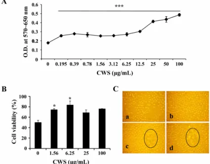
Fig. 1. Effect of cell wall skeleton of Mycobacterium bovis bacillus Calmette-Guerin (BCG-CWS) on the proliferation/
viability of spleen cells. The spleen cells were cultured at a concentration of 4 × 10⁵ cells/200 µL/well in 96-well culture plates and treated with 0~100 µg/mL BCG-CWS. After 3 days, (A) Proliferation assay using MTT solution, and (B) Trypan blue exclusion test were performed. Data are mean ± SD from three or four individual wells. *,*** indicate p < 0.05, 0.001, respectively, compared to control. (C) Proliferating clusters (black circles) in BCG-CWS-treated spleen cells. Ca: Control, Cb: CWS 1 µg/mL, Cc: CWS 5 µg/mL, Cd: CWS 25 µg/mL. Cell morphology was observed using an inverted microscope and the image was obtained by a digital camera. × 100.
결 과
비장세포의 분열능력과 생존율에 대한 BCG-CWS의 효과
비장세포의 분열능력과 생존율에 대한 BCG-CWS의 효과를 알아보기 위해, MTT assay와 trypan blue exclusion test를 사용하였다. 0~100 µg/mL의 BCG-CWS 를 처리한 비장세포를 3일간 배양한 후, MTT assay를 실시하였다. 이 결과, 3.12~100 µg/mL의 CWS를 처리한 비장세포의 증식률이 증가되었다(Fig. 1A). 비장세포의 생존율에 관한 CWS의 효과를 알아보기 위하여 trypan blue staining을 실시하였고, 6.25 µg/mL의 농도에서 가 장 높은 생존율을 보였다(Fig. 1B). 비장세포의 증식률 에 대한 또 다른 증거를 얻기 위하여 증식된 비장세포 의 cluster를 inverted microscope로 확인하여 디지털카메 라로 촬영을 하였다(Fig. 1C). BCG-CWS에 농도의존적
으로 비장세포의 cluster가 증가되었다. 이와 같은 결과 는 BCG-CWS가 비장세포에 자극효과가 있음을 말해준 다. T 림프구와 B 림프구 각각에 대한 BCG-CWS의 효 과를 알아보기 위해 세포표면염색을 하였고, CD19+ B 림프구가 CD4+ 그리고 CD8+ T 림프구보다 더 높은 분 열능력을 보였다(Fig. 2).
비장세포의 사이토카인 생산량에 대한 BCG-CWS의 영향
BCG-CWS가 림프구 성장에 관여하는 사이토카인의 생산에 관여하는 지 알아보기 위해, BCG-CWS가 처리 된 비장세포의 세포배양액에서 IL-2와 IL-4를 측정하였 다. IL-2의 생산량은 BCG-CWS의 농도와 비례하여 증 가하였다(Fig. 3). 그러나 동일한 세포배양액에서 IL-4는 측정하였지만 감지한계(detection limit) 이하였다(결과 미 제시).
Fig. 2. Selective proliferation of CD19+ B lymphocytes by BCG-CWS treatment. Spleen cells were cultured at a concentration of 5 × 10⁶ cells/5 mL/well in 6-well culture plates with medium alone or 1 µg/mL or 5 µg/mL BCG-CWS for 3 days. After harvesting, the cells were labeled with surface marker-specific antibodies. The numbers indicate the percentage of positive cells of a specific subset.
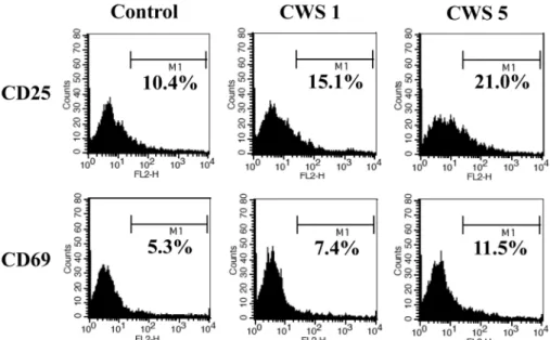
BCG-CWS에 의한 비장세포의 면역관련 표면마커 발 현 변화
림프구의 활성에 대한 BCG-CWS의 영향을 알아보기 위하여, BCG-CWS가 처리된 비장세포에서 CD25, CD69 표면인자의 발현을 평가했다. 5 µg/mL의 BCG-CWS를 처리한 비장세포에서 두 표면인자의 발현이 증가하는 것으로 나타났다(Fig. 4). 따라서 BCG-CWS는 IL-2와 같 은 림프구의 활성을 증가시키는 사이토카인에 대해 민 감하게 반응할 수 있도록 해당된 표면의 수용체분자를 증가시켜준다.
BCG-CWS에 의한 비장세포의 세포자멸사관련분자 발현의 변화
BCG-CWS가 비장세포의 세포자멸사를 얼마나 방어 하는 지 알아보기 위해 BCG-CWS가 처리된 비장세포 에서 bcl-2와 bcl-xL의 발현정도를 Western blot analysis 를 통하여 알아보았다(Fig. 5). BCG-CWS는 bcl-2와 bcl- xL 양쪽 분자를 모두 증가시켰고 이를 통해 비장세포의 세포자멸사를 높은 수준에서 억제할 수 있을 것으로 추 정된다.
Fig. 3. Spleen cells were cultured as described in Fig. 1 and treated with the indicated concentrations of BCG- CWS. After 3 days of treatment, the supernatants were collected and the amount of interleukin (IL)-2 was quantified. Data are mean ± SD from four individual wells.
ConA: Concanavalin A, LPS: Lipopolysaccharide.
Fig. 4. Enhanced expression of activation markers on spleen cells treated with BCG-CWS. Spleen cells were cultured as described in Fig. 2. Following BCG-CWS treatment, spleen cells were harvested and stained for CD25 or CD69, activation markers of lymphocytes. The number of histograms indicates the percentage of highly expressed cells.
Fig. 5. Upregulated expression of the anti-apoptotic molecules bcl-2 and bcl-xL following BCG-CWS treatment.
Spleen cells were setup as described in Fig. 2 and Western blot analysis was performed.
OVA 항원에 의해 면역된 비장세포의 분열능력과 cytokine 생산
항원특이적 면역반응에서 BCG-CWS의 효과를 알아 보기 위해, 마우스 마리당 20 µg의 OVA와 20 µL의 alum 를 복강 내 주사하였다. 2주 후, 비장세포를 채취한 후 CFSE로 염색하고 배양하였다. OVA를 투여하지 않은 마 우스와 투여한 마우스의 비장세포에 BCG-CWS를 처리 하여 4일 동안 배양하였다. MTT assay를 통하여 비장
세포의 분열능력을 측정하였다(Fig 6A). 면역된 비장세 포는 CWS 농도에 따라 분열능력이 높아진 것으로 나 타났다. 비장세포의 CFSE수준을 유세포분석으로 확인 한 결과, OVA자극에 의해 면역된 비장세포에서 BCG- CWS에 의해 분열능력이 증가한 것으로 나타났고(Fig.
6B), 이는 Fig. 2에서 나타난 것처럼 특히 CD19+ B 림 프구가 증가된 것으로 추정된다. 또한 세포매개성 면역 반응에 관여하는 IFN-gamma도 BCG-CWS의 농도에 비 례하여 증가되는 사실을 확인하였다(Fig. 7).
고 찰
가장 많이 사용되고 있는 adjuvant인 alum을 포함하 여, 현재까지 개발된 adjuvant는 독성과 안정성, 면역유 도 효과 등에서 문제점을 가지고 있다. 따라서 안전하고 안정적인 면역반응을 균형 있게 일으킬 수 있는 adjuvant 의 개발이 필요하다. Mycobacterium bovis bacillus Calmette-Guerin(BCG)는 결핵환자에 백신으로 안정적으 로 사용되었으며 [7], BCG-CWS는 암환자에 adjuvant로 서 사용이 시도되고 있다. 본 연구에서는 adjuvant로서 사용되는 BCG-CWS가 주요 면역세포인 비장세포에 어 떤 효과를 나타내는지 알아보았다.
비장은 T 림프구, B 림프구를 포함해 자연살해세포, 대식구와 수지상세포 등으로 구성되어 있으나, 실험에 쓰인 비장세포는 준비과정에서 대식세포와 같은 부착세 포를 제거하여 대부분 림프구로 구성된 세포를 사용하 였다. BCG-CWS는 비장세포의 분열능력과 생존능력을 향상시켰으며 특히 CD19+ B 림프구가 분열능력이 크게 Fig. 6. Proliferation of ovalbumin (OVA)-immunized spleen cells by BCG-CWS treatment (A) Proliferation assay using MTT solution, in which OVA-immunized spleen cells were treated with 0~5 µg/mL BCG-CWS for 4 days. Data are mean
± SD from four individual wells. **,*** indicate p < 0.01, 0.001, respectively, compared to control whereas ## indicates p <
0.01 compared to in vitro OVA restimulation control. (B) OVA-immunized spleen cells stained with carboxyfluorescein succinimidyl ester (CFSE) were cultured in 96-well culture plates with medium alone, 1 µg/mL or 5 µg/mL BCG-CWS for 4 days. After harvesting, the cells were analyzed by flow cytometry. The numbers of histograms are percentage of proliferating cells.
Fig. 7. Increased production of Interferon (IFN)-gamma by OVA-immunized spleen cells with BCG-CWS treatment.
OVA-immunized spleen cells were cultured described in Fig. 6 and treated with or without the indicated concentrations of BCG-CWS. After 4 days of treatment, the supernatants were collected and the amount of IFN- gamma was quantified. Data are mean ± SD from four individual wells. ** indicate p < 0.01 compared to control whereas ## indicates p < 0.01 compared to in vitro OVA restimulation control.
향상되었고 CD4+ 또는 CD8+ T 림프구는 분열능력 향상 이 나타나지 않았다. BCG-CWS에 의해 자극된 비장세 포에서 CD19+ B 림프구가 특히 증가되는 사실로 볼 때 BCG-CWS가 체액성 면역반응을 영향을 줄 수 있을 것 으로 추측된다. 그러나 BCG-CWS가 IgG의 Isotype의 생 산을 증가시키는 지, 또는 항원특이적인IgG의 생산을 증 가시킨 것인지 여부는 확실하지 않다.
림프구는 면역기능을 나타내는 작동세포로 여러 면역 반응에 주요한 기능을 수행하기 위해서는 효과적인 면 역반응을 나타내도록 클론확장을 포함한 분열증식을 해 야 한다 [21, 23]. IL-2는 면역세포의 증식과 분화를 촉 진시키는 사이토카인이다. BCG-CWS는 림프구를 활성 화 시키고 IL-2 수용체의 알파사슬인 CD25의 세포표면 발현을 증가시켜 비장세포가 CD25를 통하여 IL-2에 민 감하게 작용하도록 해준다. 또한 BCG-CWS는 림프구에 서 IL-2의 생산도 증가시켰다. 이러한 결과는 BCG-CWS 가 IL-2 수용체의 알파사슬인 CD25의 발현과 IL-2의 생 산량을 증가시켜 클론확장을 비롯한 림프구의 분열능력 을 증가시킬 수 있는 능력이 있음을 보여주는 것이다.
CD69는 림프구의 초기활성화마커이며 [24], BCG-CWS 는 림프구에서 CD69의 발현도 증가시켰다.
림프구의 분열증식이 면역반응에 중요하다는 뜻은 그 만큼 작동세포로 작용할 수 있는 림프구가 양적으로 중 요하다는 의미이기 때문에, 림프구의 생존능력도 면역 반응에 중요한 측면이 된다. 배양된 비장세포는 생존에 필요한 요인이나 성장요소들이 결핍되기 시작하면 세포 자멸사의 상태로 들어가기 때문에 본 연구에서 BCG- CWS를 처리하거나 처리하지 않은 비장세포의 생존능 력을 비교, 평가하였다. BCG-CWS가 림프구의 생존율 을 유의하게 상승시킨다는 사실을 확인한 후, 그 세부 작용기전을 확인하기 위해 anti-apoptotic molecules인 bcl- 2과 bcl-xL의 발현 정도를 확인하였다. 세포자멸사는 그 관련분자 들에 의해 엄격하게 조절된다 [8]. Bcl-2와 bcl- xL은 세포의 미토콘드리아에서 개시되는 세포자멸사를 방어하는 분자로 알려져 있다 [5]. BCG-CWS에 의해 bcl-2와 bcl-xL의 발현이 증가된 사실은 BCG-CWS가 림 프구의 분열능력뿐 아니라 생존율도 상승시켜 작동세포 를 양적인 측면에서 효과적인 면역작용을 일으킬 수 있 는 능력이 있는 것으로 판단된다.
BCG-CWS의 adjuvant로서의 또 다른 효능을 알아보 기 위해서 표준항원의 일종인 OVA를 사용하여 항원특 이적인 면역반응을 유도하였다. OVA특이적으로 면역된 비장세포에 BCG-CWS를 농도 별로 처리하여 배양한 결 과, OVA에 면역된 비장세포에 BCG-CWS를 처리한 샘 플에서 더 높은 분열능력을 나타내었고 특히 OVA로 시 험관 내 재자극을 받은 세포의 분열능력이 더욱 증가된
것을 확인하였다. 즉, 면역화된 세포에서 BCG-CWS는 OVA 특이적인 면역반응을 향상시킬 수 있다는 것을 확 인하였다. 이러한 실험결과를 통해 BCG-CWS가 adjuvant로서 사용될 때 비장세포의 면역기전을 더욱 활 성화시킬 수 있을 것이라 예상할 수 있다.
BCG-CWS가 B 림프구의 분열능력을 향상시키고 T 림프구의 분열능력에는 큰 영향을 주지는 못 하였지만, BCG-CWS는 암환자와 알러지환자의 치료와 세균백신 의 adjuvant로 사용되었을 때에 Th1 세포에 영향을 미 치는 것으로 알려졌다 [3, 29]. Interferon(IFN)-gamma는 T 림프구 및 자연살해세포에 의해 생산되는 사이토카인 으로 선천면역반응과 세포매개 적응면역반응 모두에서 중요한 역할을 한다 [19]. 본 연구에서는 BCG-CWS가 비장유래 T 림프구에 어떤 영향을 주는 지 알아보기 위 해, OVA 항원에 특이적으로 면역된 비장세포에 BCG- CWS를 처리한 후, IFN-gamma의 생산을 알아보았다.
BCG-CWS는 농도에 비례하여 IFN-gamma의 생산량을 증가시켰고, OVA로 시험관 내 재자극 받은 세포에서 그 생산량이 더욱 증가되어 나타났다. 이는 BCG-CWS가 비장세포에서는 T 림프구의 분열능력을 향상시키지는 못 했지만, 활성화 기능이 있음을 확인할 수 있었으며 또한 BCG-CWS에 의해 생성된 IFN-gamma은 여러 경 로를 통해 면역활동을 자극시킬 것이다 [16].
본 연구에서는 기존 백신 adjuvant의 단점들을 보완하 기 위해 연구되고 있는 후보물질 중 하나인 BCG-CWS 의 비장세포에 대한 효과를 검증하였다. 실험결과를 종 합해 보았을 때, 비장세포에서 BCG-CWS는 명백히 B 림프구의 분열능력을 증가시켰으며, 림프구의 생존율을 향상시켰다. IL-2와 IFN-gamma와 같은 주요한 사이토 카인의 생산능력을 향상시켰고 CD25와 CD69와 같은 면역활성에 관련된 표면인자의 발현을 높였다. 이러한 결과는 BCG-CWS가 비장세포에 작용해 체액성 면역과 세포성 면역반응을 나타낼 수 있음을 보여준다. 향후 실 험을 통해 BCG-CWS가 기존 adjuvant가 가지는 단점을 어떤 면에서 보완하는 지, 표준항원이 아닌 실제 병원성 항원과 함께 적용했을 때 얼마나 효능이 있는 지 등에 대한 평가가 필요할 것으로 보인다.
감사의 글
본 연구는 농림수산식품부 생명산업기술개발사업에 의해 이루어진 것임.
참고문헌
1. Audibert FM, Lise LD. Adjuvants: current status,
clinical perspectives and future prospects. Immunol Today 1993, 14, 281-284.
2. Azuma I, Ribi EE, Meyer TJ, Zbar B. Biologically active components from mycobacterial cell walls. I.
Isolation and composition of cell wall skeleton and component P3. J Natl Cancer Inst 1974, 52, 95-101.
3. Begum NA, Ishii K, Kurita-Taniguchi M, Tanabe M, Kobayashi M, Moriwaki Y, Matsumoto M, Fukumori Y, Azuma I, Toyoshima K, Seya T.
Mycobacterium bovis BCG cell wall-specific differentially expressed genes identified by differential display and cDNA subtraction in human macrophages.
Infect Immun 2004, 72, 937-948.
4. Brennan PJ, Nikaido H. The envelope of mycobacteria. Annu Rev Biochem 1995, 64, 29-63.
5. Brunelle JK, Letai A. Control of mitochondrial apoptosis by the Bcl-2 family. J Cell Sci 2009, 122, 437-441.
6. Byon YY, Kim MH, Yoo ES, Hwang KK, Jee Y, Shin T, Joo HG. Radioprotective effects of fucoidan on bone marrow cells: improvement of the cell survival and immunoreactivity. J Vet Sci 2008, 9, 359-365.
7. Colditz GA, Brewer TF, Berkey CS, Wilson ME, Burdick E, Fineberg HV, Mosteller F. Efficacy of BCG vaccine in the prevention of tuberculosis. Meta- analysis of the published literature. JAMA 1994, 271, 698-702.
8. Danial NN, Korsmeyer SJ. Cell death: critical control points. Cell 2004, 116, 205-219.
9. Fine PE. The BCG story: lessons from the past and implications for the future. Rev Infect Dis 1989, 11, (Suppl 2), S353-359.
10. Goto N, Kato H, Maeyama J, Eto K, Yoshihara S.
Studies on the toxicities of aluminium hydroxide and calcium phosphate as immunological adjuvants for vaccines. Vaccine 1993, 11, 914-918.
11. Goto N, Kato H, Maeyama J, Shibano M, Saito T, Yamaguchi J, Yoshihara S. Local tissue irritating effects and adjuvant activities of calcium phosphate and aluminium hydroxide with different physical properties.
Vaccine 1997, 15, 1364-1371.
12. Joo HG, Goedegebuure PS, Sadanaga N, Nagoshi M, von Bernstorff W, Eberlein TJ. Expression and function of galectin-3, a beta-galactoside-binding protein in activated T lymphocytes. J Leukoc Biol 2001, 69, 555-564.
13. Kim HJ, Kim MH, Byon YY, Park JW, Jee Y, Joo
HG. Radioprotective effects of an acidic polysaccharide of Panax ginseng on bone marrow cells. J Vet Sci 2007, 8, 39-44.
14. Lambrecht BN, Kool M, Willart MA, Hammad H.
Mechanism of action of clinically approved adjuvants.
Curr Opin Immunol 2009, 21, 23-29.
15. Mannhalter JW, Neychev HO, Zlabinger GJ, Ahmad R, Eibl MM. Modulation of the human immune response by the non-toxic and non-pyrogenic adjuvant aluminium hydroxide: effect on antigen uptake and antigen presentation. Clin Exp Immunol 1985, 61, 143-151.
16. Martín-Fontecha A, Thomsen LL, Brett S, Gerard C, Lipp M, Lanzavecchia A, Sallusto F. Induced recruitment of NK cells to lymph nodes provides IFN- gamma for T(H)1 priming. Nat Immunol 2004, 5, 1260-1265.
17. Mbow ML, De Gregorio E, Valiante NM, Rappuoli R. New adjuvants for human vaccines. Curr Opin Immunol 2010, 22, 411-416.
18. Ochiai T, Sato H, Hayashi R, Asano T, Sato H, Yamamura Y. Postoperative adjuvant immunotherapy of gastric cancer with BCG-cell wall skeleton. 3- to 6- year follow up of a randomized clinical trial. Cancer Immunol Immunother 1983, 14, 167-171.
19. Paul WE, Seder RA. Lymphocyte responses and cytokines. Cell 1994, 76, 241-251.
20. Petrovsky N. Novel human polysaccharide adjuvants with dual Th1 and Th2 potentiating activity. Vaccine 2006, 24 (Suppl 2), S2-26-29.
21. Quigley M, Martinez J, Huang X, Yang Y. A critical role for direct TLR2-MyD88 signaling in CD8 T-cell clonal expansion and memory formation following vaccinia viral infection. Blood 2009, 113, 2256-2264.
22. Rhoades ER, Geisel RE, Butcher BA, McDonough S, Russell DG. Cell wall lipids from Mycobacterium bovis BCG are inflammatory when inoculated within a gel matrix: characterization of a new model of the granulomatous response to mycobacterial components.
Tuberculosis (Edinb) 2005, 85, 159-176.
23. Rush JS, Hodgkin PD. B cells activated via CD40 and IL-4 undergo a division burst but require continued stimulation to maintain division, survival and differentiation. Eur J Immunol 2001, 31, 1150-1159.
24. Sancho D, Gómez M, Sánchez-Madrid F. CD69 is an immunoregulatory molecule induced following activation. Trends Immunol 2005, 26, 136-140.
25. Straw BE, MacLachlan NJ, Corbett WT, Carter PB, Schey HM. Comparison of tissue reactions produced by Haemophilus pleuropneumoniae vaccines made with six different adjuvants in swine. Can J Comp Med 1985, 49, 149-151.
26. Trunz BB, Fine P, Dye C. Effect of BCG vaccination on childhood tuberculous meningitis and miliary tuberculosis worldwide: a meta-analysis and assessment of cost-effectiveness. Lancet 2006, 367, 1173-1180.
27. Tsuji S, Matsumoto M, Takeuchi O, Akira S, Azuma I, Hayashi A, Toyoshima K, Seya T. Maturation of human dendritic cells by cell wall skeleton of Mycobacterium bovis bacillus Calmette-Guérin:
involvement of toll-like receptors. Infect Immun 2000,
68, 6883-6890.
28. Uehori J, Matsumoto M, Tsuji S, Akazawa T, Takeuchi O, Akira S, Kawata T, Azuma I, Toyoshima K, Seya T. Simultaneous blocking of human Toll-like receptors 2 and 4 suppresses myeloid dendritic cell activation induced by Mycobacterium bovis bacillus Calmette-Guérin peptidoglycan. Infect Immun 2003, 71, 4238-4249.
29. Yamamura Y, Azuma I, Taniyama T, Sugimura K, Hirao F, Tokuzen R, Okabe M, Nakahara W, Yasumoto K, Ohta M. Immunotherapy of cancer with cell wall skeleton of Myocabacterium bovis-Bacillus Calmette-Guérin: experimental and clinical results. Ann N Y Acad Sci 1976, 277, 209-227.