79
<원례보저
>
Norepinephrine induces MAIL mRNA expression in primary cultured hepatocytes through IL-1 β released from non-parenchymal cells
Hyeon-Cheol Kim, Bae Dong Jung*
College of Veterinary Medicine and Institute of Veterinary Science, Kangwon National University, Chunchon 200-701, Korea
(Accepted: March 25, 2010)
Abstract : The molecule possessing ankyrin-repeats induced by lipopolysaccharide (MAIL) protein is a novel member of the Ikappa
βfamily. In the present study, we examined the effect of norepinephrine (NE) on MAIL mRNA expression in primary cultured mouse hepatocytes and non-parenchymal liver cells. MAIL mRNA expression in hepatocytes and non-parenchymal liver cells was not directly influenced by NE. However, MAIL mRNA expression in hepatocytes was significantly induced by incubation with a culture medium of non-parenchymal liver cells, treated with NE. Pretreatment with an interleukin (IL)- 1 receptor antagonist significantly attenuated the stimulatory effect of the medium. Moreover, exogenous IL-1
βinduced MAIL mRNA expression in hepatocytes, while IL-6 and tumor necrosis factor
αdid not.
The concentration of IL-1
βin the medium of non-parenchymal liver cells was significantly increased after NE-treatment. These results suggest that NE can induce MAIL mRNA expression in hepatocytes through IL-1
β, released from non-parenchymal liver cells.
Keywords : hepatocytes, interleukin-1, MAIL, non-parenchymal liver cells, norepinephrine
Introduction
Interleukin (IL)-1 is a crucial cytokine which promotes the proliferation and differentiation of lymphocytes, induces fever and production of other cytokines, such as IL-6, in an acute phase response to inflammation [13, 18]. Moreover, the non-inflammatory stress potentiated pro-inflammatory cytokines level independently of endotoxemia, tissue damage or inflammation [2, 17].
Our previous study revealed that non-inflammatory stress increases IL-1
βin the liver, and that stress- induced IL-1
βexpression is elicited by catecholamine from the sympathetic nervous system [6]. In addition, we have demonstrated that norepinephrine (NE) enhances IL-6 mRNA expression in hepatocytes and potentiates IL-1
βproduction in non-parenchymal cells such as Kupffer cells in rat [7].
The molecule possessing Ankyrin-repeats induced by Lipopolysaccharide (MAIL) is identified as a novel nuclear Ikappa
β(Ik
β) protein in mice and the MAIL mRNA expression is significantly induced by lipopoly-
saccharide (LPS)-injection in several organs, such as liver, lung, and spleen [8]. A recent study demonstrated that MAIL is induced by pro-inflammatory cytokines such as interleukin (IL)-1, IL-6, and tumor necrosis factor (TNF)
αin cultured B-lymphocytes and monocytes / macrophages [11]. Furthermore, MAIL expression increased IL-6 production in cultured fibroblasts [8]. However, the precise mechanism associated with MAIL and pro-inflammatory cytokines expression in inflammatory response remains unknown.
According to our earlier publications, pro-inflam- matory cytokines could have potentiated MAIL mRNA expression not only during inflammatory injury, but also during non-inflammatory stress in the liver. To examine this issue, we evaluated the effect of NE on MAIL mRNA expression in primary cultured mouse hepatocytes. We found that MAIL mRNA expression in hepatocytes was not directly influenced by NE.
However, MAIL mRNA expression in hepatocytes was significantly increased by IL-1
βderived from NE- treated non-parenchymal liver cells.
*Corresponding author: Bae Dong Jung
College of Veterinary Medicine, Kangwon National University, Chunchon 200-701, Korea
[Tel: +82-33-250-8674, Fax: +82-33-244-2367, E-mail: [email protected]]
Material and methods
Materials
NE was purchased from Sigma Chemicals (USA).
Recombinant human IL-1
β, IL-6, and TNF
αwere bought from Genzyme (USA). Recombinant human IL- 1 receptor antagonist was obtained from Innogenetics (Belgium).
Animals
Male C57BL/6 mice (28-32 g; SLC, Japan) were housed with a 12 : 12 h light-dark cycle (light on: 7:00 h-19:00 h), and given free access to laboratory chow and water. All animal experiments were performed in accordance with the National Institutes of Health Guidelines for Use of Laboratory Animals.
Culture of mice hepatocytes and non-parenchy- mal liver cells
Hepatocytes and non-parenchymal liver cells were isolated from the mice liver by the collagenase perfusion method with modification [5, 12]. Briefly, the mice were anesthetized with pentobarbital sodium (50 mg/kg, i.p.), and the liver was perfused through the portal vein, initially with HEPES-buffered Hanks' solution (pH 7.4) containing 0.4 mM EGTA, and 5 mM glucose, equilibrated with 95% O
2- 5% CO
2at 37
oC and then with collagenase (0.05%, type IV; Wako Pure Chemical, Japan). After digestion, the cells were separated by low-speed centrifugation (30 × g). The pellet, which mainly consisted of hepatocytes, was subsequently centrifuged with Percoll at 400 × g to remove the residual non-parenchymal liver cells. After being washed thrice with phosphate-buffered saline (PBS), the hepatocytes were plated at a density of 6
× 10
5cells on 60-mm plates in 5 mL of Williams E medium (Gibco BRL, USA) supplemented with 5%
fetal calf serum (FCS), 100 IU/mL penicillin, 100
µg/
mL streptomycin sulfate, 1
µM insulin (Sigma Chemicals, USA) and 1
µM dexamethasone (Wako Pure Chemical, Japan). After 3 h incubation at 37
oC, the medium was replaced with fresh medium and cultured for a further 48 h at 37
oC.
For non-parenchymal liver cell culture, the supernatant after the low-speed centrifugation as described earlier, was pooled and centrifuged at 30 × g to remove the residual hepatocytes. After centrifugation for 20 min at 400 × g, the pellet of the non-parenchymal liver cells
was resuspended in Williams E medium and cultured for 3 h at 37
oC. At 3 h after plating, the nonadherent cells were removed and cultured for a further 24 h in Williams E medium supplemented with 5% FCS.
Stimulation of cells and sampling
Hepatocytes cultured for 48 h were treated with various stimulants. After 2 h of stimulation, the cells were washed with PBS and scraped into the TRIzol solution (Gibco BRL, USA) for RNA extraction. When antagonists were used, they were added to the culture medium 10 min before the addition of the stimulants.
In a separate series of experiments, non-parenchymal liver cells were cultured for 24 h, and then cultured in a fresh medium in the presence of various concen- trations of NE. After 2 h, the medium was collected for stimulation of hepatocytes, and then the cells were scraped into TRIzol. The hepatocytes were incubated with this culture medium diluted with an equal volume of fresh Williams E medium for 2 h, and then extracted with TRIzol.
Northern blot analysis
Expression of MAIL mRNA was determined by northern blot analysis. Total RNA (30
µg for hepato- cytes and 10
µg for non-parenchymal liver cells) was extracted and resolved in 1% agarose gel, stained with ethidium bromide, and transferred to a nylon membrane (Amersham, UK). The cDNA probe (common to MAIL-L and MAIL-S) corresponding to nucleotide 1,329 to 1,709 (accession number, AB020974) of the published sequence of mouse MAIL [9] were labeled as 32P-dCTP, using a multiprime DNA labeling kit (Amersham, UK). The membrane was hybridized with the labeled probe at 42°C for 20 h in the presence of 0.2 mg/mL salmon sperm DNA (Sigma Chemicals, USA), and then washed twice at 42
oC, for 20 min, with 2 × SSC (1 × SSC: 0.15 M NaCl/0.015 M sodium citrate)/
0.1% (w/v) SDS, and subsequently washed twice at
52
oC for 20 min with 0.1 × SSC/0.1% (w/v) SDS. The
radioactivity present on the membrane was analyzed
with a bioimage analyzer (BAS1000; Fuji Photo Film,
Japan). The level of MAIL mRNA was expressed as
relative to those of glyceraldehydes 3-phosphate dehy-
drogenase (G3PDH) mRNA. The cDNA probe for
G3PDH was also prepared by polymerase chain reaction
(PCR) using specific primers and a control template
(Clontech, USA).
Measurement of IL-1
βconcentration
The IL-1
βconcentration in the medium of non- parenchymal liver cells was measured using an IL-1
βenzyme-linked immunosorbent assay (ELISA) kit (Immuno-Biological Laboratories, Japan) according to the manufacturer’s instructions.
Statistical analysis
Data was expressed as means
±SE. Statistical significance was evaluated using the Fisher’s protected least significant difference test.
pvalues less than 0.05 were considered to be statistically significant.
Results
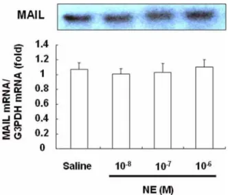
To determine whether NE induces MAIL mRNA expression in hepatocytes and non-parenchymal liver cells, primary cultured mouse hepatocytes and non- parenchymal liver cells were incubated with NE for 2 h. The MAIL mRNA levels in both types of cells were evaluated by Northern blotting. The MAIL mRNA was calculated relative to the G3PDH mRNA, which was also determined by Northern blotting. In the absence
of NE, hepatocytes showed very low MAIL expression as shown in Fig. 1. Treatment with NE did not affect the level of MAIL mRNA expression. In non- parenchymal liver cells, although basal expression of MAIL mRNA was detected, its expression was also not influenced by NE treatment (Fig. 2).
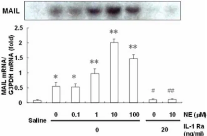
Subsequently, the effects of culture medium of non- parenchymal liver cells on MAIL mRNA expression in hepatocytes were examined, because non-paren- chymal liver cells are known to produce a variety of cytokines, such as IL-1
β, TNF
α, and IL-6 [1, 3]. When hepatocytes were incubated for 2 h with a culture medium of non-parenchymal liver cells, the MAIL mRNA expression level was significantly increased, and this expression was most significantly pronounced in culture medium of non-parenchymal cells stimulated with NE 10 µM (Fig. 3). The effects of culture medium of non-parenchymal liver cells on MAIL mRNA expression in hepatocytes were perfectly inhibited by an IL-1 receptor antagonist (Fig. 3). Thus, IL-1
βis suggested to be one of the non-parenchymal liver cell- derived factors which may effectively stimulate MAIL mRNA expression in hepatocytes. So we have examined several cytokines including IL-1
β, TNF
α, and IL-6 to test whether factors stimulate MAIL mRNA expression in hepatocytes. Fig. 4 shows changes in the expression of MAIL in hepatocytes stimulated by IL-1
β, TNF
α, Fig. 1. Effect of norepinephrine (NE) on molecule
possessing ankyrin-repeats induced by lipopolysaccharide (MAIL) mRNA expression in hepatocytes. Mouse hepato- cytes (> 98% in purity) were cultured for 48 h in William’s E medium supplemented with 5% fetal calf serum (FCS), and then various doses of NE were added to the medium, followed by incubation for 2 h. The level of MAIL mRNA expression was determined with a bioimage analyzer and expressed as a ratio to the amount of G3PDH. The results are expressed relative to the saline value (mean ± SE,
n= 6).
Fig. 2. Effect of NE on MAIL mRNA expression in non-
parenchymal liver cells. Non-parenchymal liver cells were
cultured for 24 h in William’s E medium supplemented
with 5% FCS, and then various concentrations of NE were
added to the medium, followed by incubation for 2 h. The
results are expressed relative to the saline value (mean ±
SE,
n= 6).
and IL-6. Recombinant human IL-1
βincreased MAIL mRNA expression in a dose-dependent manner, whereas
IL-6 and TNF
αdid not have any effect at all. The concentration of IL-1
βin the culture medium of non- parenchymal liver cells was estimated by using an ELISA kit, the treatment of NE significantly increased the IL-1
βlevel in a dose-dependent manner (Fig. 5).
Discussion
Our major finding is that NE enhances MAIL mRNA expression in hepatocytes through IL-1
βreleased from non-parenchymal liver cells. In addition, we have demonstrated that MAIL mRNA expression in hepatocytes and non-parenchymal liver cells was not directly influenced by NE.
MAIL, a new nuclear I
κβprotein, has long and short splicing variants, namely MAIL-L and MAIL-S [8].
MAIL-L and MAIL-S have also been reported as INAP and I
κβ-
ζ, respectively [4, 19]. MAIL mRNA expression is below detectable levels in normal mice, but it is significantly induced after LPS-injection [8].
Yamazaki
et al.[19] have reported that the ectopic expression of MAIL inhibits NF-
κβactivation in macrophage-like cells and embryonic kidney cells. The results of
in situhybridization and immunohis- tochemistry analysis revealed that MAIL is actually expressed in LPS-induced B-lymphocytes and macro- phages [11]. MAIL is also induced by inflammatory cytokines such as IL-1, IL-6, and TNF
αin various Fig. 3. Changes in the MAIL mRNA expression induced
by the culture medium of non-parenchymal liver cells.
Hepatocytes were cultured for 48 h in William’s E medium supplemented with 5% FCS and then stimulated with a culture medium of non-parenchymal liver cells diluted with an equal volume of fresh William’s E medium for 2 h. Non- parenchymal liver cells were cultured for 24 h and then incubated with increasing concentrations of NE for 2 h.
Saline indicated MAIL mRNA expression in hepatocytes without addition of the culture medium of non-parenchymal liver cells. To determine whether the interleukin (IL)-1 receptor antagonist (Ra) affect MAIL mRNA expression, the hepatocytes were preincubated in William’s E medium containing the IL-1 Ra (20 ng/mL) for 20 min, and then the culture medium of non-parenchymal liver cells treated with or without NE (10 M) was added. The results are expressed relative to the saline value (mean ± SE,
n= 6).
*p
< 0.05,
**p< 0.01
vs.saline,
#p< 0.05
vs.NE (0
µM),
##p< 0.01
vs.NE (10
µM).
Fig. 4. Changes in the MAIL mRNA expression induced by IL-1
β, TNF
α, and IL-6. Hepatocytes were stimulated with various doses of each cytokine for 2 h. The results are expressed relative to the saline value (mean ± SE,
n= 6).
*p< 0.05,
**p< 0.01
vs.saline.
Fig. 5. Effects of NE on IL-1
βproduction in cultured non-
parenchymal liver cells. Non-parenchymal liver cells were
stimulated with various doses of each NE for 2 h. The
results are expressed relative to the saline value (mean ±
SE,
n= 4).
*p< 0.05,
**p< 0.01
vs.saline.
cultured cells [11]. In the present study, northern blot analysis reveals that IL-1
βcan increase MAIL mRNA expression in cultured hepatocytes by using a cDNA probe common to MAIL-L and MAIL-S. So we can conclude that IL-1
βmay be involved in the process of MAIL expression changes in hepatocytes, but the important roles of MAIL in LPS-induced inflammatory response remains to be further studied.
The numerous literatures show that subjection to non-inflammatory stress may cause a marked elevation of plasma catecholamine and corticosterone levels [15].
Our previous reports have indicated that non-invasive stress such as oscillation and immobilization increases the mRNA expression of IL-1
βand IL-6 in the liver [6, 10]. Therefore, it is possible that hepatocytes and non-parenchymal cells may be the prerequisite for inflammatory injury and non-inflammatory stress. Liao
et al.