• 검색 결과가 없습니다.

Anomalous Arterial Supply to Normal Basal Segment of the Right Lower Lobe: Endovascular Treatment with the Amplatzer Vascular Plug

N/A
N/A
Protected

Academic year: 2021

Share "Anomalous Arterial Supply to Normal Basal Segment of the Right Lower Lobe: Endovascular Treatment with the Amplatzer Vascular Plug"

Copied!
4
0
0

로드 중.... (전체 텍스트 보기)

전체 글

(1)

Anomalous Arterial Supply to Normal Basal Segment of the Right Lower Lobe:

Endovascular Treatment with the Amplatzer Vascular Plug

Ji Hyun Kim, M.D.

1

, Sin Seung Kim, M.D.

1

, Kyung Sun Ha, M.D.

1

, Jungi Bae, M.D.

2

and Yonggeun Park, M.D.

1

1

Division of Pulmonology, Department of Internal Medicine,

2

Department of Radiology, Cheongju St. Mary’s Hospital, The Catholic University of Korea College of Medicine, Cheongju, Korea

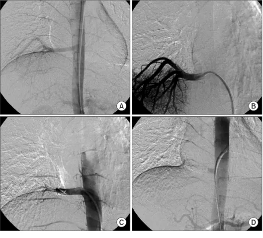
Pulmonary systemic arterialization to normal basal lung without sequestration is a rare congenital anomaly. In this rare abnormality, arterialization of the left lower lobe is the most common type. In general, surgical treatments have been performed. Recently, for reducing the complications and risks of surgery, embolization is mainly attempted by using coils. We report a case of 22-year-old male patient with a 10 mm anomalous arterial supply to his normal lung, which is being successfully treated by transcatheter embolization when using the Amplatzer Vascular Plug that has been adapted for the treatment of high-flows and large artery occlusions.

Keywords: Pulmonary Artery; Respiratory System Abnormalities; Embolization, Therapeutic

In general, surgical treatments have been performed, and recently, the embolization has been attempted.

We report the case of a 22-year-old man who was being presented with recurrent hemoptysis due to large systemic arterializations on basal segment of the right lower lobe of the lung, without sequestration. A therapeutic transarterial embo- lization using vascular plug was successfully performed. After embolization, he suffered from post-embolization syndromes such as pulmonary infarction with pneumonia. After 2 weeks of antibiotics therapy, he was discharged when the pneumo- nia improved. For the following 28 months, there have not been any hemoptysis and other complications. Our case rep- resent that large pulmonary anomalous systemic artery can be treated by transarterial embolization when using vascular plug.

Case Report

A 22-year-old man was being referred to our hospital due to hemoptysis. He experienced hemoptysis 16 months ago.

The non-contrast outside chest computed tomography (CT) missed the anomaly thereafter. Otherwise, his past medical Copyright © 2014

The Korean Academy of Tuberculosis and Respiratory Diseases.

All rights reserved.

Introduction

Pulmonary systemic arterialization to the normal basal lung without sequestration is a rare congenital anomaly. In this rare abnormality, arterialization of left lower lobe is the most common type. The involved lung has normal bronchial distri- bution, that does not include true sequestration.

CASE REPORT

http://dx.doi.org/10.4046/trd.2014.76.6.295

ISSN: 1738-3536(Print)/2005-6184(Online) • Tuberc Respir Dis 2014;76:295-298

295

Address for correspondence: Yonggeun Park, M.D.

Division of Pulmonology, Department of Internal Medicine, Cheongju St. Mary’s Hospital, The Catholic University of Korea College of Medicine, 173-19 Juseong-ro, Sangdang-gu, Cheongju 360-568, Korea

Phone: 82-43-219-8000, Fax: 82-43-219-8000 E-mail: [email protected]

Received: Dec. 20, 2013 Revised: Jan. 13, 2014 Accepted: Jan. 15, 2014

cc

It is identical to the Creative Commons Attribution Non-Commercial

License (http://creativecommons.org/licenses/by-nc/3.0/).

(2)

JH Kim et al.

296 Tuberc Respir Dis 2014;76:295-298 www.e-trd.org

history and family history were not remarkable. Physical and laboratory findings were within normal limits. A chest radio- graph demonstrated no active lung lesions. Chest CT with contrast enhancement showed a ground-glass attenuation and consolidation in the right lower lobe posterior basal seg- ment and a large anomalous artery from the distal descend- ing thoracic aorta supplying basal segment of right lower lobe (Figure 1). The diameter of the feeding artery was about 10 mm. Bronchoscopy revealed that the major bronchial branches were normal and at the right 2nd carina there were bleeding evidences and woozing on saline washing (Figure 2).

Angiography showed about 10 mm diameter feeding artery to basal segment of right lower lobe, enlarged arteriovenous ves- sels and draining vein. On diagnosis of anomalous systemic arterial supply to the basal segments of right lower lobe from descending aorta without sequestration, the therapeutic em- bolization was performed with Amplatzer Vascular Plug (two 12 mm; AGA Medical, Golden Valley, MN, USA) (Figure 3).

After the embolization, the patient suffered from fever.

At 2 days after embolization, chest X-ray revealed hazy and nodular opacities at right lower lobe (RLL). With a suspected diagnosis of post-embolization syndrome with pneumonia, the patient was prescribed to empirical antibiotics. Although the fever subsided 5 days later, his chest X-ray showed pleural effusion in the right lung (Figure 4). A diagnostic thoracentesis revealed amber colored fluids, which was found to be neutro- phil-predominant exudates. At 14 days after the antibiotics ap- plication, he was discharged when symptoms and conditions had improved, but pleural effusion sustained. Twenty-two days later, his chest CT revealed vascular plugs, anomalous systemic artery from distal descending aorta, together with decreased size of subsegment veins, posterior subpleural con- solidation, RLL that seemed pulmonary infarction and mild right pleural effusion. After 1 month of follow-up, chest X-ray indicated no lung lesions.

He recovered well after embolization for 2-year follow-up without complications (Figure 5).

Discussion

Anomalous systemic arterial supply to normal segments of the lung is a rare congenital anomaly. Anomalous systemic arterializations of right lower lobe is even more rare. Some authors mentioned this anomaly within the spectrum of pulmonary sequestration such as Pryce et al. classification

1,2

. Although it is included in the Pryce type 1 according to the original classification of pulmonary sequestration, it differs from the normal connection of the bronchial tree

3

.

The etiology of a systemic arterial supply to normal lung is unknown, the most likely theory is a persistent embryonic connection between aorta and pulmonary parenchyma due to failures of regression from the primitive aortic branches to the developing lung bud

4

.

The basal segments of left lower lobe are the most com- monly affected sites, although there are other sites which can be rarely affected. The systemic artery usually varies from the thoracic aorta, but can also arise from abdominal aorta Figure 1. (A) Computed tomography (CT) reveals a large anomalous artery from the distal descending thoracic aorta supply- ing the basal segment of right lower lobe.

(B) CT shows a ground-glass attenuation and consolidation in the posterior of the right lower lobe.

Figure 2. Bronchoscopy reveals that the normal major bronchial

branch and the right 2nd carina are bleeding evidences and wooz-

ing after saline washing.

(3)

Therapeutic embolization of pulmonary systemic arterializations to normal lung

http://dx.doi.org/10.4046/trd.2014.76.6.295 297

www.e-trd.org

or celiac axis, and very rarely from the left subclavian and internal mammary artery

5,6

. These vessels usually enter the lung at the level of pulmonary hilum or at the level of inferior pulmonary ligaments. The venous drainage is normal and is

accomplished by inferior pulmonary vein draining into the left atrium

3

.

Most patients are asymptomatic. The most common symp- tom is recurrent or massive hemoptysis during adulthood

5

. This is related to the higher pressure of systemic circulations as compared to pulmonary circulations. Other manifesta- tions include exertional dyspnea, murmur in the lower chest and congestive heart failure due to left heart overload

3

. Chest radiographs often show ill-defined opacity corresponding to the anomalous artery

7

. Therefore, incidentally, a chest radio- graphs for health screening has often found the anomaly. CT angiography can demonstrate the origin and course of aber- rant artery.

Treatment is recommended to symptomatic patients but also to asymptomatic patients owing to the risks of hemor- rhagic complications

8

.

The classic therapeutic approach is surgical resection. In recent years, because of high surgery related morbidity and mortality, (vascular plug 8-AV malformation) the endovascu- lar embolization technique has been increasingly used for the treatment of this disease. Mainly, the endovascular materials used coils

5,9-11

. However, in this case, the diameter of the feed- ing artery was about 10 mm which could be associated with high risks of incomplete occlusion, recanalization and emboli- zation of the coil

12

.

Figure 3. (A, B) Angiography showing an large anomalous systemic artery from the descending aorta which supplies the segment of the right lower lobe. (C, D) A complete occlusion of the large anoma- lous systemic artery after embolization by using the Amplatzer Vascular Plug.

Figure 4. Four days follow-up of the chest radiograph shows hazi-

ness and pleural effusions in the right lower lobe.

(4)

JH Kim et al.

298 Tuberc Respir Dis 2014;76:295-298 www.e-trd.org

The Amplatzer Vascular Plug is a self-expandable cylin- drical mesh device which is intended for the treatment of atrial septal defect and patent ductus arteriosus. Since being described, there have been sporadic case reports on its use in the literature. Vessels occluded with the device include aortopulmonary collaterals, pulmonary arteriovenous fistulas, venovenous collaterals, shunts, coronary fistulas, and certain types of patent ductus arteriosus

13

.

The Amplatzer Vascular Plug is particularly suited for the embolization of large and high-flow vessels where coil migra- tion is possible or where multiple coils may be needed

12,14

. In our case, large pulmonary anomalous systemic artery of basal segment of the right lower lobe of the lung, without the sequestration presenting recurrent hemoptysis can be treated by transarterial embolization when using vascular plug. Al- though he suffered from post embolization syndromes and complications after the embolization for 2 weeks, he had no complications for 2 years of follow-up. Our case present that large pulmonary anomalous systemic artery can be treated by transarterial embolization when using the vascular plug.

Conflicts of Interest

No potential conflict of interest relevant to this article was reported.

References

1. Pryce DM. Lower accessory pulmonary artery with intralobar sequestration of lung; a report of seven cases. J Pathol Bacte- riol 1946;58:457-67.

2. Sade RM, Clouse M, Ellis FH Jr. The spectrum of pulmonary sequestration. Ann Thorac Surg 1974;18:644-58.

3. Bhalla AS, Gupta P, Mukund A, Kumar A, Gupta M. Anoma- lous systemic artery to a normal lung: a rare cause of hemop- tysis in adults. Oman Med J 2012;27:319-22.

4. Ellis K. Fleischner lecture: developmental abnormalities in the systemic blood supply to the lungs. AJR Am J Roentgenol

1991;156:669-79.

5. Canyigit M, Gumus M, Kilic E, Erol B, Cetin H, Hasanoglu HC, et al. Aneurysm of an anomalous systemic artery supplying the normal basal segments of the left lower lobe: endovascu- lar treatment with the Amplatzer Vascular Plug II and coils.

Cardiovasc Intervent Radiol 2011;34 Suppl 2:S126-30.

6. Haraguchi S, Hioki M, Yamashita K, Yamashita Y, Matsumoto K, Shimizu K. Large anomalous systemic arterial supply to basal segments of the left lung. Jpn J Thorac Cardiovasc Surg 2004;52:395-7.

7. Yamanaka A, Hirai T, Fujimoto T, Hase M, Noguchi M, Konishi F. Anomalous systemic arterial supply to normal basal seg- ments of the left lower lobe. Ann Thorac Surg 1999;68:332-8.

8. Liu FY, Wang MQ, Fan QS, Duan F, Wang ZJ, Song P. Endovas- cular embolization of pulmonary arteriovenous malforma- tions. Chin Med J (Engl) 2010;123:23-8.

9. Jiang S, Shi JY, Zhu XH, Chen C, Sun XW, Yu D, et al. Endovas- cular embolization of the complete type of anomalous sys- temic arterial supply to normal basal lung segments: a report of four cases and literature review. Chest 2011;139:1506-13.

10. Lim HH, Huh JK, Kim JH, No HS, Choe DH, Kim CH, et al.

Therapeutic embolization of systemic arterial supply to nor- mal basal segments of the left lower lobe of the lung. Respirol- ogy 2009;14:606-8.

11. Saida T, Ninomiya H, Hojo F, Nakayama M, Yamauchi T, Saida Y. Systemic arterial supply to the normal basal segments of the left lower lobe treated by coil embolization, with long- term follow-up. Radiat Med 2006;24:365-8.

12. Kong JH, Oh TY, Kim JT, Baek KS, Chang WH. Transcatheter embolization of giant pulmonary arteriovenous malforma- tion with an Amplatzer Vascular Plug II. Korean J Thorac Car- diovasc Surg 2012;45:326-9.

13. Hwang HK, Tsai YS, Lin SM, Chen MR. Occlusion of an aber- rant artery to an intralobar pulmonary sequestration using an Amplatzer Vascular Plug. Pediatr Pulmonol 2008;43:933-5.

14. Rossi M, Rebonato A, Greco L, Stefanini G, Citone M, Spe- ranza A, et al. A new device for vascular embolization: report on case of two pulmonary arteriovenous fistulas emboliza- tion using the Amplatzer Vascular Plug. Cardiovasc Intervent Radiol 2006;29:902-6.

Figure 5. (A, B) A follow-up contrast-

enhanced computed tomography exami-

nation at 5 months demonstrates that the

Amplatzer Vascular Plug is located at the

anomalous systemic pulmonary feeding

artery without parencheymal complica-

tions

수치

Figure 2. Bronchoscopy reveals that the normal major bronchial  branch and the right 2nd carina are bleeding evidences and  wooz-ing after saline washwooz-ing
Figure 3. (A, B) Angiography showing an  large anomalous systemic artery from  the descending aorta which supplies the  segment of the right lower lobe
Figure 5. (A, B) A follow-up contrast- contrast-enhanced computed tomography  exami-nation at 5 months demonstrates that the  Amplatzer Vascular Plug is located at the  anomalous systemic pulmonary feeding  artery without parencheymal  complica-tions

참조

관련 문서