서울위생병원 1내과, 2병리과, 3흉부외과
차성철1, 김시우1, 조유진1, 박성균1, 박현근1, 김종상1, 곽재욱1, 유문빈1, 조혜제2, 이재진3
A Case of Intermediate Grade Bronchial Mucoepidermoid Carcinoma and Review in Korean Cases
Sung Chul Cha, M.D.
1, Si Woo Kim, M.D.
1, Yoo Jin Cho, M.D.
1, Sung Kyoon Park, M.D.
1, Hyun Keun Park, M.D.
1, Jong Sang Kim, M.D.
1, Jae Wook Kwak, M.D.
1, Moon Bin Yoo, M.D.
1, Hye Jae Cho, M.D.
2, Jae Jin Lee, M.D.
3Departments of
1Internal Medicine,
2Pathology and
3Thoracic and Cardiovascular Surgery, Seoul Adventist Hospital, Seoul, Korea
Bronchial mucoepidermoid carcinoma is uncommon, representing 0.2% of all lung tumors. The disease usually presents with symptoms of airway obstruction and recurrent pneumonia. It is commonly classified into two grades in Korea, low and high. We report a case of a bronchial mucoepidermoid carcinoma in a 40-year-old woman who complained of symptoms of an upper respiratory infection. The histological grade after a bronchoscopic biopsy was intermediate. A left upper lobectomy was performed as treatment. The TNM stage of this case was IA (T1N0M0). In addition, 25 cases of bronchial mucoepidermoid carcinoma from 1984 in Korea are also reviewed from the viewpoint of the relationship between the histological grade, TNM stage and clinical course of the tumor.(Tuberc Respir Dis 2008;65:41-48)
Key Words: Bronchial mucoepidermoid carcinoma, Intermediate grade, TNM stage, PET
Address for correspondence: Sung Chul Cha, M.D.
Division of Pulmonary Medicine, Department of Internal Medicine, Seoul Adventist Hospital, 29-1, Hwikyung 2- dong, Dongdaemun-gu, Seoul 130-092, Korea
Phone: 82-2-2210-3441, Fax: 82-2-2212-2673 E-mail: [email protected]
Received: Jun. 2, 2008 Accepted: Jun. 30, 2008
서 론
폐에서 흔히 발생하는 타액형 암종에는 선양낭포성암 (adenoid cystic carcinoma)과 점액표피양 암종(mucoepi- dermoid carcinoma)이 있다. 그 중 점액표피양 암종은 주 로 타액선에서 호발하며 폐나 기관지에서 원발하는 경우 는 0.1∼0.2%로 드물다1. 병리학적 기준에 근거하여 일반 적으로는 예후가 좋은 저등급(low grade)의 종양과 고등 급(high grade)의 두 등급으로 나뉜다2.
저자들은 장기간 지속되는 상기도 감염 증상으로 내원한 40세 여성 환자에서 흉부 전산화 단층촬영, 기관지내시경 검사를 통한 생검 및 양전자방출단층촬영술(positron em- ission tomography, PET)로 중간등급의 기관지 점액표피
양 암종을 확인하고 좌상엽절제술을 시행한 후에 경과 관 찰중인 1예를 경험하였기에 문헌고찰과 함께 보고하는 바 이다. 또한 현재까지 국내에 보고된 자료들을 바탕으로 조직학적인 등급과 TNM stage에 따른 임상경과의 연관관 계를 살펴보았다.
증 례
환 자:
박○○, 40세 여자주소 및 현병력:
상기 환자는 내원 1개월 전부터 시작 된 기침과 오한을 주소로 개인병원을 방문하여 상기도염 진단 하에 치료를 하였으나, 증상의 호전없이 반복되는 기침과 발열로 정밀검사 위하여 외래로 내원하였다.과거력과 가족력:
특이소견 없음.이학적 소견:
내원당시 체온은 36oC, 혈압은 100/70 mmHg, 맥박수 84회/분, 호흡수 22회/분이었으며 병색은 없었다. 결막은 창백하지 않았고 경부에서 림프절 종대는 촉지되지 않았으며, 흉부 청진에서 호흡음은 깨끗하였고 심잡음은 들리지 않았다. 복부 및 사지에는 특이소견이 없었다.Figure 1. Chest X-ray shows hazy opacities in the lingular segment of the left lung, around the left cardiac border.
Figure 2. Computed tomography scan reveals focal atele- tatic change on left lingular segment, but no mass is seen in endobronchial area.
Figure 3. Flexible bronchoscopy reveals 1.8 cm sized, soft yellowish tumor deriving from the bronchial wall. It has much mucoid like material and is bulging into the os- tium of lingular segment of bronchus (arrows).
Figure 4. Positron Emission Tomography at lower lung level. There was 18F-fluoro deoxyglucose (FDG) uptake in the bronchial area.
검사실 소견:
말초혈액검사에서 백혈구 7,850/mm3 (호중구 70.4%), 혈색소 12.0 g/dl, 혈소판 472,000/mm3 이었고 전해질검사, 요검사, 대변검사 및 혈청 생화학 검 사는 정상소견이었다. 혈청 CEA는 1.01 ng/ml로 정상 범 위 였으며, 객담검사는 음성이었다.방사선학적 소견:
내원 당시 시행한 단순 흉부 X선에 서 좌설엽에 폐침윤(pulmonary infiltration) 소견이 보였 다(Figure 1). 흉부 전산화 단층촬영에서는 좌상엽의 설구 가 부분적으로 허탈된 소견 외에는 특이소견은 보이지 않 았다(Figure 2).기관지 내시경 소견:
좌측 주기관지는 정상이었으 나, 좌상엽기관지에서 설엽의 개구부는 다량의 분비물로Figure 7. Immunohistoche- mical stain of Cytokeratin 7 (A) was positive and TTF-1 (B) was negative.
Figure 5. A yellowish solid endobronchical mass was seen, which is 1.8×3.0 cm sized and arising from the bronchial wall.
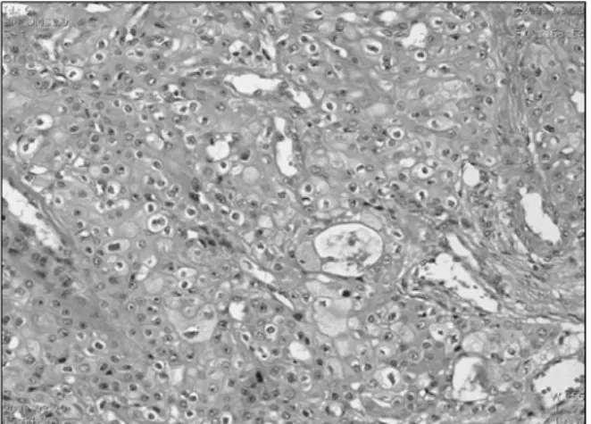
Figure 6. Histology of the tumour. The nuclear showed moderate pleomorphism. But tumor cell showed few mi- totic figures (less than 5 per 50 HPF). Well differentiated Goblet cell is seen at central portion. Necrosis is absent (H&E stain, ×400).
인해 거의 폐쇄되어 있었고 지름 1.8 cm 정도 크기의 연 황색을 띠는 매끈한 표면의 종괴가 관찰되었다(Figure 3).
양전자방출단층촬영술 소견:
수술 전 시행한 양전 자방출단층촬영술 상 좌설엽에 기관지 폐쇄에 의한 무기 폐 소견이 있었으나(Figure 4), 설엽 부위의 기관지내강으 로는 18F-fluoro deoxyglucose의 섭취는 관찰되지 않았다.수술소견:
전신마취 하에 좌측 전측방 흉부절개술을 시행하여 좌상엽절제술과 기관지 주변 및 종격동 림프절 절제술을 시행하였다. 육안적 소견 상 좌상엽기관지의 개 구부에 위치해 있었고 흉벽의 유착은 없었으며 종격동 및 기관지 주위로의 임파선 전이도 없었다.병리 조직학적 소견:
육안소견 상 기관지벽에서 돌출되어있는 황색을 띠는 1.8×3.0 cm 크기의 종괴가 관찰되 었다(Figure 5). 현미경적 소견에서는 종괴의 대부분이 기 관지내강에 주로 위치하고 있었고 일부가 기관지벽을 침 범한 소견을 보였으나, 기관지 절단면에서 보이는 경계는 깨끗하였다. 전반적으로는 편평상피가 우세하며 국소적 인 선상상피와 혼재되는 양상이었다. 세포의 핵은 균일했 고 중등도의 다형성(pleomorphism)을 보이며 뚜렷한 핵 소체를 보였지만, 세포분열은 아주 드물게 관찰되었고 조 직괴사는 관찰되지 않았다. Conlan 등3의 분류기준에 따 라서 중간등급(intermediate grade, Grade 2)의 기관지 점 액표피양 암종으로 진단하였다(Figure 6). 면역화학염색 에서는 Cytokeratin 7에 양성을 보였고 TTF-1에는 음성을
Table 1. Histology, stage and clinical course (n=22)
Case Histology Stage Treatment F/u (month) Live/Death
1 2 3 4 5 6 7*
8 9 10 11 12 13 14 15† 16 17 18 19 20 21 22 23 24 25 26
Low Low Low Low High High Low Low High High Low High Low Low Low High High - Intermediate
Low - Low Low Low - Intermediate
- - - - - - IIIA IIB IIIB IV IB IIIB
IB IB IIIB IIIB IIIB - IB IB IB IA IA IA - IA
BL BL Lo+SR
Lo Pn Conservative
Pn+Ch Lo+SR
Ch Ch Lo Ch+Ra
BL Lo Ch+Laser
Pn Conservative
- BL Lo Lo SR SR Lo BL Lo
- - - 16 3 17 34 - Loss
2 29 9 43 29 36 3 Loss
- 12 1 1 1 36 18 - 9
- - - Alive Alive Death
Alive - - Death
Alive Death
Alive Alive Death
Alive - Alive Alive Alive Alive Alive Alive Alive Alive Alive BL: bilobectomy; Lo: lobectomy; SR: sleeve resection; Pn: pneumonectomy; Ch: chemotherapy; Ra: radiotherapy.
*case of mucoepidermoid carcinoma with adenocarcinoma, †inoperable case due to location of tumor was upper trachea.
나타내어서 선암종과 감별하였다(Figure 7).
치료 및 경과:
환자는 수술 후에 합병증 없이 퇴원하였 고 재발 없이 9개월동안 외래에서 경과 관찰중이다.고 찰
기관지에서 발생하는 점액표피양 암종은 1952년 Smetana 등4에 의해 처음 보고되었고, 모든 폐종양에서는 0.1∼0.2%, 기관지선 종양의 1∼5%를 차지하는 매우 드 문 종양이다1. 발생학적으로 타액선과 유사하며, 주로 소 아에서 호발하지만, 3세에서 78세까지의 다양한 연령에서 발생할 수 있고 남녀의 발생비는 차이가 없다5.
발생원인에 관해서 Pandya 등6은 기관지 점액표피양 암종이 편측의 저형성 폐와 같은 선천성 기형을 동반하는 경우를 이야기하면서 유전적인 요소가 원인일 가능성을 제시하였고 특히, 상반전좌(reciprocal translocation)가 다
수 관찰되며 주로 t(11;19)와 연관이 있다고 보고하였다.
Tonon 등7은 t(11;19)(q14-21;p12)의 전좌로 mucoepider- moid carcinoma translocated 1-mastermind-like 2 (MECT1- MAML2)가 생기게 되고, 이것이 Notch signaling pathway 를 방해해서 암종이 발생한다고 보고하였고, Barrett 등8은 t(1;11)(p22;q13) 때문에 cyclin D1이 과표현(overex- pression)되는 것이 원인이라 제시했다.
증상은 주기관지에 호발하기 때문에 기관지 자극에 의 한 기침, 객혈, 호흡곤란 증상과 기관지 폐쇄에 의한 천명 음, 폐렴 등이 나타날 수 있으며, 인접한 폐실질에 폐렴이 나 무기폐를 동반하는 경우도 빈번하다9. 단순 흉부 X선 에서는 종양의 특성이 주로 중심부에 위치하고 크기도 작 은 경우가 많기 때문에 정상이거나 폐쇄성 폐렴이나 무기 폐의 소견을 보이는 경우가 많다. 흉부 전산화 단층촬영 은 기관지내 종양의 확인, 국소 침범, 임파절 전이의 판정 에 유효하지만 크기가 작거나 전이가 되지 않는 종양의
Table 2. Conlan's classification of mucoepidermoid carci- noma of lung
Grade Criteria
1 (low) 2 (intermediate)
3 (high)
Well formed glandular structures, abundant cytoplasm and regular nulcei
Resemble Grade 1 tumors but are less well differen-tiated and more hyperchromatic nuclei
Infrequent mitotic figure
Poorly differentiated, Prominent nuclear atypism and scanty cytoplasm, reverse N/C ratio*
Frequent mitotic figure
*nuclear to cytoplasmic ratio.
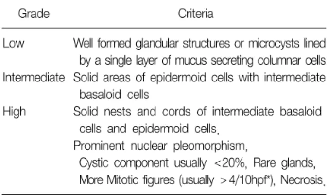
Table 3. Classification of mucoepidermoid carcinoma of salivary gland
Grade Criteria
Low Intermediate High
Well formed glandular structures or microcysts lined by a single layer of mucus secreting columnar cells Solid areas of epidermoid cells with intermediate basaloid cells
Solid nests and cords of intermediate basaloid cells and epidermoid cells.
Prominent nuclear pleomorphism,
Cystic component usually <20%, Rare glands, More Mitotic figures (usually >4/10hpf*), Necrosis.
*high power field.
경우에는 위음성을 나타내므로 유효성이 적다. 기관지 내 시경은 종괴를 직접 볼 수 있는 장점이 있고 조직 채취도 가능하여 정확한 진단에 도움이 된다.
양전자방출단층촬영술은 일반적으로 폐병변을 검사하 는데 유용하지만, 현재까지 기관지 점액상피양 암종과 관 련된 보고는 국내외를 통틀어서도 몇 예에 불과하다10. Kinoshita 등11이 보고한 바에 의하면 흉부 전산화단층촬 영에 비해서 양전자방출단층촬영술이 진단과 치료방침 결정에 있어서 더 유익하며, 특히 저등급에 비해 고등급에 서 18F-fluoro-deoxyglucose의 부분적인 섭취 증가가 더 욱 뚜렷하게 관찰되었다고 보고된 바가 있다.
기관지 점액상피양 암종의 조직학적인 특징 중 하나는 구성세포들인데, 점액분비세표, 상피양세포 그리고 중간 형세포가 혼재된 양상을 보인다. 특히 상피양세포의 상대 적인 구성비율은 저등급에서 20% 정도이고, 중간등급에 서 35∼40% 정도 고등급에서는 50% 이상으로 고등급에 서 가장 높다12. 반대로 점액세포의 비율은 저등급에서 고 등급으로 갈수록 낮다. 1989년 Heitmiller 등2은 괴사, 핵 의 다형성 그리고 활동적인 유사분열을 고등급에서만 나 타나는 특징으로 제시하였다.
하지만, 두 단계의 조직학적인 분류기준에 관해서는 아 직까지 논란의 여지가 있다. 본 예에서는 조직학적인 등 급을 두 단계로 분류할 수 없는 중간등급으로 판정하였는 데, 이러한 경우가 외국에서는 적지 않게 있었지만12,13, 국 내에서는 현재까지 1예(Table 1)가 있었고, 그나마, 분류 기준도 정확하게 설명되어있지는 않았다. 현재까지 적용 되어온 저등급과 고등급의 양측성 분류는 1979년 Klac- smann 등14과 1980년 Carter 등15이 제시한 기준을 따르는
데, 본 증례와 비교해 볼 때는 상대적으로 편평세포가 우 세한 점과 핵에서 중등도 이상의 다형성을 보인 점은 고등 급의 기준을 만족하였으나, 조직괴사가 없고 유사분열의 활성도가 낮았다는 점에서는 저등급에 해당해서 단순히 고등급또는 저등급으로의 명확한 분류는 불가능했다. 반 면에 1978년 Conlan 등3이 제시한 분류기준(Table 2)을 적용할 때에는 세포 분화도와 핵의 다형성 및 핵과세포질 의 비율 등이 고등급과 저등급의 중간정도를 보이는 점과, 괴사가 없고, 유사분열의 활성은 거의 보이지 않는 점에 근 거해서 중간등급(Grade 2)으로 정확하게 분류할 수 있었다.
점액표피양 암종은 기관지에서 발생하는 것과 발생학 적, 조직학적인 성상은 같지만14, 타액선에서 더 흔하게 발 생하기 때문에 조직학적인 분류기준에 관해서는 주로 타 액선에서 연구되었다. 현재 타액선 암종에 통상적으로 적 용되는 조직학적 분류는 3단계(Table 3)이며, 2단계 분류 에 비해서 등급의 재현성에 있어서 정확하다고 알려져 있 다16. 주목할 점은 타액선 점액표피양 암종의 중간등급은 저등급이나 고등급과 비교할 때 림프절 전이여부, 종양의 병기, 재발과 생존율에 있어서 유의한 차이점이 있었다는 점이다17. 따라서 기관지에서 발생하는 암종에서도 3가지 등급으로 분류기준을 적용하는 것이 이론적으로 합당하 고, 그 기준에 따라서 임상경과 및 예후를 평가하는 것이 타당하리라 사료된다.
면역조직화학염색은 타 종양과의 감별에 유용하다.
TTF-1에 음성을 보이고 cytokeratin 5,6,7에는 양성, cyto- keratin 20에는 음성을 보이는 점이 선암종과의 차이점이 어서 두 종양을 쉽게 감별할 수 있다18.
치료로는 외과적 절제가 원칙이다12. 예후가 좋은 저등
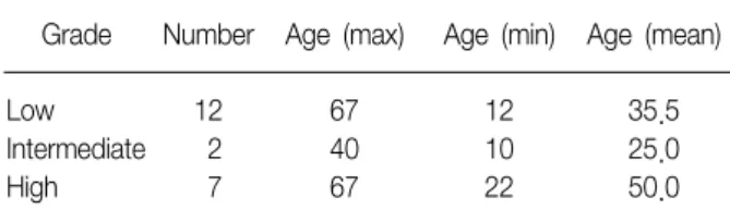
Table 4. Grade and age (n=21)
Grade Number Age (max) Age (min) Age (mean) Low
Intermediate High
12 2 7
67 40 67
12 10 22
35.5 25.0 50.0 급의 경우에 크기에 따라서 기관지 성형술이나 폐엽절제
술을 통한 완전제거로 충분하지만, 중간등급이나 고등급 의 경우는 주위조직의 침범, 재발과 전이가 흔하기 때문에 임파절 생검을 포함한 광범위의 폐절제술을 시행하며 술 후 방사선 요법이나 항암화학요법도 시행한다3,13. 본 증례 의 경우는 종양의 위치가 설엽의 개구부에 위치하였지만, 폐실질 내로의 침범을 완전히 배제할 수 없어서 기관지 성형술에 적합하지 않았으며, 기관지 내시경을 통해 얻어 낸 조직이 저등급이 아닌, 중간등급으로 판정되어 완전한 절제가 필요하였기 때문에 폐엽절제술을 시행하였다. 그 리고 TNM stage는 IA로서 초기병기에 해당하였기 때문에 화학요법이나 방사선치료는 고려하지 않았던 경우이다.
최근에는 새로운 항암화학요법으로 EGFR-TKI (epider- mal growth factor receptor-tyrosine kinase inhibitor)가 등장하였는데, Han 등19이 보고한 바에 의하면 점액표피 양 암종을 앓고 있는 환자의 40% 이상에서 EGFR TK의 돌연변이나 증식이 발견 되며, EGFR TKI는 좋은 치료반 응을 나타냈다고 한다.
예후는 일반적으로 모든 등급에서 선양낭성암종보다는 양호하다. 예후를 결정짓는 인자로 조직학적 등급과 TNM stage가 중요하다. 등급이 낮을수록 기관지에 국한되고 주 위의 폐실질로 침범하지 않는 경우가 많은 반면에 등급이 높을수록 종양내 괴사 및 임파선과 폐실질로의 침범도 흔 하고 예후도 나쁘다. 등급이 낮은 경우 5년 생존율은 80
∼87%이고 고등급의 경우에는 31%로 낮게 보고되고 있 다20. TNM stage와 관련해서 Chin 등12은 초기 병기(병기 IA, IB, IIB)의 환자(10년 생존율이 87.5%)들이 후기병기 (IIIB, IV)의 환자(1년 생존율 28.6%, 2년 생존율 0%)들에 비해 예후가 좋았다고 보고한 바가 있다.
점액표피양 암종의 전이는 국소 전이 및 임파선을 통해 이루어지며, 폐문부 임파절 전이가 된 경우에 예후가 가장 불량하다.
국내에서 보고된 점액표피양 암종에 대한 고찰
1984년 이후로 국내에서 보고된 25예와 본 1예를 조직 학적 등급과 TNM stage에 따른 병기, 임상적 경과를 중심 으로 정리하였다(Table 1)21-36.
발생 연령에 있어서는 9세에서 67세까지 다양하게 분 포하였고, 조직의 등급은 26예 중에 21예에서만 확인이 가능하였다. 등급에 따른 연령은 저등급의 평균연령이 35.5세였고, 고등급에서는 50.0세였다(Table 4).
등급별 임상경과에서 차이를 보였는데, 저등급에서는 12예 중에 수술이 불가능했던 1예와 선암종을 동반했던 1예를 제외한, 10예 중 8예가 폐엽절제술을 시행 받았고 10예 중 2예가 수상절제술을 받았으며, 12예 모두 생존하 였다. 반면에 고등급에서는 총 7예 중 5예는 사망하거나 추적관찰이 되지 못했으며, 2예만이 폐절제술을 시행 받 았고 수술을 받은 2예만이 생존하였다. 문헌상 확인할 수 있었던 평균적인 생존기간은 저등급과 고등급 각각에서 32개월과 3개월로 비교적 큰 차이를 보였다.
TNM stage에 따른 임상경과에서는 경과를 확인할 수 없었던 5예를 제외한 13예 중, 초기 병기(IIB이하) 10예는 모두 수술을 받았고 모두 생존하였지만, 후기 병기(IIIA이 상) 3예에서는 1예만 수술을 받았고, 나머지 2예 모두 사 망하였다.
자료의 불충분함과 환자수가 적다는 한계는 있었지만 현재까지 보고된 바와 같이 국내 보고에서도 조직학적 등 급과 TNM stage에 따른 임상경과의 차이가 컸음을 확인 할 수 있었다. 추후에 정확하고 많은 자료를 바탕으로 연 구가 된다면 국내에서도 등급, 병기, 임상경과 및 예후간 의 유의한 통계학적 자료도 나올 수 있을 것이라 추정되는 바이다.
요 약
기관지에서 발생하는 점액표피양 암종은 폐에서 발생 하는 암종의 0.2%를 차지할 정도로 드물다. 주로 기관지 폐쇄에 의한 증상이나 반복적인 폐렴의 임상양상을 보인 다. 조직학적인 기준에 따라서 일반적으로 저등급과 고등 급으로 나누어지고 예후도 다르다. 저자들은 반복되는 기 침과 열을 주소로 내원한 40세 여성에서 발생한 흔하지 않은 중간등급의 기관지 점액상피양 암종 1예를 경험하였 기에 문헌고찰과 함께 보고하고 현재까지 국내에 보고된 자료들을 바탕으로 조직학적인 등급과 TNM stage에 따른 임상경과의 차이를 보고하는 바이다.
참 고 문 헌
1. Colby T, Koss M, Travis W. Tumors of the lower respi- ratory tract. 3rd series. Washington, DC: Armed Forces Institute of Pathology; 1995.
2. Heitmiller RF, Mathisen DJ, Ferry JA, Mark EJ, Grillo HC. Mucoepidermoid lung tumors. Ann Thorac Surg 1989;47:394-9.
3. Conlan AA, Payne WS, Woolner LB, Sanderson DR.
Adenoid cystic carcinoma (cylindroma) and mucoepi- dermoid carcinoma of the bronchus. Factors affecting survival. J Thorac Cardiovasc Surg 1978;76:369-77.
4. Smetana HF, Iverson L, Swan LL. Bronchogenic carci- noma: an analysis of 100 autopsy cases. Mil Surg 1952;111:335-51.
5. Liu X, Adams AL. Mucoepidermoid carcinoma of the bronchus. Arch Pathol Lab Med 2007;131:1400-4.
6. Pandya H, Matthews S. Case report: Mucoepidermoid carcinoma in a patient with congenital agenesis of the left upper lobe. Br J Radiol 2003;76:339-42.
7. Tonon G, Gehlhaus KS, Yonescu R, Kaye FJ, Kirsch IR.
Multiple reciprocal translocations in salivary gland mu- coepidermoid carcinomas. Cancer Genet Cytogenet 2004;152:15-22.
8. Barrett W, Heaps LS, Diaz S, Sharma P, Arbuckle S, Smith A. Mucoepidermoid carcinoma of the bronchus in a 15-year-old girl with complex cytogenetic re- arrangement involving 11q and over-expression of cy- clin D1. Med Pediatr Oncol 2002;39:49-51.
9. Niggemann B, Gerstner B, Guschmann M, Paul K, Wit J, Mau H, et al. An 11-yr-old male with pneumonia and persistent airway obstruction. Eur Respir J 2002;19:
582-4.
10. Ishizumi T, Tateishi U, Watanabe S, Maeda T, Arai Y.
F-18 FDG PET/CT imaging of low-grade mucoepi- dermoid carcinoma of the bronchus. Ann Nucl Med 2007;21:299-302.
11. Kinoshita H, Shimotake T, Furukawa T, Deguchi E, Iwai N. Mucoepidermal carcinoma of the lung detected by positron emission tomography in a 5-year-old girl.
J Pediatr Surg 2005;40:E1-3.
12. Chin CH, Huang CC, Lin MC, Chao TY, Liu SF.
Prognostic factors of tracheobronchial mucoepider- moid carcinoma: 15 years experience. Respirology 2008;
13:275-80.
13. Matsuzaki Y, Shibata K, Yoshioka M, Inoue M, Sekiya R, Onitsuka T, et al. Successful treatment of bronchial mucoepidermoid carcinoma in an 11-year-old boy by bronchoplasty: report of a case. Surg Today 1996;26:
64-7.
14. Klacsmann PG, Olson JL, Eggleston JC. Mucoepider- moid carcinoma of the bronchus: an electron micro- scopic study of the low grade and the high grade variants. Cancer 1979;43:1720-33.
15. Carter D, Eggleston JC. Tumors of the lower respiratory tract. Atlas of tumour pathology. Washington, DC:
Armed Forces institute of Pathology; 1980.
16. Brandwein MS, Ivanov K, Wallace DI, Hille JJ, Wang B, Fahmy A, et al. Mucoepidermoid carcinoma: a clin- icopathologic study of 80 patients with special refer- ence to histological grading. Am J Surg Pathol 2001;25:835-45.
17. Hicks MJ, el-Naggar AK, Flaitz CM, Luna MA, Batsakis JG. Histocytologic grading of mucoepidermoid carcino- ma of major salivary glands in prognosis and survival:
a clinicopathologic and flow cytometric investigation.
Head Neck 1995;17:89-95.
18. Shilo K, Foss RD, Franks TJ, DePeralta-Venturina M, Travis WD. Pulmonary mucoepidermoid carcinoma with prominent tumor-associated lymphoid prolife- ration. Am J Surg Pathol 2005;29:407-11.
19. Han SW, Kim HP, Jeon YK, Oh DY, Lee SH, Kim DW, et al. Mucoepidermoid carcinoma of lung: potential target of EGFR-directed treatment. Lung Cancer 2008;
61:30-4.
20. Molina JR, Aubry MC, Lewis JE, Wampfler JA, Williams BA, Midthun DE, et al. Primary salivary gland-type lung cancer: spectrum of clinical presentation, histo- pathologic and prognostic factors. Cancer 2007;110:
2253-9.
21. Song IS, Cho KH, Lee HG. Mucoepidemoid tumor of the bronchus. Korean J Thorac Cardiovasc Surg 1984;
17:740-6.
22. Ann B, Cha HD, Kwon YM, Kang JM. Childhood bron- chial mucoepidermoid tumor: a case report and review of the literature. Korean J Pediatr 1985;28:102-6.
23. Byun HS, Oh BS, Lee DJ. Bronchial mucoepidermoid carcinoma: 1 case report. Korean J Thorac Cardiovasc Surg 1988;21:941-7.
24. Kim JH, Kim YB, Kim CS, Kim DS, Kim RH, Kim CH, et al. A report of two cases of mucoepidermoid carcinoma. Tuberc Respir Dis 1993;40:58-66.
25. Jeong JM, Song JY, Hong JR, Kim YJ, Kim MS. A case of mucoepidermoid carcinoma in pulmonary tuber- culosis patient. Tuberc Respir Dis 1994;41:429-34.
26. Yim JY, Son HY, Park KR, Lee KH, Shin MS, Chang JH, et al. A case of bronchial mucoepidermoid carcinoma. Tuberc Respir Dis 1997;44:1132-9.
27. Kim CM, Sohn JW, Yang SC, Yoon HJ, Shin DH, Park SS, et al. A case of bronchial mucoepidermoid carcino- ma associated with adenocarcinoma. Tuberc Respir Dis 1997;44:677-83.
28. Kim YJ, Park JY, Shin MC, Bae MS, Kim JS, Chae SC, et al. A clinical review of mucoepidermoid carcinoma of the lung in Korea. Tuberc Respir Dis 1992;45:
311-21.
29. Choi YH, Kim TS, Shin JS, Hwang JJ, Sohn YS, Kim HJ, et al. Low grade mucoepidermoid carcinomas of the lung. Korean J Bronchoesophagol 1998;4:225-30.
30. Son JA, Koo HH, Lee SI, Kim JG. Mucoepidermoid car- cinoma of the bronchus in a 9-year-old-child. J Korean Pediatr Soc 1998;41:837-40.
31. Lee SY, Lim MR, Koo SE, Kim Jh, Hong SJ. A case of mucoepidermoid carcinoma of bronchus. Pediatr Aller-
gy Respir Dis 2002;12:160-5.
32. Yun SW, Kim DK, Park CR, Park SI. Mucoepidermoid carcinoma of the bronchus in a 10-year-old child.
Korean J Thorac Cardiovasc Surg 2002;35:760-3.
33. Yoon KC, Park YT. Mucoepidermoid carcinoma of the lung. Korean J Thorac Cardiovasc Surg 2004;37:92-4.
34. Ryoo JY, Kim YS, Kim WS, Chang WI, Joo M. Mucoepi- dermoid carcinoma of the right lower lobe bronchus:
a case report. Korean J Thorac Cardiovasc Surg 2004;
37:955-8.
35. Lee SJ, Cho SM, Koo KM. A case of bronchial mucopei- dermoid carcinoma in a 11-year-old Child. Dongguk J Med 2004;11:389-94.
36. Park YH, Kang JW, Kim KW, Kim ES, Jee HM, Sohn MH, et al. Mucoepidermoid carcinoma in a 12-year-old boy. Pediatr Allergy Respir Dis 2007;17:420-4.