대구가톨릭대학교 의과대학 생리학교실 박윤엽, 이영만
Effects of Aspirin on the Pathogenesis of Acute Lung Injury in Rats Subjected to Hemorrhage
Yoon-yub Park, M.D., Young Man Lee, M.D.
Department of Physiology, Catholic University of Daegu, Daegu, Korea
Background : For unknown reasons, the serum ferritin concentrations are higher in patients with acute lung injury.
A pretreatment with aspirin reduces the acute lung injury in rats subjected severe hemorrhage, and increases the rate of ferritin synthesis in vitro. This study investigated the effect of aspirin on the serum ferritin changes in rats subjected to severe hemorrhage.
Methods : Hemorrhagic shock was induced by withdrawing blood (20 ml/kg of B.W.) through the femoral artery for 5 min. The rats were pretreated with aspirin (10 mg/kg, i.v.) 30 min before hemorrhage.
Results : The protein content and leukocyte count in the bronchoalveolar lavage fluid, lung tissue myeloperoxidase activities were significantly higher after hemorrhage. The aspirin pretreatment prevented these changes. The serum and lavage fluid ferritin concentrations were elevated higher after hemorrhage. These were also attenuated by the aspirin pretreatment.
Conclusion : The changes in the serum and lung lavage ferritin level might be closely related to the severity of hemorrhage‐induced acute lung injury. Therefore, the serum and lavage ferritin concentrations can be a useful biomarker for patients with precipitating conditions. (Tuberc Respir Dis 2006; 60: 83-91)
Key words : Hemorrhage, Acute lung injury, Aspirin, Ferritin
*이 연구는 2003년도 대구가톨릭대학교 의과학연구소 연구비의 지원으로 이루어졌음
Address for correspondence : Yoon-yub Park, M.D., Department of Physiology, School of Medicine, Catholic University of Daegu 3056-6 Daemyung Dong, Nam Gu, Daegu, 705-718, Korea
Phone : 82-53-650-4475 Fax : 82-53-621-4106 E-mail : [email protected]
Received : May. 26. 2005 Accepted : Dec. 26. 2005
서 론
Ferritin은 체내 철(iron) 함량을 조절하는데 중요 한 역할을 한다1,2. Ferritin은 철 결합능력이 매우 높 아서 철에 의한 독성을 제거하고 세포성장에 필수적 인 철을 저장할 수 있는 두 가지 기능을 가지고 있다.
Ferritin과 같은 단백질과 결합하지 않은 유리 철(free iron)은 Fenton 반응을 통해 활성 산소기를 발생을 촉진시켜서 산화성스트레스(oxidative stress)를 악화 시킨다3.
여러 실험에서 ferritin의 산화성스트레스 차단작용
이 보고되었는데, Balla 등4은 heme에 의해 혈관내피 세포에서 ferritin이 생성되어 세포 보호작용을 나타 낸다고 하였으며, Hybertson 등5은 xanthine oxidase 와 purine으로 유발된 폐손상이 ferritin 투여로 억제 된다고 하였다. Park 등6,7은 출혈성 쇼크로 유발된 급 성 폐손상시 혈장 ferritin이 증가하였는데, 철이 결핍 된 식이를 한 쥐에서는 폐손상과 ferritin 증가가 억제 되었다는 보고를 하여 ferritin이 급성 폐손상에 중요 한 역할을 할 것으로 보고하였다. 임상적으로도 유사 한 결과가 보고된 바 있는데, 급성호흡곤란증후군 (acute respiratory distress syndrome, ARDS)이 발 생할 가능성이 있는 환자의 혈장 ferritin을 측정한 결 과 나중에 실제로ARDS가 발생한 환자의 ferritin치 가 ARDS가 발생하지 않은 군에 비해 크게 높아서 ARDS를 예측할 수 있는 생체지표로 활용될 수 있다 고 하였다8,9.
최근 Pham 등10은 ferritin이 활성 산소기 발생을 억제하는 작용이 있어서 NF-kB가 가지고 있는 항산 화작용 및 세포자멸사(apoptosis)를 억제하는 기전에
중요하게 작용한다고 보고하였다. 그러므로, 염증반 응 시 항산호작용과 관련있는 생체 내 ferritin의 변화 를 알아보는 것은 매우 의미 있다고 생각한다.
비스테로이드성 항염증제로 널리 사용되고 있는 아스피린은 산화성스트레스로 발생하는 과산화수소 로부터 세포를 보호하는 효과가 있는데, 세포질내의 독성을 나타내는 철 이온을 제거하여 산화성스트레 스에 대한 혈관내피세포의 저항성을 증가시킨다고
한다11,12. 여러 실험조건에서는 아스피린이 폐손상을
방지 또는 감소시킨다고 한다12-15. 그러나, 아스피린 과 그 유도체인 살리실산은 동일하게 nuclear factor- kappa B (NF-κB)를 억제시켜서 항산화 효과를 나타 내지만 이부프로펜은 그런 효과가 없었으며16,17, Ei- sele 등18은 이 가운데 아스피린만 혈관내피세포에서 ferritin 생성을 증가시키는 작용이 있어서 더 큰 항산 화 및 혈관보호 효과를 나타낸다고 하였다. 그러므로 다른 비스테로이드성 항염증제와 다른 특성을 가진 아스피린이 생체 내에서 ferritin 생성에 어떤 영향을 미치는 지를 알아보는 것도 아스피린의 작용을 이해 하는데 도움을 줄 것으로 생각된다.
기존의 실험을 통하여 아스피린이 출혈성 쇼크로 나타나는 급성 폐손상을 억제하는 효과가 있다는 결 과를 보고15하였는데, 출혈성 쇼크 후 혈장 ferritin이 어떻게 변화하는지 기존에 보고된 mepacrine 처치시 의 ferritin 변화에 대한 결과7와 비교해보면 아스피린 의 항염증작용을 이해하는데 도움이 될 것으로 생각 되어 본 연구를 시행하였다.
대상 및 방법
실험동물은 체중 350g 내외의 Sprague-Dawley 종 흰쥐를 사용하였다. 실험동물은 ketamin (80 mg/kg) 과 xylazine (16 mg/kg)을 복강내 주사하여 마취하 고, 혈압측정 및 출혈을 시키기 위하여 헤파린 처리한 생리적 식염수(heparin: 100 U/ml)을 채운 polyethy- lene catheter (PE-50, Clay-Adams)를 양쪽 대퇴동 맥에 삽입하였다. 수술 후 catheter를 pressure trans- ducer (model P23)에 연결한 후 polygraph (model
79, Grass Instruments) 상에서 평균동맥압(mean ar- terial pressure; MAP)을 기록하였다.
실험군은 sham 군, 출혈군(Hemo), mepacrine (Mepa+Hemo) 및 아스피린(ASA+Hemo) 전처치 후 출혈군으로 분류하였다. Sham 군은 출혈군과 동일하 게 수술하고 출혈은 시키지 않았으며 나머지 과정은 출혈군과 동일하게 처리하였다. 출혈 30분 전 me- pacrine (60mg/kg; Sigma) 처치군은 복강내로 약물 을 주입하였고, 아스피린 (10mg/kg; Sigma) 처치군 은 대퇴정맥으로 약물을 주입하였으며 출혈군은 체 중 당 동일한 양의 생리적 식염수를 주입하였다.
출혈은 withdrawal pump (model 22, Harvard Ap- paratus)를 이용하였으며, 분당 4 ml/kg의 속도로 5 분간 총 체중 kg 당 20 ml의 혈액을 대퇴동맥에 연결 한 catheter를 통하여 출혈시켰다.
이상의 처치 후 혈장 ferritin 농도를 측정하기 위하 여 출혈 전 및 출혈 후 30분 간격으로 100㎕의 동맥혈 을 채취 하였으며, 출혈후의 폐손상 정도와 mepac- rine 및 아스피린이 이에 미치는 효과를 알아보기 위 하여 다음의 지표들을 측정하였다.
폐세척액내 단백함량과 백혈구의 산정
혈관 내피세포의 손상에 따른 단백질 및 백혈구의 유출 확인과 ferritin 정량을 위하여 기관지 폐포세척 액(bronchoalveolar lavage fluid)을 채취하였다. 폐세 척은 8 ml의 생리적 식염수를 기도로 주입하고 흡인 하는 과정을 3회 반복하여 약 6 ml의 폐세척액을 얻 었다. 이 폐세척액을 1500 rpm에서 10분간 원심분리 하여 세포성분을 가라앉히고, 상등액은 Brown 등19 의 방법에 따라 bicinchoninic acid (Sigma)를 이용하 여 단백함량을 정량하였다. 세포층은 0.2 ml의 생리 적 식염수에 부유시키고 hemocytometer를 이용하여 백혈구 수를 산정하였다.
혈장 및 폐세척액의 ferritin 정량
실험에서 얻은 폐세척액 및 혈장의 ferritin은 RAMCO Laboratories 사(Houston, TX)의 sandwich ELISA kit을 이용하여 정량하였다.
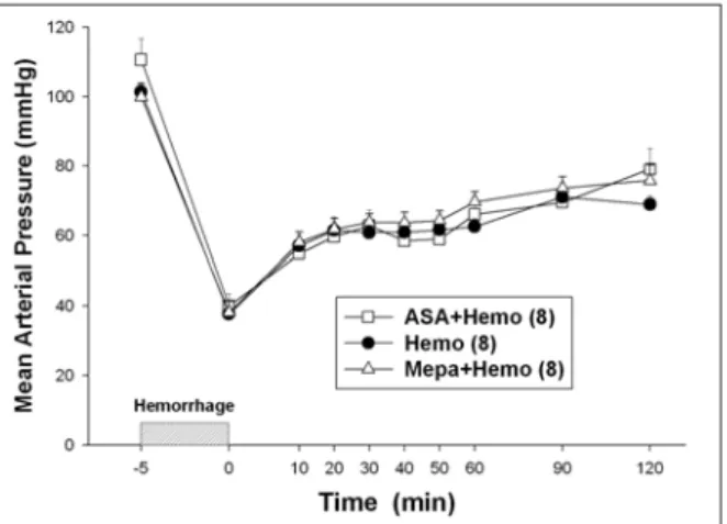
Figure 1.Changes in mean arterial pressure follo- wing hemorrhage. Each point is the mean±SEM for the number of determinations shown in parentheses.
Figure 2. Two hours after hemorhage, the hemo- rrhaged rats had increased bronchoalveolar lavage leukocyte numbers compared to sham-treated rats.
Pretreatment with mepacrine (Mepa) or aspirin (ASA) decreased the leukocyte counts. The data shown is the mean±SEM for the number of determinations shown in parentheses.
**p<0.01, ***p<0.001 compared to Sham.
##p<0.01, ###p<0.001 compared to Hemo.
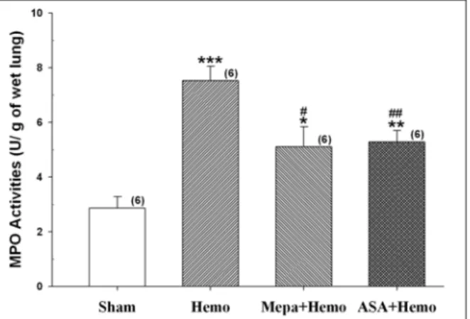
폐장내 myeloperoxidase 활성도의 측정
호중구의 폐장내 침윤 정도를 알아보기 위하여 Goldblum 등20의 방법에 따라 myeloperoxidase (MPO) 활성도를 측정하였다. 좌측폐장을 제거한 후 MPO 측정전까지 -70℃에 보관하였다가 MPO 측정 시 4.0 ml의 potassium phosphate 완충용액(20mM, pH7.4)에 조직을 녹이고 Polytron PT 1200 homoge- nizer (Kinematica AG, Switzerland)를 이용하여 조 직을 분쇄한 뒤 4℃에서 18,000 rpm (Beckman, J2- M1 high speed centrifuge, USA)으로 30분간 원심분 리하였다. 상등액을 버리고 침전물을 4.0 ml의 0.5%
hexadecyltrimethylammonium bromide (50 mM po- tassium phosphate, pH 6.0)에 다시 부유시킨 후 90초 간 sonication 하였다. 이후 60℃의 항온수조에서 2시 간 반응시켜 조직내 MPO 분해효소를 불활성화 시키 고 폐장 분쇄액을 4℃, 20,000 rpm에서 2분간 원심분 리하여 상등액을 분리하였다. 상등액 0.1 ml를 취하 여 o-dianisidine (0.168 g)이 함유된 과산화수소 용액 (500 μM) 2.9 ml와 반응시키고 시간의 변화에 따른 흡광도의 변화를 측정하여 기울기를 구하고 여기에 13.5를 곱한 값에 폐장의 무게로 나누어 MPO 활성도 (단위: U/g of wet lung)를 계산하였다.
통계 처리
모든 실험결과는 평균±표준오차로 나타내었고, 각
man-Keuls multiple comparison test를 이용하여 p 값이 0.05보다 작은 경우를 통계적으로 유의하다고 판정하였다.
결 과
1. 출혈 후 평균동맥압의 변화
평균동맥압 (mean arterial pressure, MAP)은 출혈 군 (Hemo)은 출혈 전 101±3 mmHg에서 출혈 후 38±
2 mmHg로 감소되었다. Mepacrine 처치군 (Mepa+
Hemo)은 출혈 전 100±4 mmHg에서 출혈 후 38±3 mmHg로, 아스피린 처치군 (ASA+Hemo)은 출혈 전 111±6 mmHg에서 출혈 후 40±3 mmHg로 감소되었 는데 각 군 간에 유의한 차이는 없었다. 평균동맥압은 출혈 후 점차적으로 회복되었으며 출혈 2시간 후 출 혈군은 69±2 mmHg, mepacrine 처치군은 76±4 mmHg, 아스피린 처치군은 79±6 mmHg로 회복되는 정도도 각 군이 비슷하였다 (Figure 1).
2. 아스피린이 급성 폐손상에 미치는 효과
출혈 후 급성 폐손상을 확인하기 위하여 폐세척액 내 백혈구수를 측정한 결과 출혈을 시키지 않은 대조 군(Sham)의 0.50±0.05 (millions/two lungs)에 비해 출혈군은 1.10±0.15 (millions/two lungs)로 증가하였
Figure 3.Rats subjected to hemorrhage had increa- sed lung tissue myeloperoxidase (MPO) activities compared to Sham rats. These changes were atte- nuated by pretreatment with mepacrine (Mepa) or aspirin (ASA). The data shown is the mean±SEM for the number of determinations shown in parentheses.
*p<0.05, **p<0.01, ***p<0.001 compared to Sham.
#p<0.05, ##p<0.01 compared to Hemo.
Figure 4.Two hours after hemorhage, the hemo- rrhaged rats had increased bronchoalveolar lavage protein concentrations compared to sham-treated rats. These changes were prevented by pretreatment with mepacrine (Mepa) or aspirin (ASA). The data shown is the mean±SEM for the number of determi- nations shown in parentheses.
***p<0.001 compared to Sham.
###p<0.001 compared to Hemo.
Figure 5.Rats subjected to hemorrhage had increa- sed bronchoalveolar lavage ferritin concentrations compared to sham-treated rats. Pretreatment with mepacrine (Mepa) or aspirin (ASA) decreased the increase of lavage ferritin concentrations. The data shown is the mean±SEM for the number of determi- nations shown in parentheses.
*p<0.05 compared to Sham.
#p<0.05, ##p<0.01 compared to Hemo.
다 (p<0.001, Figure 2). Mepacrine 처치군과 아스피 린 처치군은 각각 0.20±0.02, 0.47±0.04 (millions/two lungs)로 출혈군에 비해 백혈구 증가가 유의하게 억 제되었다. 폐장 조직내 호중구 침윤정도를 알아보기 위하여 측정한 MPO 활성도의 결과도 유사한 결과를 얻었는데, 출혈군은 7.52±0.52 (U/g of wet lung)로
대조군의 2.85±0.43 (U/g of wet lung) 보다 증가하였 다 (p<0.001, Figure 3). Mepacrine 처치군과 아스피 린 처치군은 대조군보다는 증가하였으나 각각 5.10±
0.73, 5.28±0.41 (U/g of wet lung)로 출혈군에 비해 유의하게 감소하였다.
출혈 후 2시간에서 폐세척액 내 전체 단백함량을 측정하였다 (Figure 4). 출혈군은 129±10 (㎍/ml)으로 대조군의 56±5 (㎍/ml) 보다 크게 증가하였다 (p<
0.001). Mepacrine 처치군과 아스피린 처치군은 각각 55±4, 59±2(㎍/ml)로 대조군에 비해 차이가 없었으 며, 출혈군에 비해서는 단백함량 증가가 억제되었다 (p<0.001).
3. 출혈 후 폐세척액 및 혈장 ferritin의 변화 그림 5는 폐세척액의 ferritin 농도를 나타낸 것이 다. 출혈시키지 않은 대조군에서는 ferritin 농도가 75±3 (ng/ml)이었는데 출혈군은 154±30 (ng/ml)으로 두 배 이상 증가하였다 (p<0.05). 이는 mepacrine 처 치(75±4 ng/ml)와 아스피린 처치(52±8 ng/ml)로 완 전히 차단되었다. 출혈 전 채혈한 혈액의 ferritin 농 도는 각 군간의 유의한 차이는 없었으며, 대조군은 2 시간 동안 유의한 변화가 나타나지 않았다 (Figure 6). 출혈 군은 출혈 전 55±6 (ng/ml)에 비해 출혈 30
Figure 6.The serum ferritin concentrations of Sham rats did not change through the 2-hour period fo- llowing hemorrhage. In contrast, the serum ferritin concentrations of hemorrhaged rats increased prog- ressively following hemorrhage. By comparison, the serum ferritin concentrations of mepacrine or aspi- rin-treated hemorrhaged rats were decreased compa- red to the values of hemorrhaged rats without pre- treatment. Each point is the mean±SEM for the num- ber of determinations shown in parentheses.
*p<0.05, **p<0.01, ***p<0.001 compared to their pre-hemorrhage baseline values.
분 후부터 유의하게 증가하여(59±4 ng/ml) 2시간 후 에는 출혈 전의 3배 이상으로 증가하였다(173±19 ng/ml). Mepacrine 처치군과 아스피린 처치군은 혈 장 ferritin이 출혈 90분 후부터 출혈 전에 비해 유의 하게 증가하는 양상을 보였으나, 출혈군에 비해서는 증가하는 정도가 크게 둔화되었으며 mepacrine 보다 는 아스피린 처치시 더욱 억제되었다.
고 찰
본 실험을 통하여 출혈성 쇼크 후 폐세척액내 백혈 구 수와 단백질 함량 및 폐장 MPO 활성도가 증가하 여 급성 폐손상이 나타났음을 알 수 있었다. 급성 폐 손상을 나타내주는 이런 결과들은 mepacrine과 아스 피린 전처치로 효과적으로 억제되었다. 이는 이미 보 고된 결과들과 일치한다15,21. 출혈성 쇼크는 급성 폐 손상을 유발하는 주요 원인중의 하나인데22,23, cyto- kine의 발현을 촉진하고 다양한 조직, 특히 간과 폐장 에 호중구를 침윤시켜 염증성 반응 및 장기손상을 유
인하여 혈관내 호중구 침윤과 호중구 막의 과산화수 소 형성이 크게 증가하였는데, 이와 같은 산화성스트 레스가 조직손상의 주된 원인이라고 하였다. 이 결과 와 더불어 본 실험에서 주목할 만 한 내용은 폐세척 액과 혈장의 ferritin 농도가 출혈 후 크게 증가하였으 며, 급성 폐손상을 억제할 수 있는 전처치를 한 경우 에는 ferritin 증가가 의미있게 둔화되었다는 사실이 다. 본 실험을 통한 결과로 보면 출혈 후 1시간 이내 에 혈장 ferritin이 증가하므로 ARDS 발병 가능성이 높은 환자를 대상으로 하는 조사를 해 볼 필요가 있 다고 생각한다.
ARDS는 최근까지의 연구로도 확실한 발병기전과 치료법이 개발되지 않아 사망율이 매우 높은 질환이 다25. 그간 ARDS의 치료를 위해 많은 연구가 있어왔 지만 아직 제대로 성공한 예는 보고되지 않았다. 그 이유는 발병원인이 다양한 데에도 있지만 조기진단 을 하기 어렵기 때문이라고 생각하고 있다26. ARDS 는 손상이 일어난 후 24시간 이내에 대부분 발병하고, 전체적으로는 72시간 내에 발병한다고 한다23,27,28. 그 러므로 정확한 조기진단이 ARDS를 막을 수 있는 좋 은 기회를 제공해 줄 수 있을 것으로 생각하고 많은 연구가 진행되고 있다26.
최근 몇 건의 연구에서 혈장 ferritin치가 급성 폐손 상의 조기진단에 가능성있는 표지인자로 활용할 수 있을 것이라는 보고가 있었다. ARDS 환자의 혈장 ferritin 농도는 정상인이나 ARDS 위험은 있지만 발 병하지 않은 사람의 혈장 ferritin 농도보다 매우 높게 나타났다고 하였다8,9. Park 등7은 동물실험을 통해서 도 동일한 결과를 얻었는데, 본 실험에서 사용한 것과 동일한 방법으로 mepacrine을 전 처치한 후 출혈을 시켰을 경우에는 급성 폐손상이 억제되었으며 폐세 척액 및 혈장 ferritin 농도의 증가도 둔화되었는데, 본 실험에서도 동일한 결과를 얻었다. 이 결과의 의미 는 mepacrine이나 아스피린으로 급성 폐손상의 발생 을 차단한 경우에는 이런 현상이 차단 또는 억제되므 로 급성 폐손상의 조기진단에 유용하게 사용될 수 있 다는 것을 의미한다.
생체내에는 활성산소기를 제거할 수 있는 많은 종
성스트레스를 유발하는데 유리된 활성 철이온은 이 를 더욱 악화시킨다29,30. 실험적으로도 세포내 철을 고갈시키면 산화성스트레스로부터 보호된다고 보고
하였다31,32. Park 등6의 연구결과에 의하면 철이 결핍
된 식이를 한 쥐에서는 본 실험과 동일한 정도의 출 혈을 시켰을 때 혈장 ferritin의 증가도 없었고 급성 폐손상이 억제되어서 체내 활성 철이온이 급성 폐손 상을 유발 또는 악화시킬 수 있는 한 요인이 된다고 보고하였다. 이런 반응은 transferrin 유전자가 결핍 된 마우스에서 산화성스트레스에 의한 폐손상이 줄 어들었다는 보고와도 일치한다33.
혈장 ferritin은 염증반응 또는 감염에 의해 acute- phase 단백질의 일종으로 혈장 ferritin치가 증가할 수 있는 제한점이 있지만34-36 대부분의 경우에서 체내 철 함량을 잘 반영해 주는 지표로 사용되고 있다37,38. 본 실험에서 혈중 ferritin 농도는 출혈 후 30분부터 증가하기 시작하여 2시간에는 출혈 전보다 3 배 이상 증가하였다. 이는 heme에 노출시킨 혈관내피세포에 서 ferritin 생성이 증가하는데 1-2 시간 정도 걸린다 는 보고4와도 일치한다. 출혈 후에는 간질액의 유입 등으로 혈장 단백질의 농도가 감소되는 희석효과가 나타난다7,39. 그럼에도 불구하고 혈장 ferritin치는 크 게 증가하였으므로 실제 ferritin 증가는 희석효과를 고려하면 더 많이 증가했다고 생각할 수 있다. 혈중 ferritin은 산화성 스트레스에 의한 세포손상의 결과 로 증가할 수 있다40. 출혈성 쇼크에서는 호중구에서 급속하게 활성산소가 만들어지고41-44 혈관내피세포 를 손상시켜서 조직의 ferritin이 유리될 수 있으며45, 다른 기전으로는 염증성 전구물질인 TNF-α와 IL-1 등이 증가하는데 이로인해서도 생체 보호기전의 일 종으로 ferritin 생성이 증가할 수 있다고 한다34,46.
출혈 후 폐세척액 내 단백질 및 ferritin 농도도 증 가하였는데, 폐세척액내 단백질은 혈관 상피세포의 손상으로 혈장 단백질이 폐포내로 누출된 것이다47. Park 등7의 보고에 따르면, 폐세척액내 전체 단백질 에 대한 ferritin의 비는 혈액보다 훨씬 높으며 폐세척 액내 ferritin 농도와 혈중 ferritin 농도 사이에는 상관 관계가 없다고 한다. ARDS 환자와 같이 폐에 급성 염증이 생기면 폐세척액 내에 TNF-α나 IL-1β와 같
은 cytokine이 증가하는데48-50, 이로 인해서도 ferritin 이 증가할 수 있다고 한다51. 그러므로 폐세척액의 fe- rritin은 폐에서 분비된 것이거나 손상된 조직에서 유 리된 것으로 생각된다.
이와 같은 관점에서 본다면 mepacrine과 아스피린 전처치시 혈중 ferritin의 증가가 둔화된 것은 급성 폐 손상 및 염증반응이 억제되었기 때문으로 생각할 수 있으며, 이는 혈중 ferritin 농도와 손상정도가 일치한 다는 임상결과8,9와도 일치한다.
최근 Pham 등10은 염증성 반응으로 증가하는 NF- κB의 항산화효과는 ferritin의 한 구성요소인 heavy chain의 증가를 통해서 나타난다고 하여 ferritin의 항 염증 작용을 보고하였다. 많은 실험결과들이 산화성 스트레스로 증가한 ferritin이 철이온과 결합하여 항 산화 작용을 나타내는 효과가 있다고 하지만 다른 면 도 고려할 필요가 있다. 일반적으로는 ferritin과 결합 한 철이온이 잘 유리되지 않지만 ARDS에 흔히 동반 되는 대사성 산증에서는 철이온이 유리될 수 있다고 한다52. 본 실험과 같은 출혈성 쇼크 후에도 ferritin에 서 철이온이 유리될 수 있으므로 ferritin의 정확한 효 과는 이 실험만으로는 단언하기 어렵고 추가적인 실 험으로 규명해야 할 점이라고 생각한다.
ARDS 환자에서는 파괴된 적혈구에서 유리된 혈 색소가 활성 산소기 형성에 관여하는 철이온의 주요 공급원이 될 수 있다45. 이것은 철이온을 제거시키는 deferoxamine을 처리하면 혈관 내피세포 손상을 방 지할 수 있었다는 여러 보고53,54가 있으므로 출혈 후 증가된 ferritin이 철 이온을 제거하여 세포손상 방지 에 도움을 줄 수 있을 것으로 여겨진다.
본 실험에서는 mepacrine과 아스피린 전처치시 항 산화효과를 나타내는 ferritin 증가가 억제되었음에도 불구하고 폐손상이 억제되었는데, ferritin 증가가 항 산화 작용을 나타내는 일부분이며 철이온을 흡수할 수 있는 다른 요소들도 보호작용을 나타낸다는 Orino 등55의 보고를 고려하면 이해될 수 있다.
추가적으로 이 실험의 결과에서 발견한 특이한 점 은 세포를 이용한 in vitro 실험에서는 아스피린이 ferritin 생성을 증가시키지만18,56, 쥐를 대상으로 한 본 실험에서는 오히려 ferritin 증가가 억제되었는데
이는 시간적인 차이 때문인지 다른 조절기전이 복합 적으로 작용된 결과인지는 이 결과만으로는 알 수 없 다. 한편 활성 철 이온의 제거로 세포보호 작용을 가 지는 ferritin 증가가 차단된다면 오히려 역효과를 가 져올 수도 있다고 생각할 수 있다. 이런 의문점들을 해결하기 위해서는 다양한 실험조건에서 더 많은 연 구가 필요할 것으로 생각된다.
이상의 결과를 종합하면 출혈성 쇼크에 의해 급성 폐손상이 유발되며, 폐세척액 및 혈장 ferritin은 급성 폐손상 정도와 비례해서 증가하였다. 그리고 급성 폐 손상을 억제시키는 효과를 가진 아스피린과 mepac- rine으로 전처치를 한 경우에는 ferritin 증가가 억제 되었다. 그러므로 ARDS가 유발될 가능성이 있는 환 자의 조기진단 목적으로 혈장 및 폐세척액의 ferritin 치의 변화를 활용할 수 있을 것으로 여겨지며 이를 위해서 더 많은 연구가 필요할 것으로 생각된다.
요 약
연구배경 :
아스피린이 출혈성 쇼크시 나타나는 급성 폐손상 에 미치는 효과와 혈장 및 폐세척액 내 ferritin 농도 변화를 알아보고자 본 연구를 시행하였다.
방 법 :
실험동물은 체중 350g 내외의 Sprague-Dawley 종 흰쥐를 사용하였고, 혈압측정 및 출혈을 시키기 위하 여 catheter를 양쪽 대퇴동맥에 삽입하였다. 수술 후 polygraph를 이용하여 평균동맥압을 기록하였으며, 출혈은 withdrawal pump를 이용하여 5분간 체중 kg 당 20 ml의 혈액을 출혈시켰다.
실험군은 대조군, 출혈군과 아스피린 처치군으로 분류하였다. 대조군은 출혈군과 동일하게 수술하고 출혈은 시키지 않았으며 나머지 과정은 출혈군과 동 일하게 처리하였다. 아스피린 처치군은 출혈 30분 전 대퇴정맥으로 아스피린(10mg/kg)을 주입하였고, 출 혈군은 체중 당 동일한 양의 생리식염수를 주입하였다.
출혈 2시간 후의 폐손상 정도와 아스피린이 이에 미치는 효과를 알아보기 위하여 폐장내 myelopero-
수 및 혈장 ferritin 농도와 폐세척액 내 ferritin 농도 를 측정하였다.
결 과 :
폐장내 myeloperoxidase 활성도와 폐세척액 내의 단백함량과 백혈구수는 출혈 후 대조군에 비해 유의 하게 증가하였다. 이러한 반응은 아스피린 전처치에 의하여 효과적으로 차단되었다. 혈장 및 폐세척액 내 ferritin 농도는 출혈 후 크게 증가하였는데, 아스피린 전처치로 반응이 억제되었다.
결 론 :
심한 출혈 후에 생기는 급성 폐손상은 아스피린 전 처치로 효과적으로 예방될 수 있으며, 출혈 후 증가하 는 혈장 및 폐세척액 내 ferritin 농도는 급성폐손상이 나타날 수 있는 환자에서 조기진단을 위한 생체지표 로 활용될 수 있다고 생각된다.
참 고 문 헌
1. Harrison PM, Arosio P. The ferritins: molecular pro- perties, iron storage function and cellular regulation.
Biochim Biophys Acta 1996;1275:161-203.
2. Torti FM, Torti SV. Regulation of ferritin genes and protein. Blood 2002;99:3505-16.
3. Arosio P, Levi S. Ferritin, iron homeostasis, and oxi- dative damage. Free Radic Biol Med 2002;33:457-63.
4. Balla G, Jacob HS, Balla J, Rosenberg M, Nath K, Apple F, et al. Ferritin: a cytoprotective antioxidant strategem of endothelium. J Biol Chem 1992;267:
18148-53.
5. Hybertson BM, Connelly KG, Buser RT, Repine JE.
Ferritin and desferrioxamine attenuate xanthine oxi- dase-dependent leak in isolated perfused rat lungs.
Inflammation 2002;26:153-9.
6. Park YY, Hybertson BM, Wright RM, Repine JE.
Serum ferritin increases in hemorrhaged rats that develop acute lung injury: effect of an iron-deficient diet. Inflammation 2003;27:257-63.
7. Park YY, Hybertson BM, Wright RM, Fini MA, El- kins N, Repine JE. Serum ferritin elevation and acu- te lung injury in rats subjected to hemorrhage: redu- ction by mepacrine treatment. Exp Lung Res 2004;
30:571-84.
8. Connelly KG, Moss M, Parsons PE, Moore EE, Moore FA, Giclas PC, et al. Serum ferritin as a predictor of the acute respiratory distress syndrome. Am J Respir
9. Sharkey RA, Donnelly SC, Connelly KG, Robertson CE, Haslett C, Repine JE. Initial serum ferritin levels in patients with multiple trauma and the subsequent development of acute respiratory distress syndrome.
Am J Respir Crit Care Med 1999;159:1506-9.
10. Pham CG, Bubici C, Zazzeroni F, Papa S, Jones J, Alvarez K, et al. Ferritin heavy chain upregulation by NF-kappaB inhibits TNFalpha-induced apoptosis by suppressing reactive oxygen species. Cell 2004;119:
529-42.
11. Podhaisky HP, Abate A, Polte T, Oberle S, Schroder H. Aspirin protects endothelial cells from oxidative stress: possible synergism with vitamin E. FEBS Lett 1997;417:349-51.
12. Wahn H, Hammerschmidt S. Inhibition of PMN- and HOCl-induced vascular injury in isolated rabbit lungs by acetylsalicylic acid: a possible link between neutro- phil-derived oxidative stress and eicosanoid metabo- lism? Biochim Biophys Acta 1998;1408:55-66.
13. Carpenter-Deyo L, Roth RA. Cyclooxygenase inhibi- tion in lungs or in neutrophils attenuates neutro- phil-dependent edema in rat lungs perfused with phorbol myristate acetate. J Pharmacol Exp Ther 1989;251:983-91.
14. Zanaboni PB, Bradley JD, Baudendistel LJ, Webster RO, Dahms TE. Cyclooxygenase inhibition prevents PMA-induced increases in lung vascular permeability.
J Appl Physiol 1990;69:1494-501.
15. Shin TR, Lee DU, Park YY. Aspirin reduces acute lung injury in rats subjected to severe hemorrhage.
Tuberc Respir Dis 2003;54:522-31.
16. Kopp E, Ghosh S. Inhibition of NF-kappa B by sodi- um salicylate and aspirin. Science 1994;265:956-9.
17. Yin MJ, Yamamoto Y, Gaynor RB. The anti-inflam- matory agents aspirin and salicylate inhibit the acti- vity of I(kappa)B kinase-beta. Nature 1998;396:77-80.
18. Eisele G, Schwedhelm E, Schieffer B, Tsikas D, Boger RH. Acetylsalicylic acid inhibits monocyte adhesion to endothelial cells by an antioxidative mechanism. J Cardiovasc Pharmacol 2004;43:514-21.
19. Brown RE, Jarvis KL, Hyland KJ. Protein measure- ment using bicinchoninic acid: elimination of interfe- ring substance. Anal Biochem 1989;180:136-9.
20. Goldblum SE, Wu KM, Jay M. Lung myeloperoxidase as a measure of leukostasis in rabbit. J Appl Physiol 1985;59:1978-85.
21. Jang YS, Kim SE, Jheon SH, Shin TR, Lee YM.
Phospholipase A2 contributes to hemorrhage-induced acute lung injury through neutrophilic respiratory burst. Tuberc Respir Dis 2001;51:503-16.
22. Repine JE. Scientific perspectives on adult respirato- ry distress syndrome. Lancet 1992;339:466-9.
23. Hudson LD, Milberg JA, Anardi D, Maunder RJ. Cli- nical risks for the development of the acute respira- tory distress syndrome. Am J Respir Crit Care Med 1995;151:293-301.
24. Hierholzer C, Harbrecht B, Menezes JM, Kane J, MacMicking J, Nathan CF, et al. Essential role of in- duced nitric oxide in the initiation of the inflamma- tory response after hemorrhagic shock. J Exp Med 1998;187:917-28.
25. Ware LB, Matthay MA. The acute respiratory distre- ss syndrome. N Engl J Med 2000;342:1334-49.
26. Connelly KG, Repine JE. Markers for predicting the development of acute respiratory distress syndrome.
Annu Rev Med 1997;48:429-45.
27. Pepe PE, Potkin RT, Reus DH, Hudson LD, Carrico CJ. Clinical predictors of the adult respiratory distre- ss syndrome. Am J Surg 1982;144:124-30.
28. Fowler AA, Hamman RF, Good JT, Benson KN, Baird M, Eberle DJ, et al. Adult respiratory distress syn- drome: risk with common predispositions. Ann Intern Med 1983;98:593-7.
29. Halliwell B, Gutteridge JM. Oxygen toxicity, oxygen radicals, transition metals and disease. Biochem J 1984;219:1-14.
30. Weiss SJ. Tissue destruction by neutrophils. N Engl J Med 1989;320:365-76.
31. Gannon DE, Varani J, Phan SH, Ward JH, Kaplan J, Till GO, et al. Source of iron in neutrophil-mediated killing of endothelial cells. Lab Invest 1987;57:37-44.
32. Schraufstatter I, Hyslop PA, Jackson JH, Cochrane CG. Oxidant-induced DNA damage of target cells. J Clin Invest 1988;82:1040-50.
33. Yang F, Coalson JJ, Bobb HH, Carter JD, Banu J, Ghio AJ. Resistance of hypotransferrinemic mice to hyperoxia-induced lung injury. Am J Physiol 1999;
277:L1214-23.
34. Rogers JT, Bridges KR, Durmowicz GP, Glass J, Auron PE, Munro HN. Translational control during the acute phase response: ferritin synthesis in respon- se to interleukin-1. J Biol Chem 1990;265:14572-8.
35. Tran TN, Eubanks SK, Schaffer KJ, Zhou CY, Linder MC. Secretion of ferritin by rat hepatoma cells and its regulation by inflammatory cytokines and iron.
Blood 1997;90:4979-86.
36. Gabay C, Kushner I. Acute-phase proteins and other systemic responses to inflammation. N Engl J Med 1999;340:448-54.
37. Cook JD, Lipschitz DA, Miles LE, Finch CA. Serum ferritin as a measure of iron stores in normal su- bjects. Am J Clin Nutr 1974;27:681-7.
38. Walters GO, Miller FM, Worwood M. Serum ferritin concentration and iron stores in normal subjects. J
Clin Pathol 1973;26:770-2.
39. Zweifach BW. Mechanisms of blood flow and fluid exchange in microvessels: hemorrhagic hypotension model. Anesthesiology 1974;41:157-68.
40. Jacobs A, Worwood M. Ferritin in serum: clinical and biochemical implications. N Engl J Med 1975;292:
951-6.
41. McCord JM. Oxygen-derived free radicals in post- ischemic tissue injury. N Engl J Med 1985;312:159- 63.
42. Anderson BO, Moore EE, Moore FA, Leff JA, Terada LS, Harken AH, et al. Hypovolemic shock promotes neutrophil sequestration in lungs by a xanthine oxi- dase-related mechanism. J Appl Physiol 1991;71:
1862-5.
43. Tan S, Yokoyama Y, Dickens E, Cash TG, Freeman BA, Parks DA. Xanthine oxidase activity in the cir- culation of rats following hemorrhagic shock. Free Radic Biol Med 1993;15:407-14.
44. Schwartz MD, Repine JE, Abraham E. Xanthine oxi- dase-derived oxygen radicals increase lung cytokine expression in mice subjected to hemorrhagic shock.
Am J Respir Cell Mol Biol 1995;12:434-40.
45. Balla J, Nath KA, Balla G, Juckett MB, Jacob HS, Vercellotti GM. Endothelial cell heme oxygenase and ferritin induction in rat lung by hemoglobin in vivo.
Am J Physiol 1995;268:L321-7.
46. Kwak EL, Larochelle DA, Beaumont C, Torti SV, Torti FM. Role for NF-kappa B in the regulation of ferritin H by tumor necrosis factor-alpha. J Biol Chem 1995;270:15285-93.
47. Holter JF, Weiland JE, Pacht ER, Gadek JE, Davis WB. Protein permeability in the adult respiratory distress syndrome: loss of size selectivity of the alve- olar epithelium. J Clin Invest 1986;78:1513-22.
48. Suter PM, Suter S, Girardin E, Roux-Lombard P,
Grau GE, Dayer JM. High bronchoalveolar levels of tumor necrosis factor and its inhibitors, interleukin-1, interferon, and elastase, in patients with adult respi- ratory distress syndrome after trauma, shock, or sep- sis. Am Rev Respir Dis 1992;145:1016-22.
49. Shenkar R, Abraham E. Mechanisms of lung neutro- phil activation after hemorrhage or endotoxemia: ro- les of reactive oxygen intermediates, NF-kappa B, and cyclic AMP response element binding protein. J Immunol 1999;163:954-62.
50. Abraham E, Carmody A, Shenkar R, Arcaroli J. Neu- trophils as early immunologic effectors in hemorrha- ge- or endotoxemia-induced acute lung injury. Am J Physiol Lung Cell Mol Physiol 2000;279:L1137-45.
51. Tsuji Y, Miller LL, Miller SC, Torti SV, Torti FM.
Tumor necrosis factor-alpha and interleukin 1-alpha regulate transferrin receptor in human diploid fibrob- lasts: relationship to the induction of ferritin heavy chain. J Biol Chem 1991;266:7257-61.
52. Reif DW. Ferritin as a source of iron for oxidative damage. Free Radic Biol Med 1992;12:417-27.
53. Balla G, Vercellotti GM, Eaton JW, Jacob HS. Iron loading of endothelial cells augments oxidant damage.
J Lab Clin Med 1990;116:546-54.
54. Olakanmi O, McGowan SE, Hayek MB, Britigan BE.
Iron sequestration by macrophages decreases the po- tential for extracellular hydroxyl radical formation. J Clin Invest 1993;91:889-99.
55. Orino K, Lehman L, Tsuji Y, Ayaki H, Torti SV, Torti FM. Ferritin and the response to oxidative stress.
Biochem J 2001;357:241-7.
56. Oberle S, Polte T, Abate A, Podhaisky HP, Schroder H. Aspirin increases ferritin synthesis in endothelial cells: a novel antioxidant pathway. Circ Res 1998;82:
1016-20.